


Introduction
Despite being relatively rare, tibial spine avulsion fractures mostly affect skeletally immature patients between the ages of 8 and 14 due to hyperextension or rotational injuries, often related to sports or road-traffic trauma [1]. An increasing incidence among adults has been reported, particularly after high-energy trauma, where concomitant collateral ligament or meniscal injuries may occur [2]. Untreated or inadequately managed displaced fractures, particularly Type III and Type IV injuries, may lead to non-union, malunion, flexion deformity, and persistent knee instability [3].
Tibial spine avulsions were first described by Poncet in 1875 and were subsequently classified by Meyers and McKeever in 1959, with modification by Zaricznyj in 1987 [4]. Magnetic resonance imaging (MRI) provides detailed evaluation of associated ligamentous, meniscal, and chondral injuries [5]. Management depends on displacement and reducibility; conservative treatment is generally reserved for minimally displaced fractures, whereas displaced or unstable injuries require surgical fixation [6].
Traditional fixation options include cannulated screws, Herbert screws, K-wires, and suture anchors, each with specific advantages and limitations [7]. Suture pull-through techniques have gained popularity because they avoid implant prominence and hardware removal while allowing fixation through the anterior cruciate ligament (ACL) substance [7]. Published studies have reported favorable functional recovery and early mobilization with suture-based fixation [8]; however, the present study was not designed to compare fixation methods.
The present study evaluated arthroscopic ACL tibial spine avulsion fixation using a suture pull-through double-tunnel technique, with emphasis on functional outcomes, knee stability, radiological union, complications, and return to pre-injury activity.
Materials and Methods
Study design and setting
A prospective observational study was conducted in the Department of Orthopaedics, N.D. Desai Medical College, and included a consecutive sample of 26 eligible patients with tibial spine avulsion fractures.
Ethical approval: The manuscript records approval from Dr. N. D. Desai Faculty of Medical Science and Research Medical Centres, Nadiad (No. Dr NDDFMSR/IEC/2026/03/05; dated May 02, 2026). Written informed consent for participation and use of anonymized clinical/radiographic material should be confirmed in the final submitted version. The approval date must be reconciled with the verified study period before submission.
Patient selection
Inclusion criteria
-
Skeletally mature patients aged 18–60 years
-
Meyers and McKeever Type II, III, or IV fractures (modified by Zaricznyj)
-
Closed injuries suitable for arthroscopic fixation.
Exclusion criteria
-
Concomitant meniscal or chondral injury requiring additional treatment
-
Associated proximal tibial fracture
-
Previous surgery on the affected knee
-
Inadequate muscle power or inability to comply with rehabilitation.
Four patients had Type II, 19 had Type III, and three had Type IV injuries. The mean interval from injury to fixation was approximately 1 week. All patients underwent arthroscopic suture pull-through double-tunnel fixation.
Surgical technique
All procedures were performed under spinal anesthesia with the patient supine. A thigh support and pneumatic tourniquet were used, and standard anteromedial and anterolateral arthroscopic portals were established.
Pre-operative preparation
-
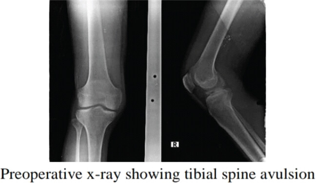
Plain radiographs confirmed the tibial spine avulsion fracture (Fig. 1)
-
MRI was used to define fracture morphology and screen for associated intra-articular injury
-
The joint was irrigated with normal saline to clear the hemarthrosis and loose debris.
Fracture reduction and fixation
-
Diagnostic arthroscopy: A 30° 4-mm arthroscope was introduced to assess the fracture and intra-articular structures
-
Fracture reduction: The fragment was reduced with the knee at approximately 40° of flexion and maintained with a probe or temporary K-wire when required
-
Suture passage: A 45° suture lasso carrying a steel wire was passed through the anteromedial portal near the ACL tibial insertion. The shuttle wire was retrieved and used to pass a non-absorbable FiberTape loop through the ACL fibers
-
Tunnel creation: Through a 3-cm oblique anteromedial tibial incision, a tibial tunnel guide set at 55° was positioned. A 4-mm drill pin was used to create anteromedial and anterolateral tibial tunnels under arthroscopic guidance
-
Final fixation: A flexible wire loop was passed through each tunnel. The suture limbs were crossed through the opposite tunnels to create a crisscross construct; reduction was confirmed arthroscopically, and the sutures were tied over a 1-cm cortical bone bridge with the knee at approximately 30° of flexion.
Post-operative assessment and rehabilitation
-
Fixation stability was confirmed by arthroscopic visualization and intraoperative fluoroscopy
-
The knee was immobilized in an ACL brace in full extension for 3 weeks
-
Weeks 1–3: Isometric quadriceps exercises, straight-leg raises, and non-weight bearing
-
Weeks 4–6: Hinged brace, progressive range of motion, and gradual weight bearing
-
Full weight bearing was permitted by 8 weeks according to clinical progress
-
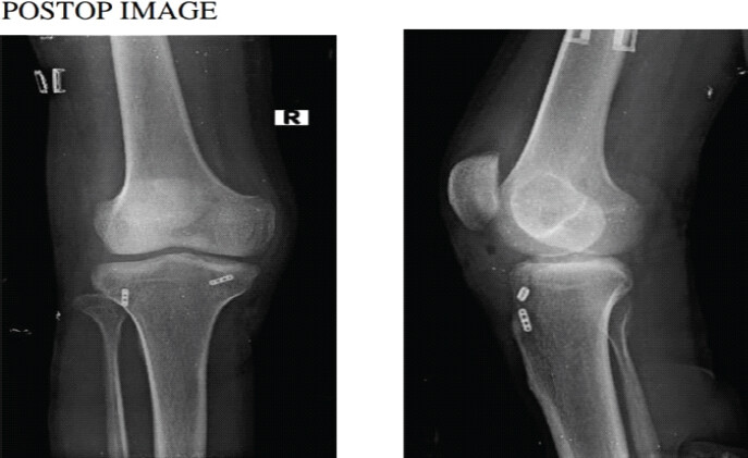
Radiographs at 3 months were used to assess union and alignment (Fig. 2).
Outcome measures and statistical analysis
The Lysholm Knee Score, Tegner Activity Scale, radiological union, return to activity, extension deficit, residual instability, fixation failure, and revision surgery were recorded. Continuous variables are presented as mean ± standard deviation and categorical variables as number and percentage. Patient-level pre-operative and post-operative Tegner scores were compared using the paired Wilcoxon signed-rank test. A two-sided P < 0.05 was considered statistically significant. Individual post-operative Lysholm scores were not available; therefore, a paired statistical test for Lysholm improvement could not be calculated. Post-operative knee stability was not quantified with KT-1000/KT-2000 arthrometry or another instrumented laxity measure, and standardized pivot-shift grading was not available. Radiographic assessment documented union and alignment, but quantitative measurements of residual displacement, articular step-off, or tunnel position were not available. Post-operative MRI assessment of ACL continuity or tension was not performed.
Results
The study included 26 patients (18 males and eight females) with a mean age of 30 years (range 18–60 years). Sports-related trauma accounted for 10 injuries and motorcycle accidents for 16 injuries (Table 1).
Fracture distribution was Type II in four patients, Type III in 19 patients, and Type IV in three patients (Table 1).
Patient demographics and fracture characteristics
| Parameter | Value |
|---|---|
| Total patients | 26 |
| Male patients | 18 |
| Female patients | 8 |
| Mean age | 30 years (range 18–60) |
| Injury mechanism | Sports: 10; motorcycle accidents: 16 |
| Fracture type | Type II: 4; type III: 19; type IV: 3 |
The patient-level pre-operative Lysholm score averaged 40.8 ± 2.2, while the reported post-operative mean was 97. Because individual post-operative Lysholm values were unavailable, the magnitude and statistical significance of paired Lysholm improvement could not be independently calculated. The Tegner score improved from 2.88 ± 0.77 to 5.96 ± 0.77 (Wilcoxon P < 0.001) (Tables 2 and 3).
Functional and radiological outcomes
| Outcome | Pre-operative | Post-operative/follow-up | P-value |
|---|---|---|---|
| Lysholm knee score | 40.8±2.2 | 97 | Not calculated: Individual post-operative Lysholm scores were unavailable |
| Tegner activity scale | 2.88±0.77 | 5.96±0.77 | <0.001 |
| Fracture union at 3 months | — | 26/26 (100%) | — |
| Return to pre-injury activity | — | 22/26 (approximately 85%) within 4–6 months | — |
Patient-level pre-operative Lysholm and Tegner activity scale scores
| Patient No. | Pre-operative Lysholm score | Pre-operative Tegner score | Post-operative Tegner score |
|---|---|---|---|
| 1 | 40 | 3 | 6 |
| 2 | 45 | 2 | 7 |
| 3 | 42 | 3 | 6 |
| 4 | 39 | 2 | 5 |
| 5 | 41 | 4 | 7 |
| 6 | 43 | 3 | 6 |
| 7 | 38 | 2 | 5 |
| 8 | 42 | 3 | 6 |
| 9 | 37 | 2 | 5 |
| 10 | 44 | 4 | 7 |
| 11 | 40 | 3 | 6 |
| 12 | 39 | 2 | 5 |
| 13 | 41 | 3 | 6 |
| 14 | 42 | 4 | 7 |
| 15 | 43 | 3 | 6 |
| 16 | 40 | 2 | 5 |
| 17 | 37 | 3 | 6 |
| 18 | 39 | 2 | 5 |
| 19 | 42 | 4 | 7 |
| 20 | 38 | 3 | 6 |
| 21 | 44 | 2 | 5 |
| 22 | 40 | 3 | 6 |
| 23 | 41 | 4 | 7 |
| 24 | 39 | 3 | 6 |
| 25 | 42 | 2 | 5 |
| 26 | 43 | 4 | 7 |
| Mean±SD | 40.81±2.19 | 2.88±0.77 | 5.96±0.77 |
SD: Standard deviation
All patients achieved radiological union by 3 months. Twenty-two of 26 patients (approximately 85%) were recorded as having returned to their pre-injury activity level within 4–6 months (Table 2). A validated return-to-sport or return-to-activity testing battery and prespecified objective criteria for successful return were not available; accordingly, this outcome should be interpreted descriptively.
Two patients developed a mild post-operative extension lag, which resolved with supervised physiotherapy. No patient required revision surgery, and no non-union, fixation failure, or clinically significant residual knee instability was reported during the available follow-up. Given the cohort size of 26 patients and the short follow-up, these observations do not exclude uncommon complications or later instability/reinjury.
Discussion
Tibial spine avulsion injuries, traditionally associated with adolescents, are increasingly recognised in adults after high-energy motor-vehicle and sports trauma [9]. Stable fixation is intended to restore knee stability and minimise functional deficit. Suture fixation has the practical advantage of avoiding implant prominence and possible secondary hardware removal, but the present uncontrolled study cannot establish superiority or equivalence compared with screw fixation, suture-anchor fixation, or other techniques [7].
Several fixation methods, including K-wires, screws, staples, suture anchors, and transosseous sutures, have been described. Suture-based methods have produced favorable outcomes in published series [10]. In the present cohort, all fractures united by 3 months, the reported post-operative Lysholm score was 97, and the Tegner Activity Scale improved significantly. These early findings are broadly consistent with reports of favorable functional recovery after suture fixation [11]. However, the lack of individual post-operative Lysholm values prevents paired statistical assessment of that score, and the absence of a comparator precludes conclusions about relative efficacy.
Two patients developed a transient extension lag that responded to supervised physiotherapy. Mild post-operative range-of-motion deficits have been reported after tibial spine fixation [12]. Prolonged immobilization and delayed rehabilitation may increase the risk of stiffness [1]. Although a rehabilitation schedule was prescribed in this study, adherence, physiotherapy intensity, and patient compliance were not objectively monitored; these factors may have influenced functional recovery.
Residual laxity, non-union, reinjury, and arthrofibrosis remain clinically relevant concerns [13]. Comparative reports have described favorable functional outcomes after suture fixation relative to screw fixation [14, 15], but the current study did not include a comparator and did not measure instrumented post-operative laxity, standardized pivot-shift grade, MRI-based ACL integrity, or quantitative radiographic reduction parameters. Therefore, the absence of clinically significant residual instability in this series should not be interpreted as proof of restored ACL biomechanics or long-term functional continuity.
Study limitations
This study has several important limitations. First, the sample size was small (26 patients), reducing statistical power and limiting the ability to identify uncommon complications or perform reliable subgroup analyses. Second, the single-Centre design restricts external validity. Third, there was no control or comparator group using screw fixation, suture anchors, or another established method; consequently, superiority, equivalence, or comparative safety of the double-tunnel technique cannot be inferred. Fourth, follow-up was limited to approximately 3–6 months, which is insufficient to evaluate recurrent instability, reinjury, late fixation-related problems, long-term ACL function, or post-traumatic osteoarthritis. Fifth, the manuscript contains a major chronology discrepancy between the stated study period and the documented 2026 ethics approval; this must be verified against institutional records before submission. Sixth, individual post-operative Lysholm scores were unavailable, preventing paired analysis of Lysholm improvement. Seventh, post-operative stability was not quantified using KT-1000/KT-2000 arthrometry or another instrumented laxity test, and standardized objective pivot-shift grading was not available. Eighth, post-operative imaging confirmed union and alignment but did not include quantitative measures of fragment displacement, articular step-off, or tibial tunnel position. Ninth, return to pre-injury activity was insufficiently defined and was not based on a validated objective return-to-sport testing protocol; moreover, only 22 of 26 patients returned to their previous activity level. Tenth, exclusion of patients with concomitant meniscal or chondral injuries requiring treatment creates selection bias and may underestimate functional limitations and complications encountered in routine practice. Eleventh, Meyers and McKeever Types II, III, and IV fractures were pooled without fracture-type-specific outcome analysis, despite the possibility that displaced and comminuted patterns respond differently. Twelfth, rehabilitation adherence, physiotherapy intensity, and patient compliance were not objectively monitored. Thirteenth, the cohort was underpowered to establish a low complication rate for uncommon events such as fixation failure, non-union, or significant instability. Fourteenth, post-operative MRI or another objective assessment of ACL continuity, tension, or healing was not performed. Finally, the observational design, short-term follow-up, and limited objective stability assessment mean that the clinical relevance should be interpreted cautiously. Larger multicenter comparative studies with longer follow-up, standardized imaging, instrumented laxity testing, fracture-type stratification, and validated return-to-activity criteria are required.
Conclusion
In this 26-patient single-Centre observational cohort, arthroscopic suture pull-through double-tunnel fixation was associated with radiological union and favorable early functional outcomes, and most patients returned to their recorded pre-injury activity level. These findings are preliminary because the study lacked a comparator, had short follow-up, and did not include instrumented laxity testing, quantitative imaging-based reduction assessment, or post-operative MRI evaluation of ACL integrity. Larger prospective comparative studies are required before the technique can be considered demonstrably superior, reliably low-risk, or durable over the long term.
Clinical Message
For displaced ACL tibial spine avulsion fractures in skeletally mature patients, the double-tunnel suture pull-through technique is a hardware-free fixation option that showed favorable early outcomes in this small cohort; its comparative efficacy and long-term stability remain to be established.
Conflict of Interest:
Nil
Source of Support:
Nil
Consent:
The authors confirm that informed consent was obtained from the patient for publication of this article
References
- Salvato D, Green DW, Accadbled F, Tuca M. Tibial spine fractures: State of the art J ISAKOS 2023;8:404-11. Google Scholar | PubMed
- Urreiztieta HC, Spence DB, Bankhead CP, Richter DL. Treatment strategies for concomitant ligament injury in anterior cruciate ligament-injured athletes, a narrative review Ann Joint 2025;10:41 doi:10.21037/aoj-25-28 Google Scholar | PubMed | CrossRef
- Talha H, Hamoutahra A. The management of tibial spine avulsion fracture in a skeletally immature patient by using staple fixation: A case report Cureus 2026;18:e102958. Google Scholar | PubMed
- Bell D, Worsley C. Anterior cruciate ligament avulsion fracture Radiopaedia.org 2021 [updated 2026 Apr 12; cited 2026 Aug 27] Available from: https://radiopaedia.org/articles/anterior-cruciate-ligament-avulsion-fracture doi:10.53347/rID-14304 Google Scholar | PubMed | CrossRef
- Stokes DJ, Sanchez RA, Williams BT, Strassman AK, Shinsako KK, DiFelice GS, et al. Tibial spine avulsion fracture fixation using a re-tensionable all-suture construct Arthrosc Tech 2024;13:102983. Google Scholar | PubMed
- Bondi M, Presicce S, Minuti N, Pizzoli A. Management of Tibial Spine Avulsion Fractures: From MRI-Based Classification to Arthroscopic Repair J Orthop Study Sports Med 2025;3 (1) 1-8 doi:10.52793/JOSSM.2025.3(1)-30 Google Scholar | PubMed | CrossRef
- Jain S, Modi P, Dayma RL, Mishra S. Clinical outcome of arthroscopic suture versus screw fixation in tibial avulsion of the anterior cruciate ligament in skeletally mature patients J Orthop 2023;35:7-12. Google Scholar | PubMed
- Elnewishy A, El Menawy Z, Zahed M, Elmesalmi M, Elnaggar N, Ahmed F. Suture fixation versus screw fixation in pediatric tibial eminence fractures: A systematic review and meta-analysis of clinical outcomes and reoperation rates Cureus 2025;17 12 e99853 doi:10.7759/cureus.99853 Google Scholar | PubMed | CrossRef
- Rivera Troia F, Perez Lopez CJ. Tibial spine avulsion fracture in an adult: An uncommon occurrence with surgical implications Cureus 2025;17:e83261. Google Scholar | PubMed
- Laddha M, Bhardwaj L, Farinelli L, Meena A. Good functional outcomes and radiological union following arthroscopic knotless anchor fixation of tibial spine avulsion fractures: A prospective clinical study J Exp Orthop 2026;13 1 e70692 doi:10.1002/jeo2.70692 Google Scholar | PubMed | CrossRef
- Chang CJ, Huang TC, Hoshino Y, Wang CH, Kuan FC, Su WR, et al. Functional outcomes and subsequent surgical procedures after arthroscopic suture versus screw fixation for ACL tibial avulsion fractures: A systematic review and meta-analysis Orthop J Sports Med 2022 Apr 6 10 (4) 23259671221085945 doi: 10.1177/232596712210→5 PMID: 35400137 Google Scholar | PubMed | CrossRef
- Jääskelä M, Turati M, Lempainen L, Bremond N, Courvoisier A, Henri A, et al. Long-term outcomes of tibial spine avulsion fractures after open reduction with osteosuturing versus arthroscopic screw fixation: A multicenter comparative study Orthop J Sports Med 2023;11 (6) 23259671231176991 doi:10.1177/23259671231176991 Google Scholar | PubMed | CrossRef
- Sapienza M, Torrisi P, Mirto F, Amico M, Russo A, Costa GG, et al. Management of type II tibial spine fractures in children and adolescents: a systematic review J Orthop Surg Res 2026;21 (1) 278 doi:10.1186/s13018-026-06797-1 Google Scholar | PubMed | CrossRef
- Shankar R, Sherawat R, Sengupta A, Verma N, Garg H, Buddhist H. Clinical outcome of arthroscopic reduction and fixation by pull through suture technique in tibial spine fractures Cureus 2025;17 3 e79902 doi:10.7759/cureus.79902 Google Scholar | PubMed | CrossRef
- Qu H, Meng QJ, Sun Q, Du D, Zhang Q. Arthroscopic fixation for tibial eminence fractures: A clinical retrospective study of cannulated screws versus transosseous anchor knot fixation techniques with suture anchors Knee 2022;35:105-113 doi:10.1016/j.knee.2022.02.002 Google Scholar | PubMed | CrossRef
© 2026 Journal of Orthopaedic Case Reports - Published by Indian Orthopaedic Research Group
About the Authors
-

Dr. Parth Jayeshkumar Patel
-

Dr. Harshlaxen Ashvinsinh Rajpardhi
-

Dr. Krishna Himmatbhai Goti
-

Dr. Vinit Jayeshkumar Panchiwala
-

Dr. Mohammadshahil Vahora
-

Dr. Hayati Bharatbhai Visani
Related Articles in Journal of Orthopaedic Case Reports
 September 1, 2026 Fixed Flexion Deformity due to Displaced Hoffa Fracture: A Case Report
 September 1, 2026 Comparing Bridge-Enhanced Anterior Cruciate Ligament Repair to Standard Autograft for Anterior Cruciate Ligament Reconstruction: Biomechanical and Clinical Outcomes
 September 1, 2026 Comparative Analysis of Functional Outcomes of Accelerated versus Conventional Rehabilitation after Anterior Cruciate Ligament Reconstruction Surgery
 September 1, 2026 Bridge-Enhanced Anterior Cruciate Ligament Repair versus Autograft Reconstruction in Anterior Cruciate Ligament Surgery