[box type=”bio”] What to Learn from this Article?[/box]
Double plating is a viable option to adequately stabilize long standing diaphyseal humeral nonunion with extensive osteolysis and cortical resorption following plate fixation.
Case Report | Volume 5 | Issue 4 | JOCR Oct-Dec 2015 | Page 50-53| Sreenivasulu Metikala, Prabhudheer Bhogadi. DOI: 10.13107/jocr.2250-0685.345 .
Authors: Sreenivasulu Metikala[1], Prabhudheer Bhogadi[2]
[1] Orthopaedic trauma, Sri Venkateswara Orthopaedic Hospital, 1/100 & 1/101, George Reddy Street, Yerramukkapalli, Kadapa, A.P., India – 516004.
[2] Department of Orthopaedics and Traumatology, Osmania Medical College and General Hospital, Hyderabad, Telangana, India – 500012
Address of Correspondence
Dr. Sreenivasulu Metikala,
Sri Venkateswara Orthopaedic Hospital, 1/100 & 1/101, George Reddy Street, Yerramukkapalli, Kadapa, A.P. India – 516004.
E mail – orthoseenu@yahoo.com
Abstract
Introduction: Nonunion following surgical stabilization of humeral shaft fractures, although infrequent, remains a challenge as limited surgical options are available. The difficulties in re-fixation are due to osteolysis produced by the loose implant components and disuse osteopenia of the entire bone segment. We share our experience in the management of a longstanding diaphyseal nonunion of humerus following titanium LCP fixation.
Case Report: A 58 years old woman presented with 20 months old nonunion following titanium LCP fixation of her closed humeral shaft fracture, done elsewhere. The interesting intraoperative findings, noteworthy, are about the extensive metallosis and the gross cortical defect measuring 10cm x 1cm x 1cm, corresponding to the foot print of the previous plate with exposed medullary canal. It was managed by debridement, dual plate fixation using 9 holed and 12 holed stainless steel LCPs in an orthogonal fashion and autologous bone grafting. The nonunion healed in 5 months and she regained all the movements except for terminal 10° of elbow extension and 15° of shoulder abduction at her final follow up of 30 months. According to Stewart and Hundley classification the final result was found to be good.
Conclusion: We recommend the judicious use of long and short plates in 90-90 orientation along with autogenous bone grafting in the management of a long standing humeral shaft nonunion having extensive cortical resorption following surgical stabilization by plating.
Keywords: Humerus fracture; nonunion; metallosis; cortical resorption; dual plating; bone graft.
Introduction
Nonunion of diaphyseal fractures of humerus is an uncommon complication. It has up to 8% incidence for fractures treated non-operatively, however, the same increases up to 13% if treated either by plating or nailing [1]. Failure to unite after surgical treatment may be due to poor contact between the bone ends, inadequate stabilisation, devitalisation of fracture fragments, infection, osteopenia and bone defects [1,2]. We describe our experience about using two plates in 90-90 construct along with autologous bone grafting to treat a long standing diaphyseal nonunion with extensive metallosis and cortical bone loss following surgical stabilisation by a titanium LCP (Locking Compression Plate).
Case report
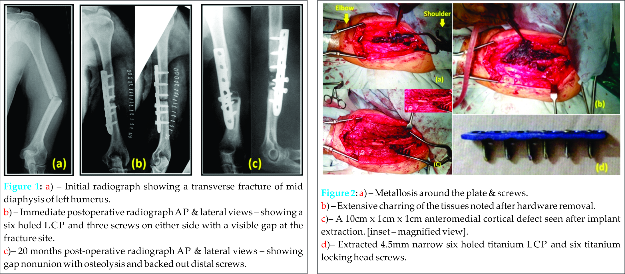
A 58-years-old right hand dominant retired teacher presented to our out-patient clinic with nonunion of left humerus shaft following surgical stabilisation by plating, done 20 months ago. It was a closed injury to begin with, which resulted from a road traffic accident and the initial radiographs (Fig. 1a) showed a transverse fracture of mid diaphysis of left humerus. She was operated on the following day by open reduction and internal fixation using a six holed LCP with six screws (Fig. 1[b]), done elsewhere.She was comfortable for two months after which she developed pain, abnormal mobility with difficulty in using the operated limb for daily activities. She was using a functional brace for support. Her medical history included controlled diabetes, hypertension and hypothyroid status. She had a BMI of 36, was a nonsmoker and nonalcoholic.On clinical examination, there was a healed linear surgical scar measuring about 15 cm along the anterolateral aspect of left arm. Abnormal mobility was noted at the fracture site alongwith significant limitation of left shoulder and elbow movements. There was no distal neurovascular deficit.Current radiograph corresponding to 20 months postoperative duration revealed a gap nonunion of the shaft of left humerus with osteolysis and backed out distal screws (Fig.1c).The haematological and biochemical parameters conducted for detecting deep infection showed a negative result.
Surgery was performed under general anaesthesia in supine position with the operating left upper limb placed over a flat radiolucent side table and intravenous prophylactic antibiotics were administered. Nonunion site was approached through the previous incision. A six holed narrow titanium LCP fixed with six titanium locking head screws was noted along the anteromedial surface of the shaft of humerus. Extensive metallosis was found around the implants and the surrounding soft tissues, giving a charred appearance (Fig. 2a, b). The distal screws were backed out and the rest could be removed without difficulty. Debridement was done to excise the charred tissues. The anteromedial cortex corresponding to the foot print of the extracted plate was found to be eroded leaving a cortical defect of 10cm x 1cm x 1cm across the fracture zone with exposed medullary canal (Fig.2c). The proximal and distal fragment ends were debrided of all the soft tissue and freshened back to bleeding bone, whichr esulted in the sacrifice of a centimeter of length of each fragment. No signs of deep sepsis were found, however, tissue samples were sent for culture study and histopathology.
The primary stabilization was achieved by a nine holed 3.5mm stainless steel LCP placed along the anterolateral surface of the fragmentswith four screws on either side (Fig.3a). Cortical screws were employed first to achieve compression followed by locking head screws. Multiple cortico-cancellous slivers were harvested from the left iliac crest and were placed in a longitudinal fashion filling the cortical defect of the anteromedial humeral surface (Fig.3b). A second12 holed 3.5mm stainless steel LCPwas placed over this grafted surface, perpendicular to the previous plate and was stabilized with two locking head screws at either ends (Fig.3c). The tissues were approximated in layers and the skin was closed by eversion sutures. Supervised physiotherapy of the shoulder and elbow joints was started soon after the pain controlled, followed by muscle strengthening and passive range of motion exercises.
The immediate postoperative radiographs showed satisfactory alignment with secure fixation in both anteroposterior and lateral planes (Fig. 4a). The culture study showed no growth of microorganisms and the histological examination revealed macrophages with intracellular black particulate matter. The skin incisions were healed by primary intention. There was no distal neurovascular deficit or graft donor site morbidity. Functional range of motion of the shoulder and elbow was regained by six weeks of supervised physiotherapy. The nonunion site healed in five months(Fig. 4b). At the final follow up of 24 months, the radiograph showed consolidation of the nonunion.She regained all the movements except for the terminal 10° of elbow extension and 15° of shoulder abduction.The final shortening of her operated arm segment was two cm. According to Stewart and Hundley classification3(Table 1) the final result was found to be good.
Discussion
Nonunion can be a complication of both conservative and operative interventions of humeral shaft fractures. However, if it happens after surgical stabilization, it is notoriously difficult to treat [2].The possible reasons of nonunion in the given case could be,(a) distraction at the fracture site (Fig. 1b), which was evident in the immediate postoperative radiograph, (b)possible devitalisation due to wide exposure of bone fragments for plate fixation, (c) LCP usage with all locking head screws. Infection as a possible cause was ruled out by haematological, microbiological and histopathological examinations. Persistent nonunion of 20 months duration resulted in osteopenia, fatigue failure of implants, increased gap across the fracture zone, progressive osteolysis around the backed out screws and cortical resorption of the foot print of the plate. The available implant options for re-fixation are interlocking nail, single plate, dual plates, plate and antegrade rush rod combination and Ilizarov frame.
Treatment with intramedullary nailing is an attractive option. However, large diameter locked nails are essential to maintain both axial and rotational stability [4].Difficulty in achieving compression at the nonunion site is a potential drawback that can result in significant failure rate. Additional problems would be rotator cuff damage, shoulder pain and stiffness when inserted antegrade and the risk of iatrogenic fracture at the insertion site when placed in a retrograde fashion [4,5]. Understanding the limitations, such as the narrow medullary canal, significant bone defect and difficulty to achieve compression, a locked intramedullary implant as a fixation device was not considered in the present situation.
It may be possible to achieve stable fixation with the Ilizarov frame, even in the presence of osteopenia or bone defects [6]. However, the potential complications are pin-site infections, nerve injuries, and frame impingement over the chest wall resulting in constant discomfort with sleep disturbances.
Compression plating using a 4.5mm narrow plate with at least eight cortices of fixation on either side of the fracture and autogenous bone grafting have been considered as the gold standard in the management of humerus shaft nonunion with a reported success rate of up to 90% [1,2]. The traditional narrow 4.5mm plate was not applicable in the given situation as the humerus was thin and slender making only a 3.5mm design suitable.
Double plating is indicated for a long standing nonunion where the fixation strength achieved by a single plate is questionable due to extensive bone loss and cortical resorption. A two plate construct was found to be significantly stiffer than a single plate construct as per the biomechanical and clinical study by Rubel et al [7]. The procedure, however, is technically demanding, as the significant soft tissue dissection needed to apply two plates is likely to be associated with increased rate of infection, nonunion and nerve palsies. The torsional stability of a locked plate is three times greater than that of a conventional plate [6,7]. In situations of poor bone stock, the LCP permits application of cortical and locking head screws to achieve compression at the fracture ends and increased pull out resistance [8]. Both the plates used here belong to 3.5 mm LCPs, placed in 90-90 orientationto each other. The first nine holed anterolateral LCP achieved compression at the nonunion site and the second 12 holed LCP, placed perpendicular to the previous one, buttressed the graft strips and was intended to improve the biomechanical stability of the entire construct over a long working length.The important point noteworthy was all the 12 screws of both the plates could get new fixation points in the bone fragments without landing in the holes made by the previous implant, thus achieving superior screw purchase. Also the combination of short and long plate avoided the generation of severe stress risers at either ends [9]. Liberal use of autogenous bone graft was a must to help the biology when the bone loss was particularly severe as in the given situation.
The problem of metallosis is more common after the usage of titanium implants than the stainless steel alloys. This results in charring of the surrounding tissue in long standing cases and debridement has a potential risk of neurovascular injury. Metallosis secondary to titanium-alloy wear debris has been associated with osteolysis in arthroplasty [10]. However, metallosis following plate osteosynthesis of long bones has limited literature support [11]. Applying the same concept to the present situation, we speculated that the nonunion as a result of distraction at the fracture ends, lead to persistent motion between the titanium implant components, generating the wear debris. These titanium-alloy wear particles may have produced a net catabolic or osteolytic effect at the fracture site contributing to such an extensive cortical resorption of the entire under surface of the plate with exposure of the medullary canal.
We do agree that fixation of two plates for a single bone resulted in additional soft tissue stripping, although bare minimal. However, the quality of mechanical stability that a two plate construct provided must be kept in mind in this osteopenic situation without which the rapid functional recovery would not have been possible. Shortening of the operated arm segment by two cm noted at the final follow up was a fair compromise in a non-weight bearing bone. Success in this case was believed to be due to adequate mechanical stability achieved bythe dual LCPs in an orthogonal patterncoupled with debridement and liberal use of autogenous bone graft.
Conclusion
We recommend the judicious use of long and short plates in a 90-90 orientation along with autogenous bone grafting in the management of a long standing nonunion of humerus shaft fracture with metallosis and extensive cortical resorption following surgical stabilization by plating. Further studies are recommended to evaluate this technique in a large group of patients.
Clinical Message
Nonunion with extensive osteolysis could result from titanium implants if the fracture is stabilized with visible gap. Double plating is recommended where the fixation strength achieved by a single plate is questionable due to profound bone loss and cortical resorption. Thorough debridement followed by judicious autogenous bone grafting is of paramount importance for successful healing of a longstanding nonunion.
References
1. Healy WL, White GM, Mick CA, BrookerAF, Weiland AJ. Nonunion of the humeral shaft. Clin. Orthop.1987;219:206-213.
2. Van Houwelingen AP, McKee MD. Treatment of osteopenic humeral shaft nonunion with compression plating, humeral cortical allograft struts, and bone grafting. J Orthop Trauma 2005;19:36–42.
3. Stewart MJ, Hundley JM. Fractures of the humerus: a comparative study in methods of treatment. J Bone Joint Surg Am 1955;37:681–92.
4. Gupta RC, Gaur SC, Tiwari RC, Varma B, Gupta R. Treatment of ununited fractuof the shaft of the humerus with bent nail. Injury, 1985;16:276-280
5. Matityahu A, Eglseder WA Jr. Locking flexible nails for diaphyseal humeral fractures in the multiply injured patient: a preliminary study. Tech Hand Up Extrem Surg. 2011;15(3):172-176.
6. Patel VR, Menon DK, Pool RD, Simonis RB. Nonunion of the humerus after failure of surgical treatment. Management using the Ilizarov circular fixator. J Bone Joint Surg [Br] 2000;82-B:977-83.
7. Rubel IF, Kloen P, Campbell D, Schwartz M, Liew A, Myers E, et al. Open reduction and internal fixation of humeral nonunions: a biomechanical and clinical study. J Bone Joint Surg Am 2002;84:1315–22.
8. Martinez AA, Cuenca J, Herrera A. Two-plate fixation for humeral shaft nonunions. Journal of Orthopaedic Surgery 2009;17(2):135-8.
9. Thakur – 2006, Elements of fracture fixation, 2/e – page 94
10. Lida H, Kaneda E, Takada H, Uchida K, Kanawabe K, Nakamura T. Metallosis due to impingement between the socket and the femoral neck in a metal-on-metal bearing total hip prosthesis. A case report. J Bone Joint Surg Am. 1999;81:400-3.
Edelstein Y, Ohm H, Rosen Y. Metallosis and pseudotumor after failed ORIF of a humeral fracture. Bulletin of the NYU Hospital for Joint Diseases 2011;69(2):188-91.
| How to Cite This Article: Metikala S, Bhogadi P. Orthogonal Double Plating and Autologous Bone Grafting of Postoperative Humeral Shaft Nonunion – A Rare Case Report and Review of Literature. Journal of Orthopaedic Case Reports 2015 Oct-Dec;5(4): 50-53. Available from: https://www.jocr.co.in/wp/2015/10/01/2250-0685-345-fulltext/ |
[Full Text HTML] [Full Text PDF] [XML]
[rate_this_page]
Dear Reader, We are very excited about New Features in JOCR. Please do let us know what you think by Clicking on the Sliding “Feedback Form” button on the <<< left of the page or sending a mail to us at editor.jocr@gmail.com


