[box type=”bio”] What to Learn from this Article?[/box]
Pseudo-aneurysm of radial artery is a rare complication after internal fixation of radius shaft fracture and arises due to intra-operative trauma to artery which can present as early as 2weeks as in this case.
Case Report | Volume 7 | Issue 6 | JOCR Nov – Dec 2017 | Page 3-5| Aditya Anand Dahapute, Rohan Bharat Gala, Sanjay B Dhar, Siddharth Virani, AvaniSudhir Vaishnav. DOI: 10.13107/jocr.2250-0685.922
Authors: Aditya Anand Dahapute [1], Rohan Bharat Gala [1], Sanjay B Dhar [2], Siddharth Virani [1], AvaniSudhir Vaishnav [2]
[1] Department of Orthopaedics, Seth GordhandasSunderdas Medical College, Mumbai, Maharashtra, India,
[2] Department of Orthopaedics, D.Y. Patil Medical College, Mumbai, Maharashtra, India.
Address of Correspondence
Dr. Rohan B Gala,
Senior Registrar, Department of Orthopaedics, Seth G.S. Medical College and K.E.M. Hospital, Parel, Mumbai, Maharashtra, India.
E-mail: drrohangala17@gmail.com
Abstract
Introduction: Pseudoaneurysms of arteries are not uncommon complications of vessel handling during surgery. Early identification and management is important to prevent disastrous complications such as rupture and thrombosis.
Case Report: We describe a case of a 28-year-old male who developed a pseudoaneurysm of the radial artery after being operated by plating for a mid-shaft radius fracture. He presented 2 weeks after surgery with swelling over the forearm which was confirmed to be a pseudoaneurysm after computed tomography angiography. It was treated with surgical excision and end-to-end anastomosis.
Conclusion: A high index of suspicion must be maintained about the occurrence of this complication secondary to both trauma and surgery.
Keywords: Radial artery, pseudoaneurysm, radius fracture.
Introduction
Peripheral pseudoaneurysms, which are more commonly iatrogenic, also occur in post-traumatic cases where a localiszedhaematoma has a persistent communication with a native artery through a narrow neck [1]. They usually present as a pulsatile palpable mass. Complications such as rupture, expansion, and even thrombosis mandate the diagnosis of arterial aneurysms as soon as possible using either color flow imaging and/or Doppler waveform analysis [2, 3, 4, 5]. Percutaneous techniques using ultrasound guidance such as manual compression, thrombin injections, and also open procedures like resection with end-to-end anastomosis have been documented [6, 7, 8]. Radius shaft fractures treated by open reduction and internal fixation (ORIF) with plating are rarely associated with any vascular complications though infection, tendon irritation/rupture, and implant failure still prevail [9]. We describe a case of radial artery pseudoaneurysm in a post-traumatic case of closed radial shaft fracture treated by ORIF with plating.
Case Report
A 28/male electrician sustained a closed fracture of middle third radius after an alleged history of road traffic accident. He underwent an ORIF of radius with 3.5 mm dynamic compression plate through volar approach. Tourniquet was used during the surgery for 70 min and extremity was exsanguinated completely by lifting it for 2 min. Postoperatively, radial pulse was present with good capillary refill. There was no spike at fracture site which was impinging on artery neither any visible contusion on radial artery. Radial artery was dissected and retracted laterally and spikes were used for retraction. On the 3rd post-operative day, dressing was done. The patient presented to casualty 15 days after surgery with sudden swelling of forearm with soakage of dressing. On examination, forearm was swollen and tender with bleeding from incision site.
 |
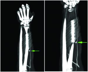
| Figure 1: Computed tomography angiogram with radial artery pseudoaneurysm at proximal end of radius plate. |
The patient was readmitted for further management and computed tomography angiogram was done. It revealed a pseudoaneurysm of the radial artery (Fig. 1). At the time of exploration, the pseudoaneurysm was located at the proximal end of radius plate. Surgical excision of the aneurysm with end-to-end anastomosis of proximal and distal end of radial artery was performed with Prolene 6–0. All clots were removed and wound was irrigated. Around 4 mm of vessel was excised. The patient was given iv heparin and pentoxiphfylline in ward. Immediate postoperatively, radial pulse was present. The patient was given above elbow slab for 3 weeks after which elbow range of motion exercises was started. At 6 months follow-up, the radial artery pulsation was well felt and Allen’s test was performed to evaluate its patency.
Discussion
Peripheral pseudo-aneurysms occur due to trauma or surgical interventions, although this occurrence is rare. There have been reports of radial artery pseudoaneurysm after implant removal [10] and after using volar plate for distal radius fracture [11]. Zitsman [12] found that all layers of an artery can be damaged by a penetrating wound of any sort resulting in an extra-arterial haematoma formation, which then organiszes, and gives origin to the fibrous walls of the pseudoaneurysm. We postulate that, in this case, it might be either due to the sharp ends of the fracture fragments and/or due to the surgical dissection which was carried out for fracture fixation. It was, however, difficult to examine the patient as he had active bleeding, and therefore, typical findings such as palpable pulsatile swelling and skin discoloration that usually aid us in our diagnosis was difficult to appreciate in this case. Some of the documented interventions in treating peripheral pseudoaneurysms are ultrasound-guided localiszed compression, ultrasound-guided thrombin injections, and open procedures like resection with end-to-end anastomosis. Although the ultrasound-guided interventions have been successful in cases where the collateral circulation is adequate, they are most preferred in cases of small, superficial aneurysms. Administrations of thrombin injections are painless, but they carry a risk of anaphylaxis as well as distal emboliszation to the site, later causing vasospastic spasms [13, 14, 15]. Some studies have documented such an injury occurring during implant removal [10] due to which we reiterate the need for meticulous techniques to identify and retract the radial artery as well the need to deflate the tourniquet in orthopaedic procedures before closure, thus avoiding vascular complications. It’s important to note that we should turn off the tourniquet and carefully find bleeding origin in any operation in the proximity of major vessels. In most cases, diagnosis is delayed due to slow progression of sign and symptoms. An important key in the diagnosis is that the surgeon should have a high degree of suspicion when the patient complaints of a mass, with paraesthesia, and pain that was inappropriate to the operation and soft tissue swelling. When the diagnosis is confirmed, the patient should be referred to a vascular surgeon.
Conclusion
Trauma and iatrogenic injury are important causes of peripheral pseudoaneurysms. The surgeon must maintain a high index of suspicion about this rare complication. Furthermore, utmost care must be taken to protect the radial artery and avoid inadvertent injury to it both during index surgery and implant removal.
Clinical Message
An orthopaedic surgeon must be aware about the incidence of pseudoaneurysms of the radial artery as a complication of occurrence and management of mid-shaft radius fractures. Radial artery to be gently handled during exposure and implant placement to prevent such complication.
References
1. McGlinchey I, Baxter GM. Technical report: An alternative mechanical technique of pseudoaneurysm compression therapy. ClinRadiol 1997;52:621-4.
2. Abu-Yousef MM, Wiese JA, Shamma AR. The “to-and-fro” sign: Duplex doppler evidence of femoral artery pseudoaneurysm. AJR Am J Roentgenol 1988;150:632-4.
3. Mills JL, Wiedeman JE, Robison JG, Hallett JW. Minimising mortality and morbidity from iatrogenic arterial injuries: The need for early recognition and prompt repair. J VascSurg 1986;4:22-7.
4. Polak JF. Peripheral arterial disease. Evaluation with color flow and duplex sonography. RadiolClin North Am 1995;33:71-90.
5. Wilkinson DL, Polak JF, Grassi CJ, Whittemore AD, O’Leary DH. Pseudoaneurysm of the vertebral artery: Appearance on color-flow dopplersonography. AJR Am J Roentgenol 1988;151:1051-2.
6. Polak JF. Peripheral Vascular Sonography: A Practical Guide. Baltimore: Lippincott, Williams and Wilkins; 1992.
7. Kang SS, Labropoulos N, Mansour MA, Michelini M, Filliung D, Baubly MP, et al. Expanded indications for ultrasound-guided thrombin injection of pseudoaneurysms. J VascSurg 2000;31:289-98.
8. Maw A, Renaut AJ. Pseudoaneurysm of the radial artery complicating excision of a wrist ganglion. J Hand Surg Br 1996;21:783-4.
9. DuchateauJ, Moermans JP. False aneurysm of the radial artery. J Hand Surg (Am) 1985;10:140-1.
10. Fung BK, Ip WY. Pseudoaneurysm of the radial artery after plate removal. Hong Kong J OrthopSurg 2001;5:138-40.
11. Dao KD, Venn-Watson E, Shin AY. Radial artery pseudoaneurysm complication from use of AO/ASIF volar distal radius plate: A case report. J Hand Surg Am 2001;26:448-53.
12. Zitsman JL. Pseudoaneurysm after penetrating trauma in children and adolescents. J PediatrSurg 1998;33:1574-7.
13. Lennox A, Griffin M, Nicolaides A, Mansfield A. Regarding percutaneous ultrasound guided thrombin injection: A new method for treating postcatheterization femoral pseudoaneurysms. J VascSurg 1998;28:1120-1.
14. Paulson EK, Nelson RC, Mayes CE, Sheafor DH, Sketch MH Jr.,Kliewer MA,et al.Sonographically guided thrombin injection of iatrogenic femoral pseudoaneurysms: Further experience of a single institution. AJR Am J Roentgenol 2001;177:309-16.
15. Quarmby JW, Engelke C, Chitolie A, Morgan RA, Belli AM. Autologous thrombin for treatment of pseudo-aneurysms. Lancet 2002;359:946.
 |
 |
 |
 |
 |
| Dr. Aditya Anand Dahapute |
Dr. Rohan Bharat Gala | Dr. Sanjay B Dhar | Dr. Siddharth Virani | Dr. Avani Sudhir Vaishnav |
| How to Cite This Article: Dahapute AA, Gala RB, Dhar SB, Virani S, Vaishnav AS. Radial Artery Pseudoaneurysm in a Post-operative Case of Midshaft Radius Fracture. Journal of Orthopaedic Case Reports 2017 Nov – Dec: 7(6): 3-5 |
[Full Text HTML] [Full Text PDF] [XML]
[rate_this_page]
Dear Reader, We are very excited about New Features in JOCR. Please do let us know what you think by Clicking on the Sliding “Feedback Form” button on the <<< left of the page or sending a mail to us at editor.jocr@gmail.com


