[box type=”bio”] What to Learn from this Article?[/box]
Stress fracture of the femoral head is an exceptional diagnosis that can be evoked when we have a chronic hip pain after, eliminating infectious and tumoral etiologies.
Case Report | Volume 7 | Issue 6 | JOCR Nov – Dec 2017 | Page 10-12| Mourad Zaraa, Heithem Sahli, Imene Yeddes, Sabri Mahjoub, Mondher Mbarek. DOI: 10.13107/jocr.2250-0685.926
Authors: Mourad Zaraa [1], Heithem Sahli [1], Imene Yeddes [2], Sabri Mahjoub [1], Mondher Mbarek [1]
[1] Department of Orthopedic Surgery, Trauma Center, Ben Arous, Tunisia.
[2] Department of nuclear medicine, Salah Azaiez Institute, Tunis, Tunisia.
Address of Correspondence
Dr. Mourad Zaraa,
Department of Orthopedic Surgery, Trauma Center, Ben Arous, Tunisia.
E-mail: mourad.zaraa@hotmail.com
Abstract
Introduction:
The stress fracture of the femoral head rarely occurs; but, it is generally encountered in case of bone insufficiency, and it is exceptional in younger individuals. The main differential diagnosis may include several diseases, namely, slipped upper femoral epiphysis, septic arthritis, osteomyelitis, and Perthes’ disease. Bone scintigraphy is very sensitive but lacks specificity. Nowadays, the magnetic resonance imaging (MRI) is the gold standard for an accurate diagnosis.
Case Report: We present the first description of this pathology in the pediatric population with the particularity of its atypical aspect on MRI through a case of stress fracture of femoral head in 12-year-old female.
Conclusion: Stress fractures may sometimes mimic malignant or infectious lesions and are easily misdiagnosed. MRI is the gold standard which may be the only modality to identify the fracture.
Keywords: Femoral head, stress fracture, diagnosis, bone scintigraphy, magnetic resonance imaging, children.
Introduction
The stress fracture of the femoral head is rarely occurs. It is generally observed in elderly women or organ transplant recipients with bone insufficiency [1, 2]. In younger individuals, these fractures may be caused by abnormal stresses applied to normal bone, like with military or marathon runners [3]. As far as we know, the following is the first description of the case of an acute onset hip pain in a healthy child in whom the diagnosis of the femoral head stress fracture was made by bone scintigraphy and magnetic resonance imaging (MRI).
Case Report
The patient is a 12-year-old female, presented to the emergency department, and complaining of a right hip pain and a limp that had progressively worsened during the past 2 weeks. She had no history of any systemic disease; she lost weight over past months; and previously, she had an injury to hips. Moreover, the oral Vitamin D supplementation was correctly done. She stated that the pain had begun after 3 weeks in a holiday camp where the physical activity was intense. On physical examination, the range of the motion of the hip was painful. Laboratory test results (white blood cells and C-reactive protein erythrocyte sedimentation rate) were normal. The radiographs of the right hip (Fig. 1) were also normal excluding the diagnosis of the slipped upper femoral epiphysis, septic arthritis, osteomyelitis, and Perthes’ disease.
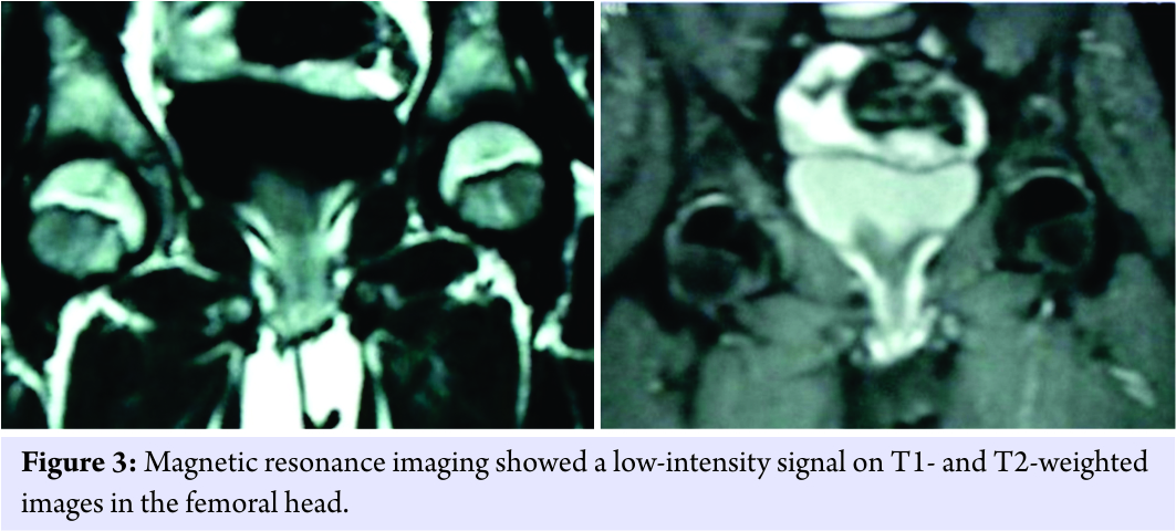
Bone scintigraphy showed an increased isotope uptake over the femoral head (Fig. 2). MRI showed, in the coronal plane with fat saturation, a low-intensity signal on T1- and T2-weighted spin echo images in the right femoral head. The lesion was linear, regular, and not enhanced with contrast injection. There was no intra-articular effusion, no soft tissue and no acetabular abnormalities. The diagnosis of stress fracture of the femoral head was established (Fig. 3).
The patient was treated conservatively with bed rest and non-weight-bearing mobilization on the left side, for 1 month. After that, a protected weight-bearing using crutch was permitted for 2 months. The evolution was favorable with a pain-free hip. After 1 year, at the last follow-up, the patient was still asymptomatic and the MRI control was requested but the patient moved and was lost from sight.
Discussion
Stress fractures are reported in three anatomical locations in the femur. The highest incidence rates occur at the femoral neck followed by the distal femoral shaft [4]. However, the fatigue stress fracture of the femoral head is a rarely encountered condition, and it is an unusual cause of acute hip pain [5]. It is generally caused by the subchondral insufficiency fractures of the femoral head due to osteoporosis or necrosis in elderly women. To the best of our knowledge, this is the first case of fatigue stress fracture in a child. These fractures are caused by abnormal stresses applied to a normal bone without a history of significant trauma and corticosteroid intake. Pain in the hip region and limping was the first reported symptoms [4]. Shortly after the onset of pain, radiographic results can be normal or nearly normal [6]. Bone scintigraphy or/and MRI should be undertaken if the result of radiography is negative. Bone scintigraphy has traditionally been used in the diagnosis of stress fractures due to their high sensitivity which has been shown to be 84–100% within 3 days of symptoms [7]. Nevertheless, this examination is not specific for stress fractures. MRI has become the gold standard due to its sensitivity which is similar to bone scintigraphy, high specificity, and the absence of radiation exposure [3]. For the femoral neck stress fractures and subchondral insufficiency fracture of the femoral head, MRI can show specific aspects [1, 2, 3]. However, in femoral head stress fractures in the young individual, MRI has no specific appearance. In our case, MRI showed a low-intensity signal on T1- and T2-weighted images in the femoral head. The diagnosis of femoral head stress fracture is generally observed in elderly women or organ transplant recipients with bone insufficiency [1, 2]. The MRI revealed the presence of a low-signal-intensity serpiginous line parallel to the articular surface which is a characteristic of the trabecular fracture and which could distinguish an insufficiency fracture from osteonecrosis. Yet, in our case, this sign was not found, probably due to the age of the patient or the early diagnosis. The main differential diagnosis for a child with groin pain may include developmental dysplasia of the hip, Perthes’ disease, slipped capital femoral epiphysis, infection, transient synovitis, pelvic muscle sprain, leukemia, tumor, and fracture [8]. Rickets, systemic diseases, and endocrine disorders must also be considered. Stress fractures can often resemble malignant lesions, osteomyelitis, or radiological abnormalities [8]. In fact, early diagnosis determines the therapeutic approach and the speed of the recovery. In our case, the treatment is based mainly on rest and restriction of weight-bearing leading to consolidation. The treatment of the femoral neck stress fractures and the subchondral insufficiency fracture of the femoral head can be functional or surgical, depending on the risk of the fracture [4, 5]; but, for the femoral head stress fractures, we think that the treatment should be functional. The literature review does not dictate any treatment because it is an exceptional pathology.
Conclusion
The femoral head stress fracture is a condition that has never been reported in children. Stress fractures may sometimes mimic malignant or infectious lesions and are easily misdiagnosed. Thus, interdisciplinary consultation, including an orthopedic surgeon, a radiologist, and a pediatrician, must be instituted to eliminate serious diseases, diagnose and treat this extremely rare stress fracture. In this way, MRI is the gold standard which may be the only modality to identify the fracture. Nevertheless, this case emphasizes the importance of good clinical observation and interpretation of radiographs in children.
Clinical Message
Good clinical observation and interpretation of radiographs and MRI are the only way to not misdiagnose malignant or infectious lesion.
References
1. Yamamoto T, Nakashima Y, Shuto T, Jingushi S, Iwamoto Y. Subchondral insufficiency fracture of the femoral head in younger adults. Skeletal Radiol 2007;36 Suppl 1:S38-42.
2. Iwasaki K, Yamamoto T, Motomura G, Mawatari T, Nakashima Y, Iwamoto Y, et al. Subchondral insufficiency fracture of the femoral head in young adults. Clin Imaging 2011;35:208-13.
3. Buttaro M, Valle A, Morandi A, Sabas M, Pietrani M, Piccaluga F. Insufficiency subchondral fracture of the femoral head. J Arthroplasty 2003;18:377-82.
4. Yamamoto T, Bullough PG. Subchondral insufficiency fracture of the femoral head and medial femoral condyle. Skeletal Radiol 2000;29:40-4.
5. Diehl JJ, Best TM, Kaeding CC. Classification and return-to-play considerations for stress fractures. Clin Sports Med 2006;25:17-28.
6. Gaeta M, Minutoli F, Scribano E, Ascenti G, Vinci S, Bruschetta D, et al. CT and MR imaging findings in athletes with early tibial stress injuries: Comparison with bone scintigraphy findings and emphasis on cortical abnormalities. Radiology 2005;235:553-61.
7. Er MS, Eroglu M, Altinel L. Femoral neck stress fracture in children: A case report, up-to-date review, and diagnostic algorithm. J Pediatr Orthop B 2014;23:117-21.
8. Fievez EF, Hanssen NM, Schotanus MG, van Haaren EH, Kort NP. Stress fracture of the femoral neck in a child: A case report. J Pediatr Orthop B 2013;22:45-8.
 |
 |
 |
 |
 |
| Dr. Mourad Zaraa | Dr. Heithem Sahli | Dr. Imene Yeddes | Dr. Sabri Mahjoub | Dr. Mondher Mbarek |
| How to Cite This Article: Zaraa M, Sahli H, Yeddes I, Mahjoub S, Mbarek M. Femoral Head Stress Fracture in Children: A Case Report. Journal of Orthopaedic Case Reports 2017 Nov – Dec: 7(6): 10-12 |
[Full Text HTML] [Full Text PDF] [XML]
[rate_this_page]
Dear Reader, We are very excited about New Features in JOCR. Please do let us know what you think by Clicking on the Sliding “Feedback Form” button on the <<< left of the page or sending a mail to us at editor.jocr@gmail.com


