[box type=”bio”] Learning Point for this Article: [/box]
In the presence of a tibial cyst associated with ACL reconstruction, always suspect communication of the cyst with the knee joint. The treatment must consider this possibility and must be aggressive in order to avoid recurrence.
Case Report | Volume 7 | Issue 6 | JOCR November – December 2017 | Page 41-45| Juan Pablo Zícaro, Agustín Rubén Molina Rómoli, Carlos Heraldo Yacuzzi, Matías Costa Paz. DOI: 10.13107/jocr.2250-0685.942
Authors: Juan Pablo Zícaro [1], Agustín Rubén Molina Rómoli [1], Carlos Heraldo Yacuzzi [1], Matías Costa Paz [1]
[1] Department of Orthopaedics, Italian Hospital from Buenos Aires, Buenos Aires, Argentina.
Address of Correspondence:
Dr. Agustín Rubén,
Juan Domingo Perón 4190 (C1199ABB), Buenos Aires, Argentina.
E-mail: agustinmolinaromoli@gmail.com
Abstract
Introduction: Anterior cruciate ligament (ACL) reconstruction is a “daily” surgery. Nevertheless, pretibial cyst formation is a very rare complication and may appear years after the reconstruction. Even more infrequent is the recurrence of this complication.
Case Report: We present a patient who developed a pretibial cyst 44 months after an ACL reconstruction and who underwent three open surgeries due to recurrence. Up to date, there are no symptoms or signals of cyst recurrence after 18 months of follow-up.
Conclusion: Initial surgical resection might have failed due to insufficient surgical treatment. In case of suspicion of articular communication, we consider that the correct treatment is the resection of the cyst, proper curettage of the tunnel walls and a thorough filling of the space with bone graft. This course of treatment may avoid a recurrence.
Keywords: Anterior cruciate ligament reconstruction, pretibial cyst, complication, recurrent.
Introduction
Anterior cruciate ligament (ACL) reconstruction is a “daily” surgery and has been improved through time with new surgical techniques and new materials. Bioabsorbable interference screws have gained ground over the classic metallic screws due to better post-operative imaging and decreased risk of graft laceration. However, new complications have appeared such as pretibial cyst formation. This is a very rare complication, but it has already been described in the literature. Nevertheless, it may appear years after the reconstruction. Even more unfrequent is the recurrence of this complication.
Case Report
In May 2011, a 34-year-old male was treated for an ACL rupture using a hamstring graft fixation associated with a partial internal meniscectomy. The hamstring grafts were fixated using a transfixation system in the femur and a biodegradable screw in the proximal tibia. The patient’s post-operative course was uneventful, and he returned to sports without further difficulties. In July 2014 (3 years and 6 months following the initial operation), a tibial cyst suddenly presented and was first treated conservatively, including physical therapy and non-steroidal anti-inflammatory drugs, until it became symptomatic. There were no signs of clinical or laboratory infection. Magnetic resonance imaging (MRI) showed a 10 mm × 25 mm cyst lesion filled with liquid (Fig. 1). 
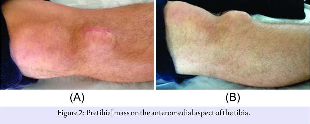
The physical examination revealed a stable knee and a complete range of motion. An approximately 50 mm × 40 mm palpable mass was evident at the anteromedial proximal tibia. MRI images were consistent with a homogeneous, fluid-filled cyst with a connection toward the tibial tunnel (Fig. 3).
On April 2015, a second open resection and an exploratory arthroscopy were performed. The cyst was approached through the previous skin incision. The mass was meticulously dissected, with care not to injure the cyst or its stalk. After the cyst was isolated from the surrounding soft tissue, it was brought back to its original placing at the tibial tunnel. The cyst and stalk were then excised altogether. Once removed, the tunnel showed no evidence of communication with the joint and the walls showed signs of sclerosis. Walls were debrided with a rongeur and curette and filled with bone plugs extracted from the lateral femoral condyle. Graft was impacted, and fascia and subcutaneous tissue were sutured. The pathology report revealed a fibrous tissue capsule compatible with a synovial cyst (Fig. 4).
The patient remained asymptomatic for 3 months, but when he returned to sport activities, a painful palpable mass was evident again in the same site. Like previous episodes, no clinical or laboratory parameters of infection were found. The X-ray and MRI revealed a new cyst recurrence, with an increase in the tunnel size compared to the previous episode.

Discussion
Among complications following ACL reconstruction, the formation of a pretibial cyst in the site of the tibial tunnel is very rare and may happen even years after surgery [1, 2, 3]. The etiology of pretibial synovial cyst is not clearly described in current literature and recurrences are quite infrequent [4, 5, 6, 7, 8, 9]. Gonzalez-Lomas et al. [10] reported in 2011, 7 cases with pretibial cyst. The average time between surgery and the appearance of the cyst was 2 years. In all of them, a biodegradable poly-L-lactic (PLLA) screw was used. Allografts were used in four cases and hamstrings in three. After a simple excision of the cyst, fragments of PLLA screw surrounded by histiocytes were found. They concluded that cyst formation was due to remnants of PLLA. Recurrence is very rare, and an incomplete resection or curettage may cause a new episode. Sekiya et al. [11] submitted in 2004, a case report of a patient with two recurrences. The initial formation of the cyst was 5 years after ACL reconstruction and the fixation used was a biodegradable screw with a hamstring graft. The procedure for cyst excision was a simple resection and closure of the fascia, and the histological study found suture remains. 1 year after the excision, the cyst recurred and was drained by aspiration. The second recurrence was 3 months later. On that occasion, they performed a larger curettage of the cyst removing remnants of the walls of the tunnel and suture. Again, the histological study showed remnants of non-biodegradable suture. This author did not fill the cavity with bone graft, suspecting the etiology of the cyst to be a foreign body reaction. Ghazikhanian et al. [3] stated in a literature review that the placement of bone graft in the tunnel can be very important to reduce the risk of recurrence, especially when cysts, is suspected to be communicant. No author described an association of this complication with graft loosening. In our institution, we perform about 200 ACL surgeries per year. During 2008–2016, 14 patients with pretibial cyst were intervened over this complication. Although we believe the etiology to be multifactorial, cysts can be defined as communicating or non-communicating. This definition may help to determine the surgical treatment. The interesting thing about this case report is that it involves only one patient who developed a recurrence and required three surgeries for a definitive treatment. We believe it was a communicating cyst. The first procedure performed at another medical center (simple excision and closure) was insufficient. The second surgery, though we provided bone graft from the lateral epicondyle, was not enough to fill the cyst tunnel, and may have been the reason for its reoccurrence. Performing a proper curettage of the walls and filling the cyst with bone allograft was necessary for a correct sealing and, up to date, a definitive treatment.
Conclusion
We presented a very unusual case of a pretibial cyst that recurred 3 times and required three surgeries for a definitive treatment. Initial surgical resection might have failed due to insufficient surgical treatment. The correct treatment in case of articular-communication suspicion is the resection of the cyst, proper curettage of the tunnel walls, and a thorough filling of the space with bone graft. This may avoid a recurrence.
Clinical Message
The formation of a pretibial cyst in the site of the tibial tunnel after an ACL reconstruction is a rare complication; however, the standard arthroscopic surgeon is not exempt from this pathology. It is important to be aggressive with surgical treatment to avoid recurrence of the cyst.
References
1. Díaz Heredia J, Ruiz Iban MA, Cuéllar Gutiérrez R, Ruiz Diaz R, Cebreiro Martinez del Val I, Turrion Merino L. Early postoperative transtibial articular fistula formation after anterior cruciate ligament reconstruction: A review of three cases. Arch Orthop Trauma Surg 2014;134:829-34.
2. Feldmann DD, Fanelli GC. Development of a synovial cyst following anterior cruciate ligament reconstruction. Arthroscopy 2001;17:200-2.
3. Ghazikhanian V, Beltran J, Nikac V, Feldman M, Bencardino JT. Tibial tunnel and pretibial cysts following ACL graft reconstruction: MR imaging diagnosis. Skeletal Radiol 2012;41:1375-9.
4. Tsuda E, Ishibashi Y, Tazawa K, Sato H, Kusumi T, Toh S. Pretibial cyst formation after anterior cruciate ligament reconstruction with a hamstring tendon autograft. Arthroscopy 2006;22:691.e1-6.
5. Simonian PT, Wickiewicz TL, O’Brien SJ, Dines JS, Schatz JA, Warren RF. Pretibial cyst formation after anterior cruciate ligament surgery with soft tissue autografts. Arthroscopy 1998;14:215-20.
6. Martinek V, Friederich NF. Tibial and pretibial cyst formation after anterior cruciate ligament reconstruction with bioabsorbable interference screw fixation. Arthroscopy 1999;15:317-20.
7. Faunø P, Christiansen SE, Lund B, Lind M. Cyst formation 4 years after ACL reconstruction caused by biogradable femoral transfixation: A case report. Knee Surg Sports Traumatol Arthrosc 2010;18:1573-5.
8. Thaunat M, Chambat P. Pretibial ganglion-like cyst formation after anterior cruciate ligament reconstruction: A consequence of the incomplete bony integration of the graft? Knee Surg Sport Traumatol Arthrosci 2007;15:522-4.
9. Sprowson AP, Aldridge SE, Noakes J, Read JW, Wood DG. Bio-interference screw cyst formation in anterior cruciate ligament reconstruction–10-year follow up. Knee 2012;19:644-7.
10. Gonzalez-Lomas G, Cassilly RT, Remotti F, Levine WN. Is the etiology of pretibial cyst formation after absorbable interference screw use related to a foreign body reaction? Clin Orthop Relat Res 2011;469:1082-8.
11. Sekiya JK, Elkousy HA, Fu FH. Recurrent pretibial ganglion cyst formation over 5 years after anterior cruciate ligament reconstruction. Arthroscopy 2004;20:317-21.
13. Ramadan KM, Shenkier T, Sehn LH, Gascoyne RD, Connors JM. A clinic-pathological retrospective study of 131 patients with primary bone lymphoma: A population-based study of successively treated cohorts from the British Columbia cancer agency. Ann Oncol 2007;18:129-35.
 |
 |
 |
 |
| Dr. Juan Pablo Zícaro |
Dr. Agustin Molina Romoli | Dr. Carlos Heraldo Yacuzzi | Dr. Matías Costa Paz |
| How to Cite This Article: Zícaro JP, Molina Rómoli AR, Yacuzzi C, Costa-Paz M. Multiple Recurrent Cyst Formation after Anterior Cruciate Ligament Reconstruction – A Case Report. Journal of Orthopaedic Case Reports 2017 Nov-Dec; 7(6): 41-45 |
[Full Text HTML] [Full Text PDF] [XML]
[rate_this_page]
Dear Reader, We are very excited about New Features in JOCR. Please do let us know what you think by Clicking on the Sliding “Feedback Form” button on the <<< left of the page or sending a mail to us at editor.jocr@gmail.com


