[box type=”bio”] Learning Point of the Article: [/box]
This case suggests that prompt surgical repair of a functionally deficient triceps tendon tear and conservative management of associated UCL injury in a young athlete may result in good prognosis and return-to-play within six months.
Case Report | Volume 8 | Issue 5 | JOCR September – October 2018 | Page 15-18| Lafi S Khalil, Khalid Alkhelaifi, Fabien Meta, Vincent A Lizzio, Ramsey Shehab, Eric C Makhni. DOI: 10.13107/jocr.2250-0685.1188
Authors: Lafi S Khalil[1], Khalid Alkhelaifi[1], Fabien Meta[1], Vincent A Lizzio[1], Ramsey Shehab[1], Eric C Makhni[1]
[1]Department of Orthopedic Surgery, Henry Ford Hospital, 2799 W. Grand Blvd, Detroit, MI 48202,United States.
Address of Correspondence:
Dr. Eric C Makhni,
Department of Orthopedic Surgery, 6777 W Maple Rd., 3rd Floor East, West Bloomfield, MI 48322,United States.
E-mail: ericmakhnimd@gmail.com
Abstract
Introduction: With the increasing number of children and adolescents participating in sports, pathologies once reserved for high-level athletes are now emerging in this younger population. Distal triceps tendon tears represent an injury infrequently seen even among older, skeletally mature athletes. We report a case of distal triceps tendon tear with concomitant ulnar collateral ligament (UCL) injury in a skeletally-immature football player.
Case Report: This is a rare case of traumatic triceps tendon tear with UCL injury in a 13-year-old male football player during a fall and hyperextension of his elbow. Management included surgical treatment of the triceps tear with suture anchors in double row technique. The concomitant UCL injury was treated conservatively.
Conclusion: This case suggests that this type of injury can occur in young athletes, but good prognosis can be expected with prompt management. Surgical repair of a functionally deficient triceps tendon tear and conservative management of associated UCL injury can result in returntoplay within 6 months.
Keywords: Triceps tendon, tendonrepair, ulnar collateral ligament.
Introduction
The number of children and adolescents participating in sports is increasing yearly, with estimations of approximately 35 million children and adolescents involved in sports, 2 million of which are participating in organized little league activities [1]. As a result, there has been an emergence of pathologies once reserved for athletes participating in high-level competition. Overuse is the likely culprit for many elbow injuries in young athletes, including damage to ligamentous structures and stress- or overuse-related fractures [2]. As athletes continue to engage in higher levels of competitive sports and younger ages, injuries that were previously only seen in older, skeletally mature athletes, are becoming more frequently encountered in this younger population. One such injury – the distal triceps tendon tear – is infrequently encountered in the adolescent population. Only 21 such tears were found between 1991 and 1996 in the National Football League, of which 15 required surgical interventions at that time [3]. In this report, we present a rare case of distal triceps tendon tear with concomitant ulnar collateral ligament (UCL) injury in a young athlete following a tackling injury during a football game. This report overviews the diagnosis, management, and successful follow-up of this injury. To the best of our knowledge, there are currently no existing reports in the literature that describes a distal triceps tendon tear accompanied by UCL tear in the pediatric population. The authors have obtained the patient’s and parent’s informed consent for print and electronic publication of the case report. None of the contributing authors have any conflicts of interest to disclose relevant to this case.
Case Report
A 13-year-old right-handed male, with no pertinent medical history or prior history of an elbow injury, presented to primary care physician three days after the patient was tackled while playing football and heard a “pop” following hyperextension of his left elbow.
Operative details
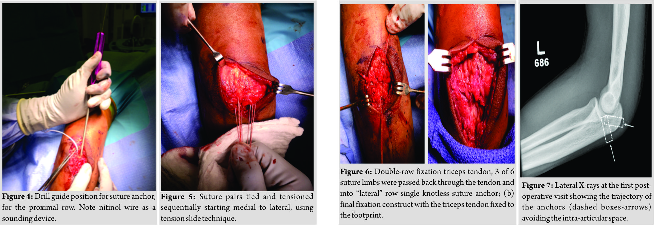
On the day of surgery, the patient’s left elbow was marked by the senior author. Details of the procedure, post-operative recovery, and risks and benefits were again discussed and agreed on by patient and family. Regional anesthesia, as well as pre-operative antibiotics, was administered. The patient was positioned in a lateral decubitus position using a bean bag, and a non-sterile tourniquet was applied (Fig. 2).A standard posterior approach to the elbow was made using a curved incision to the lateral aspect of the elbow, approximately 10–15 cm in length. Sharp dissection was carried down to the triceps avulsion. The distal end of the triceps was readily identified and seen to be detached nearly completely from the footprint at the olecranon, with only a few remaining fibers intact medially. The necrotic and fibrotic tendon edges were sharply debrided. The footprint on the olecranon was identified and lightly decorticated for tendon preparation (Fig. 3). One medial and one lateral 3.0 double-loaded suture tac (Arthrex, Naples, Florida, USA) was inserted into the footprint (Fig. 4).After drilling through the drill guide, a free nitinol wire was used as a sounder inside the drill hole to ensure that the joint surface had not been penetrated. A Krakow stitch was then placed through the distal end of the tendon with each limb of the pairs. The other suture pairs were placed into a horizontal mattress stitch through the distal tendon. With the elbow in approximately 20° of flexion, the suture pairs were sequentially tied and tensioned, starting medially and going laterally, using a tension slide technique (Fig. 5). Three suture strands were then re-passed through the tendon just proximal to the site of a fixation in corporated into a single 3.5 mm Swive Lock anchor (Arthrex, Naples, Florida, USA) that was placed 2 cm distal to the suture tac. After fixing the Swive Lock, the elbow was ranged from extension to flexion to confirm that the construct was stable (Fig. 6a, b).After closing the incision, the elbow was placed in a posterior mold splint with the elbow in 30° of flexion. Post-operative follow-up visits were planned for2 weeks, 6 weeks, 3 months, and 6 months.
Post-operative course
At the first post-operative visit, the patient’s wound was inspected and showed complete healing. The patient had full passive range of motion. We positioned the patient in a hinged elbow brace locked at 60° of flexion (0-60). X-rays were taken and showed good positioning of the anchor tracks at the olecranon without joint penetration (Fig. 7). At 6 weeks postoperatively, the patient had full active range of motion with no instability or opening to valgus stress test. The recommendation was to continue bracing for a total of 10 weeks postoperatively, followed by gradual strengthening program, without brace, and with physiotherapy referral after 10 weeks. At 3 months postoperatively, the patient had completed his physiotherapy course, and elbow strength had returned to pre-injury level relative to the healthy contralateral side. Overall function, as reported by the patient, was “100%” at this time-point. He was not complaining of any weakness or pain. At 6 months postoperatively, he had returned to sport and competition at full, pre-injury level without complications. At 12 months postoperatively, the patient reports that he has not suffered any setbacks or complications and has remained in full contact sport. The patient perceives his strength and function to be 100% relative to the contralateral side and has not complained of any weakness or pain.
Discussion
Triceps tears are quite rare considering its low incidence in the general population, and they are even more rare in the pediatric population. Our literature review produced only three cases that reported triceps tendon tears in skeletally immature patients[4, 5, 6], of which two involved the distal triceps and one involved the proximal long head of the triceps. The primary mechanism of injury for triceps tears is a fall on an outstretched arm, but they have also been reported to be caused by a direct blow to the elbow [7]. Diagnosis of this injury at the acute phase is challenging due to excessive swelling. The examiner should evaluate the elbow for weakness in resisted extension, and a palpable gap just proximal to the olecranon [8]. Diagnostic imaging should start with orthogonal X-ray views of the elbow to assess any bony injuries. If the physical exam is suggesting triceps tear, ultrasound, or MRI should be obtained to confirm the diagnosis[8, 9]. Partial triceps tears that exhibit minimal loss of function have been treated non-surgically with good results [10]; therefore, these patients should be managed accordingly. However, complete triceps tear in young, healthy athletes is considered an indication for repair[8].Good outcomes have been reported for complete triceps tear repairs in professional football players with all players returning to play [3].Published favorable outcomes, coupled with imaging evidence of a high-grade tear and associated weakness, indicated that operative management of the triceps tear would be beneficial in the case presented here. The concomitant UCL injury in this unique case required further consideration. UCL injuries of the elbow are usually found in sports that require overhead motion such as baseball, volleyball, or racquetball[11, 12].Initial management of this injury is conservative treatment consisting of NSAIDs, rest, and physical therapy. Many studies have reported on the outcomes of conservative treatment for UCL injury, with about 42%–50% of athletes returning to their sports activities[11].Our approach to this case included conservative treatment for the injured UCL and surgical intervention of the acute rupture of the distal triceps. This management allowed our patient to return to pre-injury function after 3 months and return to sport after 6 months.
Conclusion
This case describes the successful outcome of a young athlete managed for triceps tear and associated UCL injury of the elbow. A triceps tendon tear with functional deficiency and weakness can be repaired surgically with suture anchors in double row technique, while the associated UCL injury can be treated conservatively during recovery with good functional outcomes and successful return to pre-injury level of athletic participation.
Clinical Message
Triceps tendon repair with conservative management of concomitant UCL injury allows a successful return to pre-injury level of athletic participation in teenage athletes.
References
1. Greiwe RM, Saifi C, Ahmad CS. Pediatric sports elbow injuries. Clin Sports Med 2010;29:677-703.
2. Makhni EC, Jegede KA, Ahmad CS. Pediatric elbow injuries in athletes. Sports Med Arthrosc Rev 2014;22:e16-24.
3. Finstein JL, Cohen SB, Dodson CC, Ciccotti MG, Marchetto P, Pepe MD, et al.Triceps tendon ruptures requiring surgical repair in national football league players. Orthop J Sports Med 2015;3:2325967115601021.
4. Kibuule LK, Fehringer EV. Distal triceps tendon rupture and repair in an otherwise healthy pediatric patient: A case report and review of the literature. J Shoulder Elbow Surg 2007;16:e1-3.
5. Sheps D, Black GB, Reed M, Davidson JM. Rupture of the long head of the triceps muscle in a child: Case report and review of the literature. J Trauma 1997;42:318-20.
6. Zionts LE, Vachon LA. Demonstration of avulsion of the triceps tendon in an adolescent by magnetic resonance imaging. Am J Orthop (Belle Mead NJ) 1997;26:489-90.
7. Farrar EL 3rd, Lippert FG 3rd. Avulsion of the triceps tendon. Clin OrthopRelat Res 1981;161:242-6.
8. Yeh PC, Dodds SD, Smart LR, Mazzocca AD, Sethi PM. Distal triceps rupture. J Am AcadOrthop Surg 2010;18:31-40.
9. Bennett JB, Mehlhoff TL. Triceps tendon repair. J Hand Surg Am 2015;40:1677-83.
10. Mair SD, Isbell WM, Gill TJ, Schlegel TF, Hawkins RJ. Triceps tendon ruptures in professional football players. Am J Sports Med 2004;32:431-4.
11. Safran M, Ahmad CS, Elattrache NS. Ulnar collateral ligament of the elbow. Arthroscopy 2005;21:1381-95.
12. Zellner B, May MM. Elbow injuries in the young athlete – An orthopedic perspective. PediatrRadiol 2013;43 Suppl 1:S129-34.
 |
 |
 |
 |
 |
 |
| Dr. Lafi S Khalil | Dr. Khalid Alkhelaifi | Dr. Fabien Meta | Dr. Vincent A Lizzio | Dr. Ramsey Shehab | Dr. Eric C Makhni |
| How to Cite This Article: Khalil L S, Alkhelaifi K, Meta F, Lizzio V A, Shehab R, Makhni E C. Complete Rupture of the Triceps Tendon and Ulnar Collateral Ligament of the Elbow in a 13-Year-Old Football Player: A Case Report. Journal of Orthopaedic Case Reports 2018 Sep-Oct; 8(5):15-18. |
[Full Text HTML] [Full Text PDF] [XML]
[rate_this_page]
Dear Reader, We are very excited about New Features in JOCR. Please do let us know what you think by Clicking on the Sliding “Feedback Form” button on the <<< left of the page or sending a mail to us at editor.jocr@gmail.com


