[box type=”bio”] Learning Point of the Article: [/box]
Timely diagnosis of synovial lipomatosis ensures appropriate management and decreased joint morbidity.
Case Report | Volume 9 | Issue 4 | JOCR July-August 2019 | Page 22-25 | Arti Khatri, Nidhi Mahajan, Anil Agarwal, Neeraj Gupta. DOI: 10.13107/jocr.2019.v09i04.1462
Authors: Arti Khatri[1], Nidhi Mahajan[1], Anil Agarwal[2], Neeraj Gupta[2]
[1]Department of Pathology, Chacha Nehru Bal Chikitsalaya, Geeta Colony, New Delhi, India.
[2]Department of Orthopedics, Chacha Nehru Bal Chikitsalaya, Geeta Colony, New Delhi, India.
Address of Correspondence:
Dr. Dr. Nidhi Mahajan,
Department of Pathology, Chacha Nehru Bal Chikitsalaya, Geeta Colony, New Delhi – 110031, India.
E-mail: nidhi0615@gmail.com
Abstract
Introduction: Synovial lipomatosis is a rare disease entity and a very small number of cases have been reported so far. It is characterized by villous proliferation of the synovium with expansion by mature adipose tissue. The etiology is unclear, thought cases can be seen secondary to injury, inflammation, chronic degenerative changes and neoplasms. Etiopathogenesis is still unclear, however is seen secondary to injury, inflammation, chronic degenerative conditions and neoplasms.
Case Report: An 8-year-old female child presented with pain and swelling in the left knee. Radiological examination suggested of a lytic lesion in upper tibia along with reactive synovial thickening. The lytic lesion was excised and an incisional biopsy was taken from the hyperplastic synovium. Histopathological examination of the synovial tissue showed villi-like structures with mature adipose tissue expanding the synovial lining along with the presence of mild chronic inflammatory cell infiltrate. The lytic lesion showed a cartilaginous tumor comprising mineralized chicken wire matrix surrounding the chondroblasts. A final diagnosis of synovial lipomatosis with chondroblastoma was made on histopathological examination.
Conclusion: This may be the first case report in medical literature of synovial lipomatosis coexisting with chondroblastoma in an adolescent girl. It also highlights the need for its increased awareness among young radiologists and pathologists so that an early diagnosis directs correct management and prevents further joint morbidity.
Keywords: Childhood, Chondroblastoma, Lipoma, Lipomatosis, Synovium.
Introduction
Synovial lipomatosis is an uncommon intra-articular proliferative lesion of mature adipose tissue causing villous transformation of synovium [1]. Chondroblastoma is a rare benign cartilaginous tumor arising from epiphyseal plates and apophyses of long bones such as humerus, femur, and tibia [2]. Extensive literature search failed to yield any case of chondroblastoma in association with synovial lipomatosis and this case may be the first case reported in medical literature. Both synovial lipomatosis and chondroblastoma are more commonly seen in males. Further, synovial lipomatosis is seen commonly in the 4th–6th decade of life. This case is rare not only due to its rarity but also due to its presentation in a pediatric age group female.
Case Report
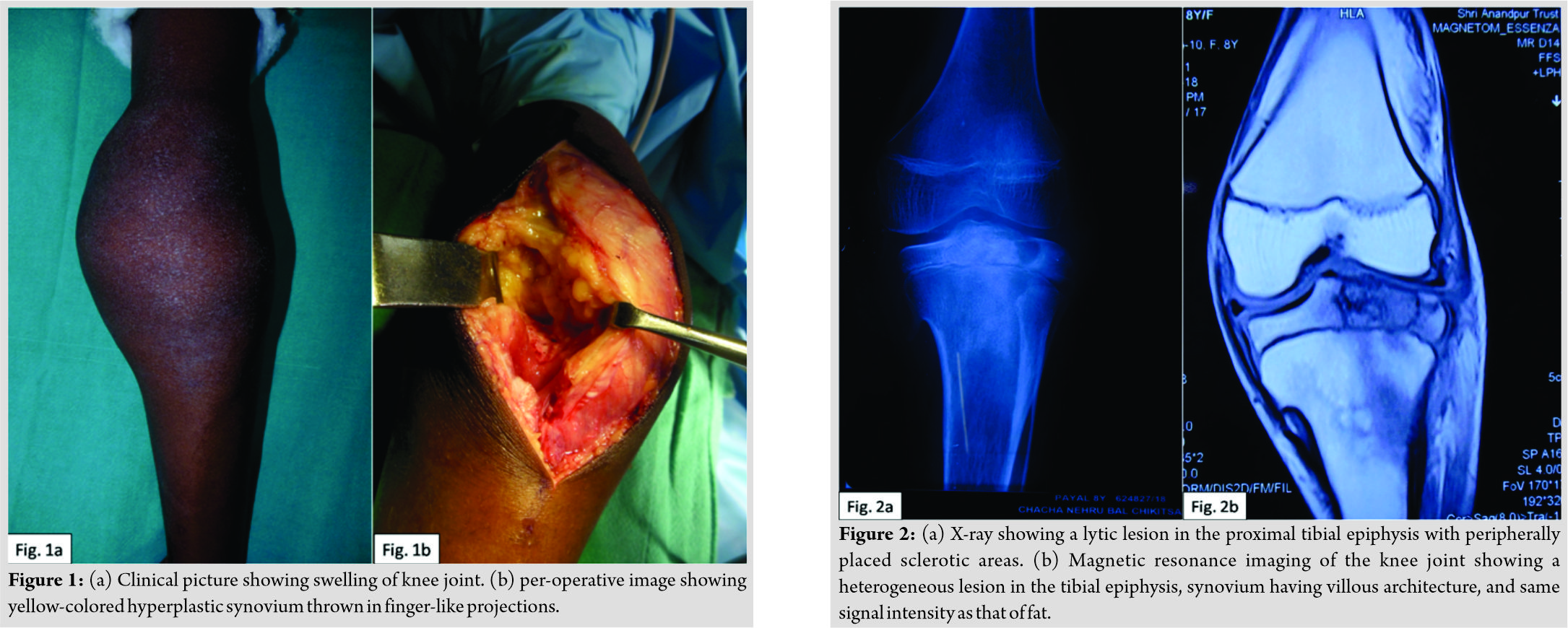
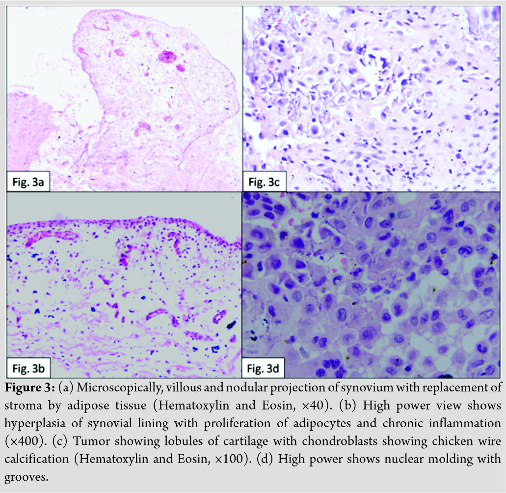
An 8-year-old girl presented to the orthopedics OPD with swelling and pain in the left knee for the past 6 months (Fig. 1a). The swelling had rapidly increased in size in the past 1 month. There was a history of difficult in walking and frequent locking of this knee. No history of trauma or any other joint involvement could be elicited. No similar history was found in siblings or mother. An X-ray of the knee joint was advised which showed a lytic lesion in the proximal tibial epiphysis with peripherally placed sclerotic areas (Fig. 2a). X-ray did not reveal any significant joint effusion and in view of marked joint swelling, a magnetic resonance imaging (MRI) of knee joint was done. The MRI showed a heterogeneous lesion in the tibial epiphysis with altered signal intensity and a sclerotic rim (Fig. 2b).
Discussion
Synovial lipomatosis is a rare benign proliferation of mature adipose tissue cells in the synovial membrane. The most common site involved is suprapatellar pouch of knee joint. It is also known as lipoma arborescens and diffuse articular lipomatosis [3]. In 1904, this entity was first reported by Albert Hoffa, hence, the name Hoffa’s disease. Later on, the more elaborate term villous lipomatous proliferation of the synovial membrane as it is not a true neoplasm [3]. It commonly affects the older adults with median age of 50 years and very rarely does it affects the pediatric age group [1]. The males are more commonly affected than females [4]. The present case is a pediatric age female who presented with massive knee joint effusion. Radiological investigations aided in reaching the diagnosis of a lytic lesion with reactive synovium. Histopathological examination revealed synovial lipomatosis possibly secondary to chondroblastoma. The etiopathogenesis of this entity is still debatable and several theories are proposed. Rao et al. in their study proposed that individuals with high body mass index (BMI) are at increased risk for synovial lipomatosis due to excess fat deposition. This has been supported by observations of lipomatosis in multiple joints of a patient with short bowel syndrome [1]. However, the present case is an 8-year-old female with normal BMI of 19 kg/m2. The literature also describes its occurrence secondary to trauma, inflammation, degenerative joint conditions, and tumor’s emphasizing its metaplastic nature [5]. The clinical features of synovial lipomatosis include swelling in the affected joint, moderate intensity pain, and ipsilateral joint effusion. The joint effusion usually results from proliferation of synoviocytes. Pain in the joint may be due to the pressure effect of proliferating synovium or due to eroded articular cartilage secondary to any primary joint disorder. When knee joint is involved, it can even cause locking, joint line tenderness, and debilitation [1]. Our patient also presented with swelling, pain, and frequent locking of the knee. The biochemical and culture analysis of the joint aspirate in such cases usually reveals no abnormality. X-ray is not diagnostic and the lesion may be missed, especially when focal and small in size. Ultrasonography shows a wavelike motion in cases of lipomatosis associated with intra-articular effusion [6]. MRI is the radiological investigation of choice for differentiating synovial lipomatosis from its closer mimics [5, 7]. Synovial lipomatosis characteristically shows fatty villous synovial proliferation without other signal intensities within the lesion on MRI [7]. Microscopically, it is characterized by diffuse involvement of the synovium with replacement by mature fat cells; however, sometimes, it can present as a solitary pseudomass [4, 7]. Another important characteristic feature is the presence of chronic mononuclear inflammatory cell infiltrate in the synovial tissue, indicating its origin secondary to chronic inflammatory conditions [4]. The present case also showed the presence of inflammatory cells along with fatty replacement of synovium. The most common radiological and histopathological differential for synovial lipomatosis is synovial or intra-articular lipoma [6]. The common site of involvement for both is knee joint but can also involve hip, lumbar spine, and tarsometatarsal joint [8]. Clinical, radiological, and histopathological examination of the excised tissue has paramount importance in differentiating synovial lipomatosis from its other very close mimics, which is highlighted in Table 1 [1, 3, 4, 7, 8]. Asymptomatic cases need no intervention; however, symptomatic synovial lipomatosis can be temporarily treated with intra-articular corticosteroids, and the definitive treatment in such cases is arthroscopic excision of the synovium. Patients with extensive synovium involvement require open synovectomy [3]. This case report also intends to highlight the importance of effective clinical, radiological, and histopathological communication to reach to a diagnosis which not only affects patient treatment but also its prognosis.
Conclusion
This article aims at increasing the awareness of this entity among radiologists and orthopedic surgeons that although synovial lipomatosis is a very rare intra-articular lesion, it should be considered as differential diagnosis in any patient with any age presenting with a chronic soft boggy joint swelling. When diagnosed and treated on time, it will decrease the morbidity and joint abnormalities.
Clinical Message
Synovial lipomatosis is a rare entity but can be seen coexistent to non-neoplastic and neoplastic lesions of bones, contributing to the morbidity of the disease. MRI is the gold standard investigation for establishing the diagnosis. Early detection on radiology ensures timely intervention and reduced future joint abnormalities.
References
1. Rao S, Rajkumar A, Elizabeth MJ, Ganesan V, Kuruvilla S. Pathology of synovial lipomatosis and its clinical significance. J Lab Physicians 2011;3:84-8.
2. Yang J, Tian W, Zhu X, Wang J. Chondroblastoma in the long bone diaphysis: A report of two cases with literature review. Chin J Cancer 2012;31:257-64.
3. De Vleeschhouwer M, Van Den Steen E, Vanderstraeten G, Huysse W, De Neve J, Bossche LV. Lipoma Arborescens: Review of an uncommon cause for swelling of the knee. Case Rep Orthop 2016;2016:5.
4. Kloen P, Keel SB, Chandler HP, Geiger RH, Zarins B, Rosenberg AE. Lipoma Arborescens of the knee. J Bone Joint Surg 1998;80:4.
5. Al-Ismail K, Torreggiani WC, Al-Sheikh F, Keogh C, Munk PL. Bilateral lipoma Arborescens associated with early osteoarthritis. Eur Radiol 2002;12:2799-802.
6. Kheok SW, Ong KO. Benign periarticular, bone and joint lipomatous lesions. Singapore Med J 2017;58:521-7.
7. Vilanova JC, Barceló J, Villalón M, Aldomà J, Delgado E, Zapater I, et al. MR imaging of lipoma Arborescens and the associated lesions. Skeletal Radiol 2003;32:504-9.
8. Poorteman L, Declercq H, Natens P, Wetzels K, Vanhoenacker F. Intra-articular synovial lipoma of the knee joint. BJR Case Rep 2015;1:20150061.
 |
 |
 |
 |
| Dr. Arti Khatri | Dr. Nidhi Mahajan | Dr. Anil Agarwal | Dr. Neeraj Gupta |
| How to Cite This Article: Khatri A, Mahajan N, Agarwal A, Gupta N. Synovial Lipomatosis with Chondroblastoma in an 8-year-old Female: A Previously Unreported Entity. Journal of Orthopaedic Case Reports 2019 Jul-Aug; 9(4): 22-25. |
[Full Text HTML] [Full Text PDF] [XML]
[rate_this_page]
Dear Reader, We are very excited about New Features in JOCR. Please do let us know what you think by Clicking on the Sliding “Feedback Form” button on the <<< left of the page or sending a mail to us at editor.jocr@gmail.com


