[box type=”bio”] Learning Point of the Article: [/box]
Osteochondroma of proximal ulna is a rare presentation that demands clinical suspicion and extraperiosteal excision is performed once it turns symptomatic.
Case Report | Volume 10 | Issue 6 | JOCR September 2020 | Page 1-4 | Divesh Jalan, Saumya Agarwal, Shivank Prakash. DOI: 10.13107/jocr.2020.v10.i06.1850
Authors: Divesh Jalan[1], Saumya Agarwal[1], Shivank Prakash[1]
[1]Department of Orthopaedics, Vardhman Mahavir Medical College and Safdarjung Hospital, New Delhi, India.
Address of Correspondence:
Dr. Saumya Agarwal,
Central Institute of Orthopaedics, Vardhman Mahavir Medical College and Safdarjung Hospital, New Delhi, 110029. India.
E-mail: saumyathecalyx.agarwal@gmail.com
Abstract
Introduction: Osteochondroma is a most common primary bone tumor which forms due to exophytic protuberance on the surface of growing bones. Proximal ulna is an atypical location for osteochondroma. The case report is a rare solitary presentation and has been only twice reported in the literature.
Case Report: This case reports a rare presentation of a 35 year old Hindu male with osteochondroma at proximal ulna which is painful with terminal restriction of the elbow joint movement. Surgical excision was performed and histopathology confirmed the diagnosis. Patient was asymptomatic with full range of movement of the elbow joint at 2 months of follow-up and there were no signs of recurrence at 2 years of follow-up.
Conclusion: Atypical presentation is a possibility; therefore, surgeon should always keep in mind the possibility of the tumor and accurately diagnose the tumor with the help of imaging modalities and biopsy.
Keywords: Osteochondroma, proximal ulna, bone tumor, elbow, excision.
Introduction
According to the World Health Organization, osteochondromas are bony projections enveloped by a cartilage cover that arise on the external surface of the bone [1]. Osteochondroma occurs in 3% of the general population and accounts for more than 30% of all benign bone tumors and 10–15% of all bone tumors [1]. Despite their predominant bony composition, their growth takes place in the cartilaginous portion [2]. Osteochondromas are commonly identified during childhood or adolescence [1]. Here, we present a case of osteochondroma in a 35-year-old male which is an unusual age for this tumor. Osteochondromas more frequently affect the appendicular skeleton (upper and lower limbs) [3]. The long bones of the lower limbs are most commonly affected, especially the knee (40%), the proximal femur, and the humerus (26%) [3]. Proximal ulna is an atypical location for osteochondroma, and this type of rare presentation has been only reported twice in the literature.
Case Report
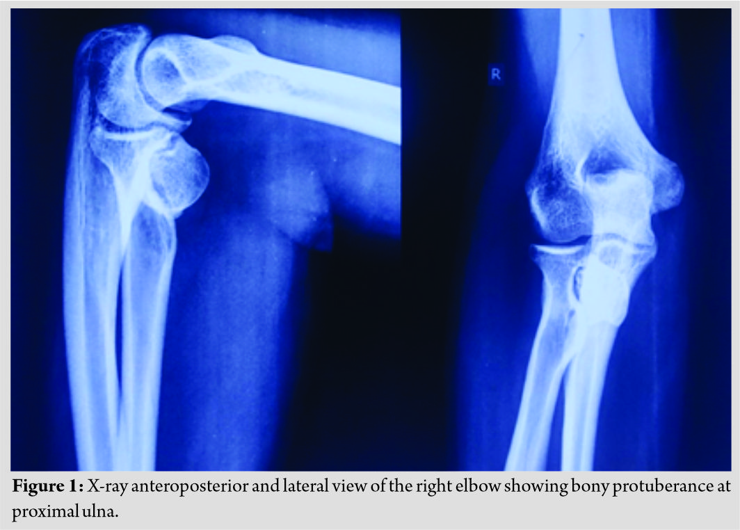
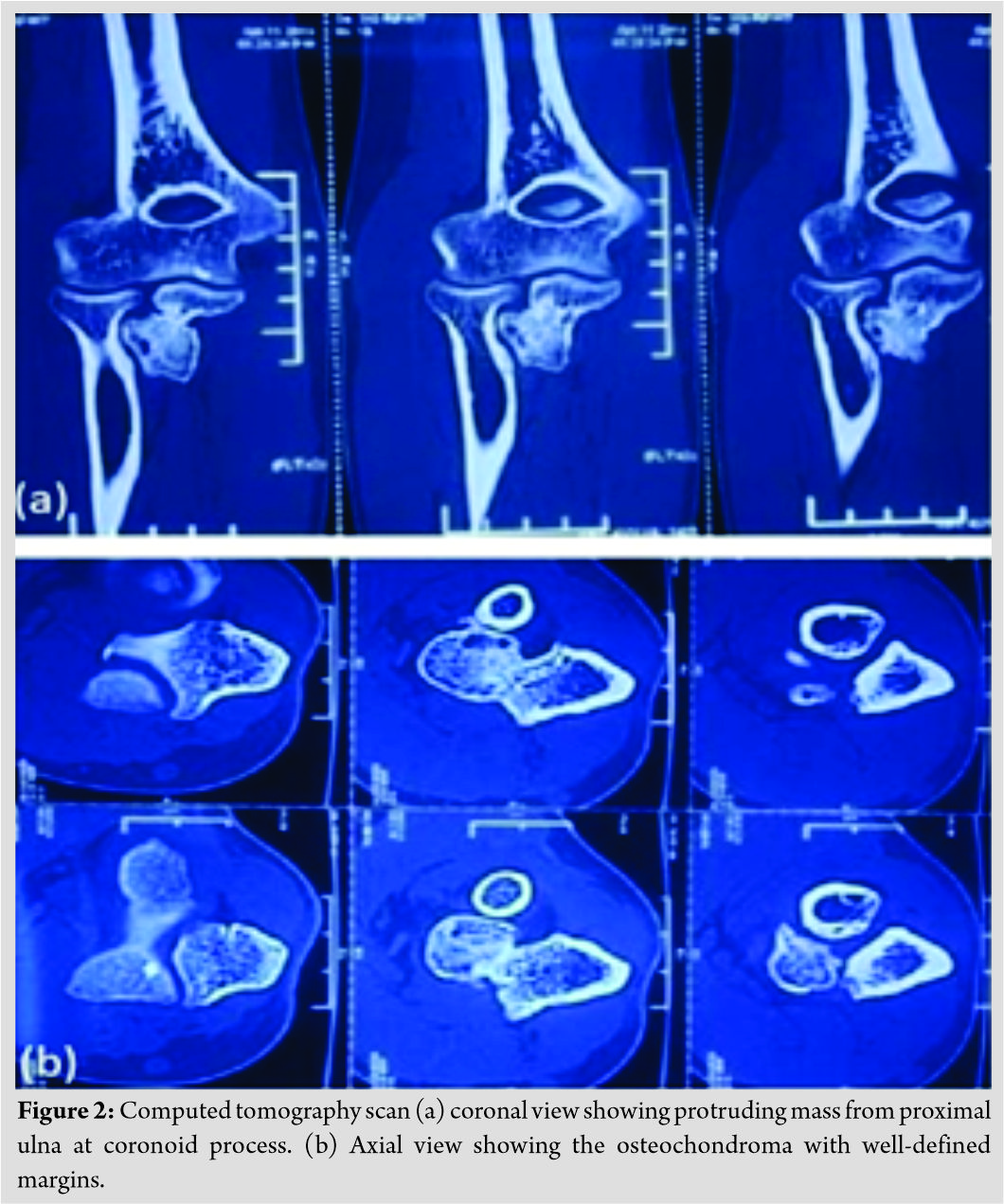
We present a case of a 35-year-old male who came to the orthopedics outdoor patient department with a complaint of swelling in the right elbow and restricted range of movement of elbow joint for 3 months. On local examination, a swelling was found, which was of 3 cm × 3 cm in size, hard, non-fluctuant, not mobile, was attached to the underlying bone, non-tender, with normal skin without any neurovascular deficit. A terminal restriction of the flexion of the right elbow joint was noticed, which was about 130 degrees and restricted pronation was found to be 65 degrees at proximal radio-ulnar joint while other movements were found to be normal. A provisional diagnosis was made based on clinical findings, radiographs, and a computed tomography (CT) scan. X-ray (anteroposterior and lateral view) (Fig. 1) of the right elbow showed a solitary external bony protuberance at proximal ulna around 5 mm distal to the coronoid process.


Discussion
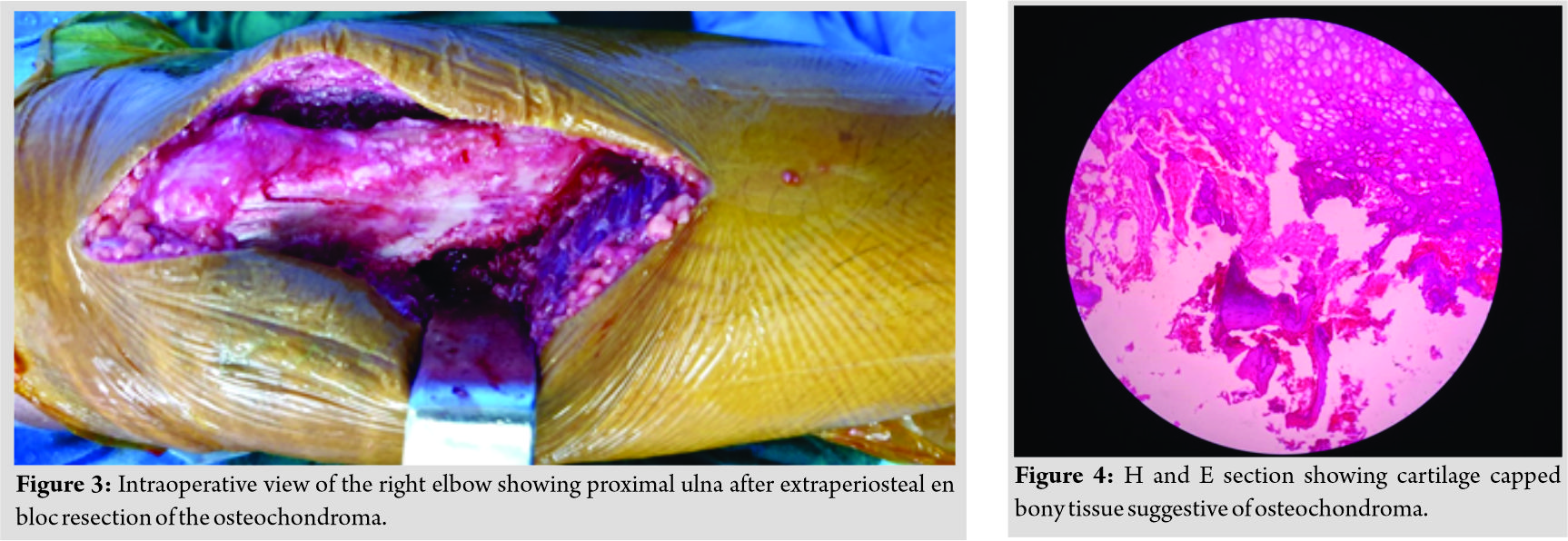
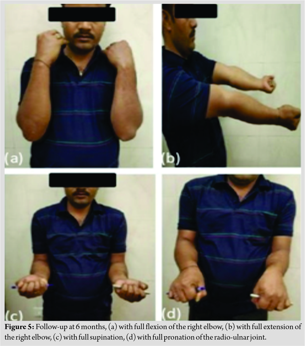
Osteochondroma is a most common primary bone tumor which manifests as two different forms, solitary osteochondromas also known as exostosis and multiple osteochondromas [3]. Solitary osteochondromas constitute 10% of all bone tumors and 35% of the benign tumors [1]. Single lesions are found in 85% of the individuals diagnosed with osteochondroma [3]. Approximately 15% of osteochondromas occur in the context of hereditary multiple osteochondromas, a disorder that is inherited in an autosomal dominant manner [4]. with a positive family history and/or mutation in one of the EXT genes (EXT1, EXT2, and EXT3) which are found in chromosomes 8, 11, and 19, respectively [5]. Despite the slight predominance of the male gender over the female gender that has been reported by some authors, it seems that there is no effective predilection according to sex [1]. The usual location is in the metaphysis and rarely in the diaphysis [6]. Flat bones such as the scapula and hip may also be involved [3]. The cause of osteochondromas remains unknown [1]. Separation of a fragment of growth cartilage followed by endochondral ossification and continuous growth leads to the formation of an outgrowth that projects from the bone surface, coated with a covering of cartilage, and the growth ceases once the skeletal maturity is reached [6]. Among solitary osteochondromas, the vast majority are asymptomatic [7]. In fact, they are usually discovered by chance. An osteochondroma can occur near a nerve or blood vessel, the most common being the popliteal nerve and artery. The affected limb can exhibit numbness, weakness, loss of pulse, or changes in color [8]. The tumor can be found under a tendon, resulting in pain during relevant movement and thus causing restriction of joint motion [9]. Rapidly increasing lesion size and local pain processes suggest that sarcomatous transformation is occurring in individuals with osteochondroma that was previously asymptomatic [1]. On radiographs the characteristic image consists of an external bone protuberance [1], and it may have a wide base (sessile) or a narrow base (pedicled or pedunculated). Singular appearances are most commonly diagnosed with a biopsy. CT shows details of the continuity of the cortical and spongy bone inside the lesion, and their relationship with the adjacent soft tissue. Axial tomographic slices facilitate the interpretation of the lesions located in anatomical sites of greater complexity [10]. Magnetic resonance well demonstrates the cortical and medullary continuity between the osteochondroma and host bone [11]. In the same way, as seen in a normal piece of bone, the cortical bone of the exostosis presents low signal intensity (hyposignal) in all sequences, whereas the medullary component continues to have the appearance of the yellow medulla [6]. Macroscopically, lesion surface is lobulated and has an abundant cartilaginous cover [2] and microscopically solitary and multiple osteochondromas are histologically similar [12]. The lesion presents three layers [1]: Perichondrium (most external), cartilage (intermediate), and bone (most internal). Differentiation from normal cartilage is generally done in relation to secondary chondrosarcoma of low-grade malignity [12]. Patients who are asymptomatic are managed conservatively, while surgical removal is indicated if the tumor causes pain or functional incapacity, either due to neurovascular compression or due to limitation of joint movement and if there is a fracture of base of the osteochondroma. A review of the literature showed only two cases of osteochondroma at proximal ulna. Hamada et al. [13] presented a case of osteocartilaginous mass involving the proximal of ulna in a 4-year-old girl with swelling, deformity, and restricted range of motion at the elbow joint and forearm with developmental dislocation of the radial head with intact neurology. She was treated with complete excision of the tumor mass and trapezoidal shortening osteotomy at radial neck, followed by oblique ulnar osteotomy. Excellent results were obtained with pain-free and full range of motion. Another case was presented by Jaganathan and Sivaprasath [14], in which there was an osteochondroma at proximal ulna with cubitus valgus deformity and tardy ulnar nerve palsy in a 12-year-old girl. She presented with a painless swelling with a full range of motion and carrying angle of 25° and ulnar clawing and Froment’s sign positive. Extra-periosteal excision was done followed by anterior transposition of the ulnar nerve. At 6 months follow up, the patient was asymptomatic with full range of motion without any neurovascular deficit. We presented a case of osteochondroma of the proximal ulna in a 35-year-old male which is not a usual age of presentation for osteochondroma. There was no associated deformity or any neurovascular deficit. We performed extra-periosteal excision, and the patient became asymptomatic with a full range of motion. Life expectancy is not affected by osteochondromas as they are benign lesions. However, there is always a risk of malignant transformation (to secondary chondrosarcoma). An extra-periosteal excision is the treatment of choice and completely removes the tumor, but if the tumor is not completely excised, then there are chances of recurrence. The overall survival of patients with sarcomatous transformation is generally good. However, those with poorly differentiated lesions have a much worse prognosis [15].
Conclusion
Osteochondromas are most commonly found in the metaphysis of long tubular bones, especially tibia or femur, and proximal humerus, but atypical presentation is a possibility; therefore, surgeon should always keep in mind the possibility of the tumor and accurately diagnose the tumor with the help of imaging modalities and biopsy. Most of the cases are asymptomatic and are advised conservative treatment, but in patients with painful swelling and restricted joint movement with or without neurovascular deficit, extraperiosteal bony excision remains the mainstay of the treatment.
Clinical Message
Osteochondroma can present at rare sites like proximal ulna, which require clinical suspicion and radiological and histopathological confirmation. Extraperiosteal excision is required when the lesion is symptomatic with pain, restriction of movement or any neurological deficit.
References
1. Khurana J, Abdul-Karim F, Bovée JV. Osteochondroma. In: Fletcher CD, Unni KK, Mertens F, editors. Pathology and Genetics of Tumours of the Soft Tissues and Bones. Lyon: IARC Press; 2002. p. 234-7.
2. Unni KK. Osteochondroma. Dahlin’s Bone Tumors: General Aspects and Data on 11, 087 Cases. 5th ed. Thomas; Springfield: 1996. p. 11-23.
3. Dorfman HD, Czerniak B. Osteochondroma. Bone Tumors. St. Louis: Mosby; 1998. p. 331-46.
4. Saglik Y, Altay M, Unai VS, Basari K, Yildiz Y. Manifestations and management of osteochondromas: A retrospective analysis of 382 patients. Acta Orthop Belg 2006;72:748-55.
5. Wu YQ, Heutink P, de Vries BB, Sandkuijl LA, van den Ouweland AM, Niermeijer MF. Assignment of a second locus for multiple exostoses to the pericentromeric region of chromosome 11. Hum Mol Genet 1994;3:167-71.
6. Murphey MD, Choi JJ, Kransdorf MJ, Flemming DJ, Gannon FH. Imaging of osteochondroma: Variants and complications with radiologic-pathologic correlation. Radiographics 2000;20:1407-34.
7. Giudici MA, Moser RP Jr., Kransdorf MJ. Cartilaginous bone tumors. Radiol Clin North Am 1993;31:237-59.
8. Van Oost J, Feyen J, Opheide J. Compartment syndrome associated with an osteocartilaginous exostosis. Acta Orthop Belg 1996;62:233-5.
9. Bovée JV, Hogendoorn PC. Multiple osteochondromas. In: Fletcher CD, Unni KK, Mertens F, editors. World Health Organization Classification of Tumours. Pathology and Genetics of Tumours of Soft Tissue and Bone. Lyon, France: IARC; 2002. p. 360-2.
10. Kenney PJ, Gilula LA, Murphy WA. The use of computed tomography to distinguish osteochondroma and chondrosarcoma. Radiology 1981;139:129-37.
11. Resnick D, Kyriakos M, Greenway GD. Osteochondroma. In: Resnick D, editor. Diagnosis of Bone and Joint Disorders. 3rd ed. Philadelphia, PA: Saunders; 1995. p. 3725-46.
12. Shah ZK, Peh WC, Wong Y, Shek TW, Davies AM. Sarcomatous transformation in diaphyseal aclasis. Australas Radiol 2007;51:110-9.
13. Hamada Y, Hibino N, Horii E. Radial head dislocation due to gigantic solitary osteochondroma of the proximal ulna: Case report and literature review. Hand (NY) 2015;10:305-8.
14. Jaganathan SJ. A rare case of osteochondroma proximal ulna with cubitus valgus deformity and tardy ulnar nerve palsy. Univ J Surg Surg Spec 2019;5:1-4.
15. de Souza AM, Bispo RZ. Osteochondroma: Ignore or investigate? Rev Bras Ortop 2014;49:555-64.
 |
 |
 |
| Dr. Divesh Jalan | Dr. Saumya Agarwal | Dr. Shivank Prakash |
| How to Cite This Article: Jalan D, Agarwal S, Prakash S. Osteochondroma of Proximal Ulna – A rare case presentation. Journal of Orthopaedic Case Reports 2020 September;10(6): 1-4. |
[Full Text HTML] [Full Text PDF] [XML]
[rate_this_page]
Dear Reader, We are very excited about New Features in JOCR. Please do let us know what you think by Clicking on the Sliding “Feedback Form” button on the <<< left of the page or sending a mail to us at editor.jocr@gmail.com


