Clinical and radiological assessment of ankle with high index of suspicion should be done in all the adolescent patients presenting with tibial shaft fractures.
Dr. Vivek Kumar David,
Department of Joint Replacement and Orthopaedics, Tata Main Hospital, Jamshedpur, Jharkhand, India.
E-mail: viv.dav17@gmail.com
Introduction: Triplane fracture of ankle is a rare adolescent injury. Its association with ipsilateral tibial shaft fracture is very uncommon in pediatric orthopedic traumatology and rarely reported in the literature till date. Timely diagnosis and management is required to optimize the outcome and avoid complications.
Case Report: This is a case of a 14-year-old male who sustained a twisting injury to his right leg during early phase of COVID-19 pandemic. He sustained a three-part lateral triplane fracture of the ankle with a concomitant displaced spiral fracture of the shaft of the right tibia. He underwent close reduction under fluoroscopy and above-knee casting for 10 weeks followed patellar tendon weight-bearing cast for 4 weeks. Both fractures healed uneventfully in 14 weeks with patient returning to full activities in 22 weeks.
Conclusion: The ankle injury in adolescent age group (12–15 years) can easily be missed in the presence of the more obvious tibial fracture and therefore, we recommend ankle assessment of all patients with tibial shaft fractures in this age group both clinically and radiologically.
Keywords: Triplane ankle fracture, tibia shaft fracture, adolescent.
Distal tibia physeal injury is the second most common physeal injuries in children associated with long bone fractures [1, 2, 3]. Tibia shaft fracture in children is the third most common long bone shaft fracture [3, 4, 5]. Triplane fractures of the distal tibia represent around 5–7% of all pediatric ankle fractures [1, 6]. However, the association of these two injury patterns in pediatric population is seldom reported [7, 8, 9]. They typically occur in the transitional phase of 12–18 months during adolescence (12–15 years) preceding physeal closure of distal tibia plate due to asymmetric progression [2, 3, 9, 10]. Orthopedic surgeons should be aware of such association of tibia fractures, especially spiral fractures, with ankle injuries. If unrecognized, it can lead to premature physeal closure, angular deformity, and degenerative changes in ankle [11, 12, 13]. However, as these injuries occur almost at the end of physeal growth, they rarely can result in growth arrest/limb length discrepancies [10, 14]. Ankle should be clinically evaluated, and a formal ankle radiography should be a routine. Computerized tomography (CT) scan of ankle should be done in all suspicious cases to exclude or identify any such patterns and plan management [3, 9]. This paper aims to report one such rare association and its outcome with conservative management and judicious follow-ups.
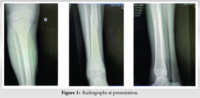
This is a case of a 14-year-old male who sustained a twisting injury to his right leg during early phase of COVID-19 pandemic. He sustained a three-part lateral triplane fracture of the ankle with a concomitant ipsilateral displaced spiral fracture of the shaft at middle-lower third junction of his right tibia (Fig. 1).

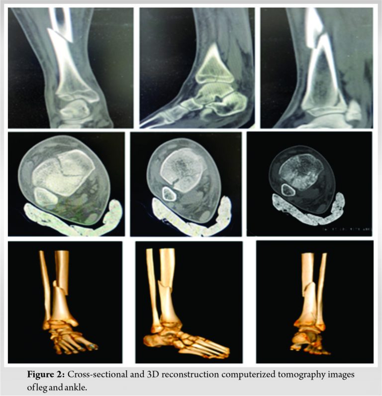
The mechanism of injury was a twisting injury with external rotation of the foot while running indoors. The ankle injury was initially missed in the emergency. The orthopedic team examined the patient the following morning and ordered a CT scan of the ankle on clinical and radiographic suspicion. CT ankle reported the triplane injury pattern (Fig. 2).

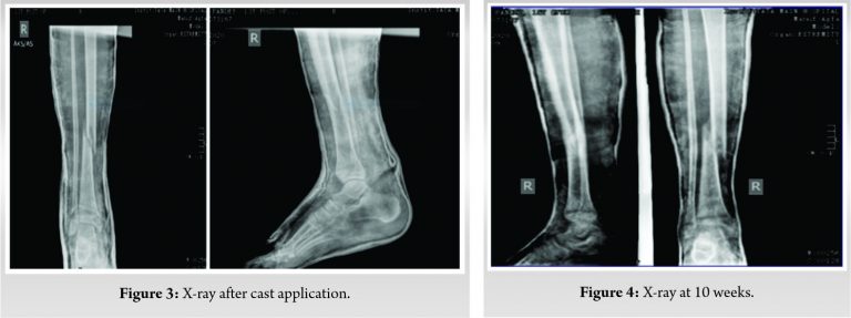
The coronal, sagittal cuts revealed 2 mm gap and axial cut revealed 3 mm gap at the articular surface without any step. Conservative plan of management was decided as the displacement of shaft fracture was <50%, varus/valgus angulation <5o, recurvatum <5 degrees and shortening <1 cm and triplane ankle fracture was without any intra-articular step. The patient underwent close reduction under fluoroscopy and above-knee casting (Fig. 3) and was discharged on day-2. Regular follow-ups were done at weekly intervals for initial 3 weeks post-discharge to check for any fracture displacement and then at 6, 10, 14, 18, and 22 weeks, 6 months, 9 months, and 1 year.

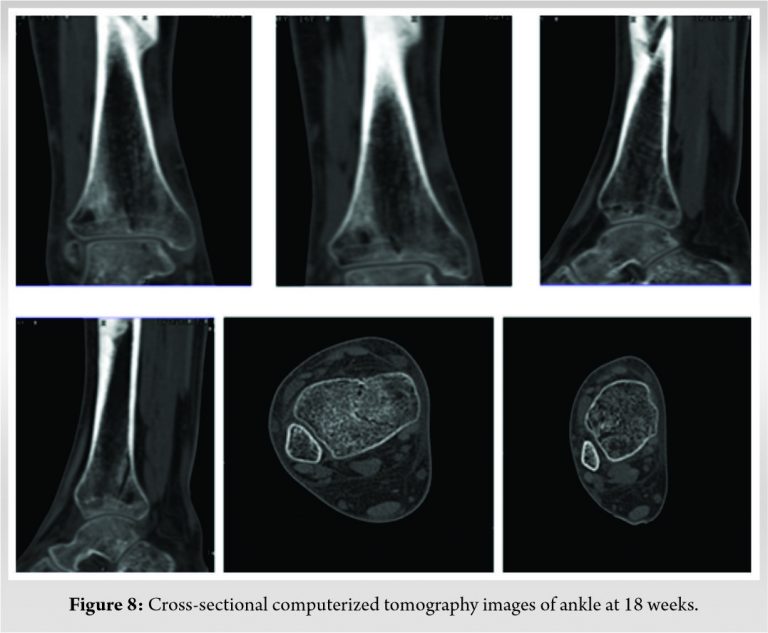
Above-knee plaster cast was converted to a patellar tendon bearing (PTB) cast at the 10th week for another 4 weeks and weight-bearing was started on PTB cast. The shaft fracture showed abundant callus at 14 weeks and follow-up X-rays (Fig. 4, 5, 6, 7).

The triplane fracture was uniting well with no disruption of the tibial plafond (Fig. 8) and an initially open anterolateral physis of the distal tibial gradually closed in the follow-up X-rays. The patient achieved a good functional recovery in 22 weeks and there was no limb length discrepancy at the end of 1 year. Evaluation was done based on modified Weber scale [15] using the pre-operative and post-operative scores for pain, walking, activity, and ankle, subtalar function and awarded clinical demerit points with scores 15/24 at 10 weeks, 10/24 at 14 weeks, 6/24 at 18 weeks, 4/24 at 22 weeks, 2/24 at 6 months, and 0/24 at 9 months.

Physeal injuries in children occur in about 15–30% of long bone fractures, with distal radius being most common and distal tibia being second common, if physeal fractures involving the phalanges are excluded [1, 2, 3]. Tibia shaft fracture in children is the third most common long bone shaft fracture in isolated injuries (after clavicle and forearm) and polytrauma patients (after femur and humerus) as well [3, 4, 5]. Triplane fractures of the distal tibia represent around 5–7% of all pediatric ankle fractures [1, 6]. The term, “complex triplane” fracture has also been utilized to describe triplane ankle injury with ipsilateral diaphyseal tibia shaft fracture [7]. This in children is infrequent and very less reported in literature [7, 8, 9]. Triplane fractures of the distal tibial segment are generally secondary to sports injuries in obese adolescents [13]. However, Tandon et al. conducted an epidemiological study of pediatric trauma in urban scenario of India and reported that majority of pediatric fractures occur in males and at home [16]. The average age of patients is approximately 13 years, although they have been reported in young children as well [6, 17]. It is a transitional period of around 12–18 months when the distal tibial physis closure progresses in a unique asymmetric pattern, from central to medial and then to the lateral. The anterolateral open physis is vulnerable to shear forces during this period at the time of injury [9, 10]. Hence, they are also referred to as transitional fractures. The mechanism of injury is due to internal rotation of leg on a plantar flexed foot fixed firmly on the ground producing torque forces which drives talus against fibula leading to tensioning of anterior inferior tibiofibular ligament and Salter-Harris type III avulsion fracture of open anterolateral physis of distal tibia (biplane fracture of Tillaux). As the forces continue, it leads to triplane pattern at ankle. Further, external rotation of the foot explains spiral/oblique pattern of tibia shaft fracture which commonly occurs at middle and distal third junction [2, 17, 18]. Kasture and Azurza, however, proposed that a more displaced tibia shaft fracture signifies initial failure at shaft followed by triplane pattern at ankle [10]. Triplane injuries are multiplanar, incorporating fracture lines in sagittal, coronal, and axial planes which correspond to Salter-Harris type II, III, and IV types, respectively, and appear as Salter-Harris type III in X-ray ankle anteroposterior view and type-II in lateral view [2, 17, 18]. Triplane fractures can be two parts, three parts, or four parts/comminuted, all reported in literature. They can be lateral triplane (fracture in sagittal plane in epiphysis, axial plane in physis, and coronal plane in metaphysis) or medial triplane (fracture in coronal plane in epiphysis, axial plane in physis, and sagittal plane in metaphysis) [6]. Triplane fractures were first described by Johnson and Fahl in 1957 [19]. In 1970, Marmor identified three distinct fragments that result after these injuries: An anterolateral epiphyseal fragment, a posterior metaphyseal fragment that was attached to the remainder of the epiphysis, and the tibial shaft when they operated on a 12-year-old girl [20]. Lynn coined the term triplane fracture in 1972 when he identified these patterns in two children [21]. After review of literature, we could find only a few articles with 20 cases published till date describing this unique complex triplane fracture and its management. Of these, 11 triplane fractures and 13 tibia shaft fractures were managed conservatively. Peiro et al. reported one case in their study in 1981 which was managed conservatively [22]. Rapariz et al. found that 48% of triplane fractures were associated with a fractured fibula and 8.5% were associated with an ipsilateral tibia shaft fracture. They published a case series in 1996 in which three cases had combined injuries and two were managed conservatively and one with external fixation [23]. Jarvis and Miyanji published a case series of six patients of these combined injuries in 2001 where the author managed these fractures conservatively with good outcome [7]. Morgan and Jimenez published a single case report in 2003 where ankle was managed conservatively, and tibia shaft was operated [18]. Rico-Pecero and Dwyer published a case report in 2009 in a 13-year-old girl with Gilbert syndrome where unreducible shaft fracture of tibia was managed with internal fixation and ankle was managed conservatively [17]. De Rover et al. also reported a case of similar fracture without the involvement of the fibula in 2011. This injury was treated operatively with good result [12]. Kasture and Azurza in 2017 reported a similar case and managed it by surgical fixation [10]. Holland et al. published a case series of five patients in 2018 where all the triplane injuries were surgically treated and four of five tibia shaft were managed conservatively. They also found an incidence of concurrence of tibia shaft fracture in triplane ankle injuries to be 8.5% [11]. Sferopoulos published an article of a similar case in international journal of radiology in 2018 wherein the patient was managed conservatively [2]. Healy et al. reported a triplane fracture associated with a proximal fibula fracture and syndesmotic injury (Maisonneuve equivalent) [24]. Sheffer et al., however in 2020, found an incidence of 2.15% of triplane injuries associated with tibia shaft fracture in a retrospective analysis of 517 fractures [25]. The treatment protocols vary with simple plaster cast immobilization, open/closed reduction with or without internal or external fixation all being described in the literature [6, 12]. Oblique or spiral tibial fractures in children are known to unite with good outcomes even with intact fibula when treated with a well-molded plaster cast. A comparative study on the clinical and radiographic outcome of triplane fractures was conducted by Ryu et al. in 2017 to find out the need of operative intervention if the intra-articular displacement is more than 2 mm. After a 2-year follow-up and assessment by the Ankle-Hindfoot scale and the modified Weber protocol score, the author did not find any statistically significant difference in the outcome of non-operative and operative groups [26]. We believe that in a normal scenario, all intra-articular fractures with a displacement of 2 mm or more should be internally fixed. However, as this case presented in early phase of COVID-19 pandemic, there was unavailability of definitive testing for SARS-CoV-2 and no surgical guidelines were framed yet. Following the institutional and departmental protocols and after thorough discussion with the patient’s family, we decided to manage our patient conservatively considering overall acceptable alignment of the limb and no articular step in the tibial plafond. The patient was judiciously followed-up, assessed and the outcome was satisfactory with patient resuming all activities pain free.
The triplane fractures can be easily overlooked in the presence of the more obvious tibial fracture and have the potential of serious sequelae such as chronic pain, deformity, and degenerative post-traumatic arthritis, if it is missed. As this injury is peculiar to the adolescent age group, we recommend ankle assessment of all patients with tibial shaft fractures in this age group both clinically and radiologically.
Careful selection of patient, establishing a good communication with the patient’s family and judicious follow-up in the surgical era, still allows conservative approach an option with good outcome.
References
- 1.Mizuta T, Benson WM, Foster BK, Paterson DC, Morris LL. Statistical analysis of the incidence of physeal injuries. J Pediatr Orthop 1987;7:518-23. [Google Scholar]
- 2.Sferopoulos NK. Concomitant tibia shaft and distal triplane fractures. Int J Radiol 2018;5:197-201. [Google Scholar]
- 3.Wuerz TH, Gurd DP. Pediatric physeal ankle fracture. J Am Acad Orthop Surg 2013;21:234-44. [Google Scholar]
- 4.Hedström EM, Svensson O, Bergström U, Michno P. Epidemiology of fractures in children and adolescents. Acta Orthop 2010;81:148-53. [Google Scholar]
- 5.Joeris A, Lutz N, Blumenthal A, Slongo T, Audigé L. The AO pediatric comprehensive classification of long bone fractures (PCCF). Acta Orthop 2017;88:129-32. [Google Scholar]
- 6.Kay RM, Matthys GA. Pediatric ankle fractures: Evaluation and treatment. J Am Acad Orthop Surg 2001;9:268-78. [Google Scholar]
- 7.Jarvis JG, Miyanji F. The complex triplane fracture: Ipsilateral tibial shaft and distal triplane fracture. J Trauma 2001;51:714-6. [Google Scholar]
- 8.Patel NK, Horstman J, Kuester V, Sambandam S, Mounasamy V. Pediatric tibial shaft fractures. Indian J Orthop 2018;52:522-8. [Google Scholar]
- 9.Jung KJ, Chung CY, Park MS, Chung MK, Lee DY, Koo S, et al. Concomitant ankle injuries associated with tibial shaft fractures. Foot Ankle Int 2015;36:1209-14. [Google Scholar]
- 10.Kasture S, Azurza K. Triplane ankle fracture with concomitant ipsilateral shaft of tibia fracture: Case report and review of literature. J Orthop Case Rep 2017;7:84-7. [Google Scholar]
- 11.Holland TS, Prior CP, Walton RD. Distal tibial triplane fracture with ipsilateral tibial shaft fracture: A case series. Surgeon 2018;16:333-8. [Google Scholar]
- 12.De Rover WB, Alazzawi S, Hallam PJ, Walton NP. Ipsilateral tibial shaft fracture and distal tibial triplane fracture with an intact fibula: A case report. J Orthop Surg (Hong Kong) 2011;19:364-6. [Google Scholar]
- 13.Tan AC, Chong RW, Mahadev A. Triplane fractures of the distal tibia in children. J Orthop Surg (Hong Kong) 2013;21:55-9. [Google Scholar]
- 14.Crawford AH. Triplane and Tillaux fractures: Is a 2 mm residual gap acceptable? J Pediatr Orthop 2012;32 Suppl 1:S69-73. [Google Scholar]
- 15.Thomas JD. Early postoperative weight-bearing and muscle activity in patients who have a fracture of the ankle. J Bone Joint Surg Am 1989;71:1430-1. [Google Scholar]
- 16.Tandon T, Shaik M, Modi N. Paediatric trauma epidemiology in an urban scenario in India. J Orthop Surg (Hong Kong) 2007;15:41-5. [Google Scholar]
- 17.Rico-Pecero J, Dwyer A. Triplane fracture of the ankle associated with homolateral tibial fracture in a teenager. A case report. Rev Española Cir Ortop Traumatol 2009;53:254-6. [Google Scholar]
- 18.Morgan JH Jr., Jimenez AL. Triplane fracture of the distal tibia with ipsilateral tibial shaft fracture: A case report. 2008;56:309-15. [Google Scholar]
- 19.Johnson EW, Fahl JC. Fractures involving the distal epiphysis of the tibia and fibula in children. Am J Surg 1957;93:778-81. [Google Scholar]
- 20.Marmor L. An unusual fracture of the tibial epiphysis. Clin Orthop 1970;73:132-5. [Google Scholar]
- 21.Lynn MD. The triplane distal tibial epiphyseal fracture. Clin Orthop 1972;86:187-90. [Google Scholar]
- 22.Peiro A, Aracil J, Martos F, Mut T. Triplane distal tibial epiphyseal fracture. Clin Orthop 1981;160:196-200. [Google Scholar]
- 23.Rapariz JM, Ocete G, González-Herranz P, López-Mondejar JA, Domenech J, Burgos J, et al. Distal tibial triplane fractures: Long-term follow-up. J Pediatr Orthop 1996;16:113-8. [Google Scholar]
- 24.Healy WA 3rd, Starkweather KD, Meyer J, Teplitz GA. Triplane fracture associated with a proximal third fibula fracture. Am J Orthop (Belle Mead NJ) 1996;25:449-51. [Google Scholar]
- 25.Sheffer BW, Villarreal ED, Ochsner MG 3rd, Sawyer JR, Spence DD, Kelly DM. Concurrent ipsilateral tibial shaft and distal tibial fractures in pediatric patients: risk factors, frequency, and risk of missed diagnosis. J Pediatr Orthop 2020;40:e1-5. [Google Scholar]
- 26.Ryu SM, Park JW, Kim SD, Park CH. Is an operation always needed for pediatric triplane fractures? Preliminary results. J Pediatr Orthop B 2018;27:412-8. [Google Scholar]


