Periosteal ganglionic cyst of long bones are uncommon presentations in orthopedic outdoors, and once diagnosed can be managed with enbloc excision
Dr. Nandlal Bharwani, Department of Orthopaedics, Dr. Sampurnanand Medical College, Jodhpur, Rajasthan, India. E-mail: nandlalbharwani21@gmail.com
Introduction: Periosteal ganglion is a form of cystic swelling commonly seen around long bones of lower extremities.
Case Report: A 55-year-old male presented to outdoor clinic with gradually increasing swelling around anteromedial aspect of the right knee joint with intermittent pain on prolonged standing and walking for 8 months. Magnetic resonance imaging was suggestive of ganglionic cyst which was confirmed later by histopathological examination.
Conclusion: Ganglionic cyst of periosteal origin is a rare entity. Complete excision is the recommended treatment option; if not performed appropriately chances of recurrence are high.
Keywords: Knee joint, periosteal ganglion cyst, cystic swelling, pes anserine bursitis, long bone
A ganglionic cyst is defined as a mass filled of gelatinous material rich in mucopolysaccharides and hyaluronic acid surrounded by fibrous tissue. These are commonly encountered near joints of wrist and foot originating from the tendon sheaths [1]. Ganglion cyst is further classified as extra-articular, intra-articular, intra-osseous, and periosteal type (rare) [2]. Periosteal myxomatous degeneration is the pathological process behind the origin of periosteal ganglionic cyst that commonly involves long bones of lower extremities [3]. Occasionally, this entity presents with extension into soft tissue, cortical erosion, and periosteal reaction [4]. For accurate diagnosis, radiological investigations such as digital radiographs and magnetic resonance imaging (MRI) along with histopathological examination (HPE) are of utmost value [3, 5]. Till date, very few literature on this rare presentation is available. We report a clinical case scenario of ganglionic cyst located in proximal tibia region without any ligamentous extension along with radiological investigations presenting as pes anserine bursitis.
A 55 year-old male patient presented to outdoor clinic with gradually increasing swelling on anteromedial aspect of the right knee joint with intermittent episodes of pain on prolonged standing and walking since 8 months. Proper history, clinical examination, digital radiographs, and routine investigations were performed. Digital radiographs (anteroposterior and lateral views) showed swelling arising from anteromedial aspect and lucency around cortex of tibia (Fig. 1). Thereafter, MRI of knee joint was performed which showed well-defined cystic lesion with internal septations arising from medial aspect of tibia with soft-tissue extension and measured around 4 × 3 cm (Fig. 2).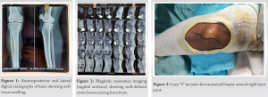 Informed consent was taken before starting the treatment. Preoperatively, examination showed cystic swelling on anteromedial aspect of the right knee with fixity to underlying bone; there was no restriction of range of motion. Thereafter, excisional biopsy was planned. The patient was laid supine in operation theater, neuraxial anesthesia was given, painting and draping were done, and tourniquet was inflated to 320 mm Hg pressure. Then, lazy-S skin incision was made around anteromedial aspect of knee, subcutaneous dissection was carried out along with complete en bloc excision which showed cystic mass arising from periosteum of proximal tibia. Intergrity of medial collateral ligament was confirmed after the procedure (Fig. 3 and 4). Thorough wash was given followed by closure in layers, range of motion and weight-bearing was allowed from next day onwards. Size of excised tissue measured around 4.2 × 3 × 1 cm. It was sent for HPE, gram’s staining, and fungal culture (Fig. 5).
 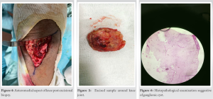 Diagnosis was confirmed with HPE reports showing gelatinous material within cyst along with presence of pseudo-synovial cells and periosteum with fibrous tissue on outer aspect of cavity suggestive of ganglionic cyst of periosteal origin (Fig. 6). Knee joint ROM was normal at final follow-up. The patient was followed for 1-year postoperatively and period was uneventful.
Around 18th century, Poncet and Olliers coined the term “periostitis albuminosa” for ganglionic cyst of periosteal origin. Although rare, it occurs most commonly around proximal tibia around pes-anserinus region, while some other locations are distal radius, ulna, ilium, and medial malleolus [6, 7]. We searched PubMed and Google Scholar with the following key words: “Periosteal ganglionic cyst” and “long bone“. We included the search results from 2012 to 2022 and included all types of studies. Out of the search results, we found five relevant case reports. Out of five cases, four of them were males [7, 8, 9, 10] and one female [11] aged 35–62 years with mean age (49.6 years). The disease duration was from 1 month to 10 years. In all the cases, long bones were involved except one in which iliac crest involvement was noted. The most common noted symptom was swelling which was progressive with time. In all cases, surgery for cyst removal was successful except in one case for which aspiration was performed. The complete details related to onset, signs, symptoms, local examination, radiological findings, HPE findings, and complete follow-up results are shown in Table 1. The studies [8, 9, 11] in review of literature showed involvement of some rare locations such femur condyle, iliac crest, and distal end radius. This entity showed slight male predominance and seen commonly around 4th–5th decade of life as shown in the present case scenario [12]. Although the etiology of this rare condition is still unknown, some suggest trauma and repetitive injury as probable causes, which leads to myxomatous degeneration of periosteum leading to formation of gelatinous fluid [3]. In the present case scenario and review of literature mentioned above, prior history was noted in only one case [11]. This leads to presentation of periosteal ganglion as diffuse cystic swelling with tenderness, soft-tissue extension, cortical erosion, and periosteal reaction [4,12]. Thus, proper clinical examination, radiological investigations, and HPE are of utmost importance for confirm at orydiagnosis. Although several treatment modalities for periosteal ganglion are available, complete excision along with the surrounding periosteum is the recommended option. In this rare entity, chances of recurrence are high if my xomatous degeneration continues in the connective tissues around surgical area or if communication between nearby joint and cyst is not removed properly [5, 13].
Ganglionic cyst of periosteal origin is considered as benign cystic lesions commonly affecting long bones of lower limbs and it has a favorable prognosis. Radiological investigations such as digital radiographs and MRI along with HPE are of value for proper diagnosis.
Whenever a case of cystic swelling is present around knee joint, differentials such as pes anserine bursitis, hematoma, aneurysmal bone cyst, and abscess should be ruled out with radiological investigations and confirmatory diagnosis are provided by HPE.
References
- 1.Gude W, Morelli V. Ganglionic cysts of the wrist. Pathophysiology, clinical picture, management. Curr Rev Musculoskelet Med 2008;1:205-11. [Google Scholar]
- 2.Perdikakis E, Skiadas V. MRI characteristics of cysts and cyst-like lesions in and around the knee: What the radiologist needs to know. Insights Imaging 2013;4:257-72. [Google Scholar]
- 3.McCarthy EF, Matz S, Steiner GC, Dorfman HD. Periosteal ganglion: A cause of cortical bone erosion. Skeletal Radiol 1983;10:243-6. [Google Scholar]
- 4.Kobayashi H, Kotoura Y, Hosono M, Tsuboyama T, Sakahara H, Koinishi J. Periosteal ganglion of the tibia. Skeletal Radiol 1996;25:381-3. [Google Scholar]
- 5.De Maesener M, De Boeck H, Shahabpour M, Hoorens A, Oosterlinck D, Van Tiggelen R. Subperiosteal ganglion cyst of the tibia: A communication with the knee demonstrated by delayed arthrography. J Bone Joint Surg 1999;81-B:643-6. [Google Scholar]
- 6.Valls R, Melloni P, Darnell A, Munoz J, Canalies J. Diagnostic imaging of tibial periosteal ganglion. Eur Radiol 1997;7:70-2. [Google Scholar]
- 7.Reghunath A, Mittal MK, Khanna G, Anil V. Tibial periosteal ganglion cyst: The ganglion in disguise. Indian J Radiol Imaging 2017;27:105-9. [Google Scholar]
- 8.Oshima J, Imai Y, Sasaki K, Sekido M. Giant ganglion cyst arising from iliac wing, an atypical site. Indian J Plast Surg 2021;54:244-5. [Google Scholar]
- 9.Vora PH, Bhavsar NM, Musa R, Trivedi A, Amin P. A case report on rare occurrence of periosteal ganglion cyst in femoral intercondylar region. J Clin Orthop Trauma 2018;9:S44-8. [Google Scholar]
- 10.Wigley C, Morris G, Evans S, Botchu R. A Case of an extraosseous pretibial ganglion cyst. Indian J Musculoskelet Radiol 2019;1:114-6. [Google Scholar]
- 11.Lim TK, Kang HJ, No SH, Chun K. Subperiosteal ganglion cyst of the distal radius. J Korean Soc Surg Hand Vol 2012;17:76-81. [Google Scholar]
- 12.Ferguson NN, Asarch A, Tschetter AJ, Stone M. Periosteal ganglia presenting as subcutaneous nodules on the tibia. JAMA Dermatol 2014;150:663-4. [Google Scholar]
- 13.Paul DB, Thomas GW. Periosteal ganglion. J Bone Joint Surg 1970;52-B:290-5. [Google Scholar]