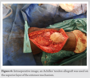
The novel combination of Poly-Tape supplemented with an Achilles’ tendon allograft has been proven in this case to provide a structurally competent reconstruction for chronic quadriceps tendon rupture.
Dr. Anabelle Permutt, 14 West Heath Avenue, NW11 7QL, London, United Kingdom. E-mail: anabellepermutt@hotmail.co.uk
Introduction: Chronic rupture of the quadriceps tendon is an uncommon but debilitating injury, seen only in 1.37/100,000 patients per annum. The problems associated with this injury are the inability to walk due to disruption of the extensor mechanism and pain. There is limited literature on the reconstruction methods for this injury. This study aims to provide a case report and review of similar cases, using artificial tape and allograft.
Case Report: A 60-year-old male patient was operated on for chronic quadriceps tendon rupture after falling on his knee with forced flexion. The surgical management in our case consisted of mobilization of the proximal quadriceps tendon and muscle belly with a V-Y tendon plasty, advancement of the tendon, and repair using the Krakow technique through intraosseous patellar tunnels, augmented with Poly-Tape (Neoligaments©) and an Achilles’ tendon allograft. This was used due to poor patient tissue quality and the extent of the defect. We also searched the literature for chronic quadriceps tendon ruptures reconstruction using poly tape or Achilles’ tendon allograft.
Conclusion: Chronic quadriceps tendon rupture repairs have relatively poor outcomes, and no surgical procedure has been proven to be the gold standard treatment. We believe the novel combination of Poly-Tape supplemented with Achilles’ tendon allograft produces a structurally competent reconstruction in patients with poor tissue quality and large defects and produced good results in our case report.
Keywords: Poly-Tape, quadriceps tendon rupture, Achilles’ tendon allograft.
Quadriceps tendon rupture is an uncommon but debilitating injury, seen in only 1.37/100,000 patients per annum [1]. The tendon is formed by the muscular junction of four muscles: The rectus femoris, the vastus lateralis, the vastus medialis, and the vastus intermedius. The quadriceps tendon forms one of the key components of the extensor mechanism. Without this, a patient is left unable to walk and has reduced stability of the knee. Patients generally present with the classic triad of diagnostic signs. These include acute pain, inability to actively extend the knee joint, and a palpable suprapatellar gap [1]. Surgical management of chronic quadriceps tendons is variable, with limited methods described in the literature. There are many risk factors for quadriceps tendon injuries, and they predominantly affect middle-aged males with a mean age of 51.1 years [1]. In addition, multiple conditions put patients at risk of quadriceps tendon injuries, such as rheumatoid arthritis, gout, chronic renal failure, and diabetes mellitus. Steroid abuse is another common predisposing condition, as corticosteroids can inhibit collagen synthesis and compromise the blood supply, weakening tendons [2]. However, trauma does occur during activity in healthy controls, usually due to eccentric loading of the knee extensor mechanism [1]. We report a case using polyethylene terephthalate tape (Poly-tape), an artificial material used to reconstruct the extensor mechanism. It is made of a woven, multifilament polyester fiber designed to be a frame for soft-tissue ingrowth and Neoligaments© formation [3]. The Poly-tape system, which has been used for ligament and tendon reconstruction, is 30 mm wide x 800 mm long and consists of an open-weave polyester mesh. The main advantages of Poly-Tape are a reduction in autograft donor site morbidity and reduced creep, compared to human tissue, which may lead to graft failure [4]. In our case, a fresh-frozen non-irradicated Achilles tendon was also used to supplement the reconstruction by augmentation.
The authors have obtained the patient’s informed consent for print and electronic publication of the case report. A 60-year-old man was seen in the clinic in May 2021 and diagnosed with a right-sided full-thickness quadriceps tendon tear. The injury occurred in January 2021 when he fell on his right knee with forced flexion. He delayed getting help, and the lockdown further compounded this during the COVID-19 pandemic. He presented in the clinic with a noticeable loss of quadriceps muscle bulk and significant swelling. The patient could not attain a full straight leg raise or walk properly. He had a medical history of hypertension, pre-diabetes, and obesity (BMI: 32.9). The senior surgeon subsequently sent the patient for an MRI, which confirmed the suspected quadriceps tendon rupture. Furthermore, the patellar tendon’s thickened and edematous appearance suggested a possible degree of underlying patellar tendinosis. In addition, the scan showed chondromalacia in the lateral patellar facet with subchondral bone changes (Fig. 1). The patient’s quality of life decreased significantly due to the injury; he reported that he could only walk limited distances with difficulty, and he felt unsteady. He also had great difficulty getting in and out of his car. Therefore, following discussion and patient informed consent, it was decided he would benefit from surgical reconstruction of the tendon rupture. An ultrasound was arranged as part of the preoperative planning to assess the gap between the proximal end of the quadriceps tendon and the proximal pole of the patella. This was only undertaken as the MRI scan had been performed earlier in the year; therefore, concern of further proximal quadriceps retraction was significant. There was a rupture of the medial and middle parts of the quadriceps tendon at the patellar insertion, with a proximal retraction of at least 82 mm in length. In addition, blunting of the distal margin of the quadriceps musculature was noted, and the patellar tendon was intact. Preoperative X-rays (Fig. 2) showed patella Baja, which can be seen to have been significantly improved in the postoperative X-rays (Fig. 3).


Partial muscle or tendon tears that lack chronic pain and with no significant functional deficit are predominantly managed nonoperatively. However, a complete rupture can have detrimental effects on the patient’s quality of life, so most surgeons would advise taking an operative route [6]. Chronic quadriceps tendon rupture repairs have relatively poor outcomes, with high re-rupture rates and levels of postoperative infection. The use of synthetic grafts, that is, Poly-tape, or allografts, could increase the success of reconstructing the extensor mechanism [7]. These injuries stand a much better chance of healing when operated on sooner. It has been recommended that surgical treatment takes place within 2–3 weeks of rupture [8]. Scuderi [9] recommended repair within 48–72 h post-injury to achieve the most successful outcome. The longer the injury is left before treatment, the higher the chance for the tendon to retract further, hindering the opportunity for successful results. However, rewarding outcomes are still possible with an extended period between injury and treatment, proved by our case report. There is no widely accepted treatment for chronic quadriceps tendon ruptures due to the injury’s rarity and the lack of reported literature. In addition, none of the repair or reconstruction methods have been consistently successful in reinstating the function of the extensor mechanism [10]. The most common treatment options are synthetic grafts, allografts, and autografts. Synthetic grafts, made from polymers, provide excellent intrinsic strength, which allows the repaired construct to act against tensile forces [11]. There are several types of synthetic grafts, such as Marlex Mesh, Leeds-Keio Ligament, and the Neoligaments Poly-Tape system; generally used for reconstruction of quadriceps and patellar tendons, rotator cuff muscles (Pitch-Patch device), and the anterior cruciate ligament (JewelACL system) [4]. Chen et al. [12] state that high tensile strength, abrasion resistance, and no immune reaction should all be basic properties of an ideal synthetic material used in artificial ligament manufacture. It should also allow the ingrowth of surrounding tissues. Poly-Tape possesses all these properties so are, therefore, a suitable scaffold for implantation. Literature has suggested that Poly-Tape may be superior to its counterparts. One study showed that Poly-Tape has the highest rate of cell attachment 1-day post-implant of the scaffold, with higher levels than other scaffolds such as X-repair and LARS ligament; cell attachment is required for the implanted scaffold to be incorporated into the body [13]. Artificial ligaments made from polyethylene terephthalate now act as the main apparatus in the reconstruction of the quadriceps tendon, including Poly-Tape and Leeds-Keio ligaments [12]. There are, however, some contraindications with using Poly-Tape, described by the manufacturer. The primary being where patients experience hypersensitivity to implant materials. Others include patients with any infection or pathological bone or soft-tissue condition and skeletally immature patients [4]. One limitation of Poly-Tape is that, due to being hydrophobic, it produces poor integration into the surrounding femur and patella bone tissue [14]. Yet, Li et al. [14] found a solution: a composite coating of 58S bioglass and hydroxyapatite, which can enhance the osseointegration of poly-tape in the bone tunnel. The Achilles’ tendon allograft provides sufficient thickness and robustness to repair the quadriceps tendon. One of the main known limitations for allografts is the risk of infection, and although this is true for ruptures post-TKA, it has been found that after a primary procedure, the risk of infection is up to 10-fold lower than reported in the literature for post-TKA [15]. Furthermore, the risk of infection does not represent a mechanical failure on the allografts’ part [16]. The other potential issue with any allograft material used for tendon or ligament reconstruction is creep, resulting in tissue elongation over time. Published literature has compared the outcomes of operative treatment using allografts versus synthetic ligaments. Artificial tape in reconstructing extensor mechanism ruptures is becoming more popular. To the best of our knowledge, the combination of Poly-Tape and an Achilles’ tendon allograft has not been reported before in the literature. The authors believe that the combination of the two grafts could lead to a more beneficial and structurally competent reconstruction, by combining the tensile strength of the allograft, and the added strength with a smaller risk of immune reaction from the Poly-Tape. The results of this technique are commendable provided a strict postoperative rehabilitation plan is followed.
As far as the literature states, there is no singular procedure yielding significantly better results. Therefore, unfortunately, there is no widely accepted treatment for chronic quadriceps tendon rupture. What has been borne out of this study, is that both the Poly-Tape and the Achilles’ tendon allograft have their benefits, and combining them can lead to a more beneficial outcome for the patient.
Managing chronic quadriceps tendon ruptures with poor tissue and significant defects can be challenging, due to being an uncommon injury and the lack of consensus in treatment. Surgical reconstruction with a combination of synthetic graft and allograft for chronic injuries can produce good functional outcomes.
References
- 1.Ciriello V, Gudipati S, Tosounidis T, Soucacos PN, Giannoudis PV. Clinical outcomes after repair of quadriceps tendon rupture: A systematic review. Injury 2012;43:1931-8. [Google Scholar]
- 2.Leopardi P, di Vico G, Rosa D, Cigala F, Maffulli N. Reconstruction of a chronic quadriceps tendon tear in a body builder. Knee Surg Sports Traumatol Arthrosc 2006;14:1007-11. [Google Scholar]
- 3.Leciejewski M, Królikowska A, Reichert P. Polyethylene terephthalate tape augmentation as a solution in recurrent quadriceps tendon ruptures. Polim Med 2018;48:53-6. [Google Scholar]
- 4.QuadsTape System; 2021. Available from: https://neoligaments.com [Last accessed on 2021 Nov 11]. [Google Scholar]
- 5.Rendon A, Schäkel K. Psoriasis pathogenesis and treatment. Int J Mol Sci 2019;20:1475. [Google Scholar]
- 6.Nag HL, Jain G, Nayak M, Goyal A. Result of delayed repair of quadriceps muscle following a sharp cut injury. BMJ Case Rep 2021;14:e239863. [Google Scholar]
- 7.Piatek AZ, Lee P, DeRogatis MJ, Boyajian DA, Issack PS. Knee osteoarthritis with chronic quadriceps tendon rupture treated with total knee arthroplasty and extensor mechanism allograft reconstruction: A case report. JBJS Case Connect 2018;8:e46. [Google Scholar]
- 8.Unlu MC, Kaynak G, Caliskan G, Birsel O, Kesmezacar H. Late repair of quadriceps tendon ruptures with free hamstring autograft augmentation and tension relief in patients with predisposing systemic diseases. J Trauma 2011;71:1048-53. [Google Scholar]
- 9.Scuderi C. Ruptures of the quadriceps tendon; study of twenty tendon ruptures. Am J Surg 1958;95:626-34. [Google Scholar]
- 10.Llombart Blanco R, Valentí A, Díaz de Rada P, Mora G, Valentí JR. Reconstruction of the extensor mechanism with fresh-frozen tendon allograft in total knee arthroplasty. Knee Surg Sports Traumatol Arthrosc 2014;22:2771-5. [Google Scholar]
- 11.Crozier-Shaw G, Mahon J, Bayer T. The use of bioabsorbable compression screws & polyethylene tension band for fixation of displaced olecranon fractures. J Orthop 2020;22:525-9. [Google Scholar]
- 12.Chen T, Jiang J, Chen S. Status and headway of the clinical application of artificial ligaments. Asia Pac J Sports Med Arthrosc Rehabil Technol 2015;2:15-26. [Google Scholar]
- 13.Smith RD, Carr A, Dakin SG, Snelling SJ, Yapp C, Hakimi O. The response of tenocytes to commercial scaffolds used for rotator cuff repair. Eur Cell Mater 2016;31:107-18. [Google Scholar]
- 14.Li H, Ge Y, Wu Y, Jiang J, Gao K, Zhang P, et al. Hydroxyapatite coating enhances polyethylene terephthalate artificial ligament graft osseointegration in the bone tunnel. Int Orthop 2011;35:1561-7. [Google Scholar]
- 15.Mortazavi SM, Schwartzenberger J, Austin MS, Purtill JJ, Parvizi J. Revision total knee arthroplasty infection: Incidence and predictors. Clin Orthop Relat Res 2010;468:2052-9. [Google Scholar]
- 16.Diaz-Ledezma C, Orozco FR, Delasotta LA, Lichstein PM, Post ZD, Ong AC. Extensor mechanism reconstruction with achilles tendon allograft in TKA: Results of an abbreviate rehabilitation protocol. J Arthroplasty 2014;29:1211-5. [Google Scholar]


