Distal leg angioleiomyomas are rare but must be included in the differential diagnoses of palpable masses in the region for timely diagnosis and treatment.
Dr. Sandeep Mohan, Department of Orthopedics, St Joseph’s Hospital, Karuvanchal, Kannur, Kerala, India. E-mail: sandeepmohan123@gmail.com
Introduction: Angioleiomyomas are rare benign tumors originating from smooth muscle cells of blood vessels. Although they can occur in various anatomical locations, angioleiomyomas of the distal leg are relatively uncommon. Due to its clinical resemblance to other soft-tissue tumors, misdiagnosis can occur leading to inadequate treatment.
Case Report: We present a case of angioleiomyoma in a 54-year-old female who presented with a palpable mass in her distal leg. The tumor was surgically excised, and histopathological examination confirmed the diagnosis of angioleiomyoma. In this article, we discuss the clinical presentation, diagnostic evaluation, and management of angioleiomyoma, with a focus on distal leg tumors. Furthermore, we provide a comprehensive review of the existing literature on angioleiomyomas, emphasizing findings and treatment outcomes reported in previous studies.
Conclusion: Angioleiomyomas are uncommon soft-tissue tumors that can mimic other more common lesions such as ganglion cysts. Hence, diagnosis requires a high index of suspicion. Surgical excision is the treatment of choice for angioleiomyoma. Complete resection is generally curative, with a low rate of recurrence.
Keywords: Angioleiomyoma, distal leg, surgical excision, review of literature.
Angioleiomyomas are infrequent benign tumors derived from the smooth muscle cells of blood vessels. Although they can manifest in various anatomical regions, angioleiomyomas located in the distal leg are relatively rare. This article presents a case of angioleiomyoma in the distal leg of a 54-year-old female, detailing the clinical features, diagnostic approaches, and management options. Due to their rare occurrence, lack of clinician awareness, and paucity of published literature, they are rarely diagnosed pre-operatively. An accurate diagnosis can prevent delays in treatment, improve the outcomes, and also help in excluding the involvement of malignancies. Enhancing the general awareness of angioleiomyomas in clinical practice will improve its treatment outcomes [1]. In addition, we review the literature on angioleiomyomas, focusing on distal leg tumors, to enhance the understanding and management of this condition.
A 54-year-old female presented with a progressively enlarging mass in her right distal leg anterior aspect for 2 years, associated with occasional pain over the past 6 months. Physical examination revealed a palpable, non-tender, subcutaneous mass measuring approximately 2 cm in diameter, located in the medial aspect of the distal leg. Ultrasonography demonstrated a well-defined hypoechoic lesion with vascularity. Considering the suspicious nature of the mass, surgical excision was performed under regional anesthesia (Fig. 1).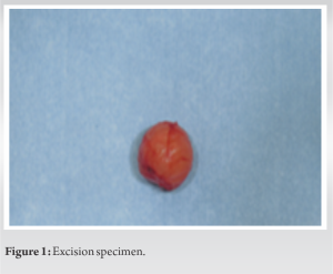 Histopathological analysis revealed a well-circumscribed encapsulated neoplasm composed of myoepithelial cells proliferating with perivascular attenuation around slit such as vascular spaces, consistent with a diagnosis of angioleiomyoma (Fig. 2 and 3). The patient recovered completely and was followed up at 6 monthly intervals for 2 years without any recurrence.
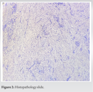

Angioleiomyoma is one of the three different forms of leiomyoma, which is also known as vascular leiomyoma. The other forms of leiomyoma include piloleiomyoma (arising from arrestor pili muscles) and genital leiomyoma (arising from the smooth muscles of the scrotum, vulva, or nipple). Angioleiomyoma is uncommon and benign. Its etiology is largely unknown but factors such as trauma, infection, hormones, and arteriovenous malformations have been known to be present in various cases [1, 2]. Histologically, they have a well-circumscribed dermal nodule surrounded by a compressed connective tissue and separated from it by clefts. Various sized veins with muscular walls are present within the nodule, and smooth muscle bundles extend tangentially from the periphery of the vessels. The lumina of the veins are rounded or slit-like and have a minimal amount of collagen [1, 3]. Pain is not always present, but if present, can be of varied character – pressure pain, sharpness, or pinching type and is affected by temperature [1, 4]. This pain has been hypothesized to be a result of local tissue anoxia or due to compression of the local neural structures [1, 5]. Distal leg angioleiomyomas, although rare, can present as palpable masses associated with pain or discomfort. Diagnostic evaluation typically involves imaging modalities such as ultrasonography, magnetic resonance imaging, or angiography. Surgical excision is considered the mainstay of treatment, providing both therapeutic and diagnostic benefits. There should be an increased index of suspicion for angioleiomyoma, especially when a freely movable subcutaneous mass is encountered in a middle-aged female patient. Treatment of such masses should involve surgical excision to relieve the symptomatology and to obtain tissue for a definitive histologic analysis. If the mass recurs, then the possibility of leiomyosarcoma should be explored [5]. In the literature review, we found that most angioleiomyoma has a peak incidence in the fourth to sixth decades of life. The solid type is more common in females, whereas the venous and cavernous types show a slight male predominance [6]. It typically presents as a solitary, firm, well-circumscribed, slow-growing, subcutaneous nodule, which is often painful. The diameter ranges from 0.2 to 4.3 cm (usually <2 cm) [7]. Simple excision is the treatment of choice and prognosis is excellent. It is recommended that patients should be followed up for 1 year after excision [8]. Cases of malignant transformation have been reported, though extremely rare. Complications, such as secondary calcification, myxoid degeneration, hyalinization, and malignant transformation, were reported [1, 9]. Due to
rarity of the lesion and extensive differential diagnosis, it is commonly misdiagnosed before histopathological study. [10]. Differential diagnoses can include lipoma, fibroma, ganglions, schwannoma, cutaneous angiolipoma, glomus tumor, etc. [1, 2].
Angioleiomyomas are infrequent benign tumors arising from smooth muscle cells of blood vessels. While distal leg angioleiomyomas are rare, they should be included in the differential diagnosis of palpable masses in this region. Surgical excision with histopathological examination remains the gold standard for diagnosis and treatment. Awareness of this entity and its clinical characteristics is crucial for accurate diagnosis and appropriate management.
Due to rarity of the angioleiomyomas in distal leg, misdiagnosis can occur unless a high index of suspicion is maintained in cases of distal leg soft-tissue tumors.
References
- 1.Bodapati VS, Sunderamoorthy D. Angioleiomyoma-rare soft tissue tumor of the foot and ankle, review of two patients and review of the literature. J Surg Case Rep 2021;2021:rjab535. [Google Scholar]
- 2.Dominguez-Cherit J, Brandariz A. Distal digital angioleiomyoma: A case report and review of the literature. Int J Dermatol 2003;42:141-3. [Google Scholar]
- 3.Requena L, Sangueza OP. Cutaneous vascular proliferations. Part III. Malignant neoplasms, other cutaneous neoplasms with significant vascular component, and disorders erroneously considered as vascular neoplasms. J Am Acad Dermatol 1998;38:143-75; quiz 176-8. [Google Scholar]
- 4.Requena L, Baran R. Digital angioleiomyoma: An uncommon neoplasm. J Am Acad Dermatol 1993;29:1043-4. [Google Scholar]
- 5.Hanft JR, Carbonell JA, Do HQ. Angioleiomyoma of the lower extremity. J Am Podiatr Med Assoc 1997;87:388-91. [Google Scholar]
- 6.Matsuyama A. Angioleiomyoma. In: World Health Organization Classification of Tumours: Soft Tissue and Bone Tumours. Lyon: IARC Press; 2020. p. 186. [Google Scholar]
- 7.Hachisuga T, Hashimoto H, Enjoji M. Angioleiomyoma. A clinicopathologic reappraisal of 562 cases. Cancer 1984;54:126-30. [Google Scholar]
- 8.Yeung CM, Moore L, Lans J, Lozano-Calderón L. Angioleiomyoma of the hand: A case series and review of the literature. Arch Bone Jt Surg 2020;8:373-7. [Google Scholar]
- 9.Gajanthodi S, Rai R, Chaudhry RK. Vascular leiomyoma of foot. J Clin Diagn Res 2013;7:571-2. [Google Scholar]
- 10.Taj H, Comba I, Vasquez J, et al. A rare case of Angioleiomyoma of the Hand. Cureus 2020;12(4): e7530. doi; 10.7759/cureus.7530. [Google Scholar]