Radial neck epiphysiolysis with delayed presentation requires open reduction and internal fixation for good functional outcome.
Dr. Shaswat Mishra, Department of Orthopaedics, Grant Government Medical College and Sir JJ Group of Hospitals, Mumbai, Maharashtra, India. E-mail: shaswatmishra1994@gmail.com
Introduction: Radial neck fractures in the pediatric age group account for 1% of all fractures in children. A completely displaced fracture with an angle >30° requires operative intervention.
Case Report: An 11-year-old boy with Judet IVb radial neck epiphysiolysis 4 weeks old was managed by open reduction and internal fixation with K-wires.
Conclusion: Anatomic reduction must be achieved in radial neck fractures to restore their function. Open reduction and internal fixation with K wires must be considered in patients with >30° angle of inclination, neglected fracture, and failure of closed reduction.
Keywords: Judet IVb, delayed presenting, radial neck, epiphysiolysis, open reduction.
Radial neck fractures account for 1% of all fractures in children and they make up to 5–10% of elbow injuries [1]. Metaphyseal fracture of the radial neck is the most common type followed by epiphyseal separation [2]. Falling onto an outstretched hand is the most common mechanism of injury, and the age interval is between 4 and 14 years with a maximum peak frequency between 9 and 12 years [3, 4]. Radial neck fractures with angle of inclination >30° require operative intervention by closed reduction, percutaneous pin reduction and fixation, elastic stable intramedullary nailing, or open reduction with internal fixation [5]. This case report describes a patient with radial neck epiphysiolysis with delayed presentation.
An 11-year-old boy presented to us with left elbow deformity and painful restricted range of motion for 4 weeks. He had a history of fall from height while playing and was managed conservatively by a traditional bone setter. The anteroposterior and lateral radiographs showed completely displaced radial neck fracture with an angle >80° suggestive of Type IVb according to Judet’s classification (Fig. 1). Under general anesthesia, an attempt of closed reduction was carried out which was unsuccessful. Using Kocher’s approach, open reduction was carried out with meticulous dissection while preserving the medial periosteal blood supply (Fig. 2). We stabilized the reduction using two transepiphyseal 1.6 mm K-wires (Fig. 3). A dorsal slab was applied with elbow in 90° flexion.
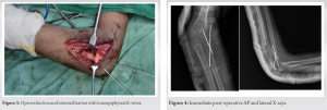 At 4 weeks, the removal of K-wires was done and step-wise rehabilitation was started. X-rays were done at immediate post-operative period, 4 weeks, 3 months, 6 months, and 1 year, respectively (Fig. 4 and 5).
 Full range of motion without any restriction in pronation and supination was achieved at the end of 1 year with excellent functional outcome according to Mayo Elbow Performance Score (Fig. 6).
Radial neck fractures are rare in children, and it is mostly seen between 9 and 12 years of age before the complete ossification of the epiphyseal cartilage. These are classified by Judet functional classification modified by Metaizeau according to which Type 1 is undisplaced, Type 2 has <30° angulation, Type 3 has an angle between 30 and 60°, Type 4a has an angle between 60 and 80°, and Type 4b has an angle >80° [6]. Preservation of elbow joint integrity is to be stressed upon to achieve good functional outcome. Proximal and distal radioulnar joint mainly helps in rotation and anatomic reduction of radial neck fractures is a must to for this movement. Management of these fractures depends on the angle of inclination. Types 1 and 2 and mainly managed conservatively. Types 3 and 4 require closed reduction with or without fixation. Open reduction is considered when the fractures cannot be reduced adequately to >45° of angulation with closed or percutaneous techniques [7]. The main principle is to achieve anatomic reduction of <30° angulation and restore radial length and alignment of radiocapitellar joint. Many publications in the literature describe radial neck fractures in acute setting. Radial neck fractures with delayed presentation have very few case reports in the literature. Majed and Baco in his case report described a 5-day-old radial neck epiphysiolysis managed with closed reduction with elastic intramedullary nailing [8]. Papageorgiou et al. reported a 120-day-old injury managed with open reduction and internal fixation with transepiphyseal K-wire and had excellent functional outcome with no signs of avascular necrosis of radial head [9]. Gutierrez-De La Iglesia et al. studied 51 patients with Judet Types 3 and 4 fractures and concluded that there were no significant differences in functional outcome and complications between closed and open reduction [10]. Turan et al. reported a case of radial neck fracture 3 weeks old managed with open reduction and internal fixation with K-wires and had good functional and radiological outcome [11]. Fracture displacement is the independent predictor of functional outcome irrespective of the technique of reduction.
Delayed presenting radial neck epiphysiolysis must be managed with open reduction and internal fixation with K-wires and the functional outcome depends mainly on the surgical dissection preserving the medial periosteal attachment of the radial neck.
We believe that good functional and radiological outcome can be achieved by open reduction with preservation of medial attachment to radial neck even in delayed or neglected radial neck fractures.
References
- 1.Brandão GF, Soares CB, Teixeira LE, de Castro Boechat L. Displaced radial neck fractures in children: Association of the Métaizeau and Böhler surgical techniques. J Pediatr Orthop 2010;30:110-4. [Google Scholar]
- 2.Radomisli TE, Rosen AL. Controversies regarding radial neck fractures in children. Clin Orthop Relat Res 1998;353:30-9. [Google Scholar]
- 3.Metaizeau JP, Lascombes P, Lemelle JL, Finlayson D, Prevot J. Reduction and fixation of displaced radial neck fractures by closed intramedullary pinning. J Pediatr Orthop 1993;13:355-60. [Google Scholar]
- 4.Lejman T, Sułko J, Kotulski D. Radius neck fracture in children. Chir Narzadow Ruchu Ortop Pol 1996;61:343-8. [Google Scholar]
- 5.Falciglia F, Giordano M, Aulisa AG, Di Lazzaro A, Guzzanti V. Radial neck fractures in children: Results when open reduction is indicated. J Pediatr Orthop 2014;34:756-62. [Google Scholar]
- 6.Steele JA, Graham HK. Angulated radial neck fractures in children. A prospective study of percutaneous reduction. J Bone Joint Surg Br 1992;74:760-4. [Google Scholar]
- 7.Schmittenbecher PP, Haevernick B, Herold A, Knorr P, Schmid E. Treatment decision, method of osteosynthesis, and outcome in radial neck fractures in children: A multicenter study. J Pediatr Orthop 2005;25:45-50. [Google Scholar]
- 8.Majed A, Baco AM. Late diagnosis and treatment of a paediatric radial neck fracture. Injury Extra 2006;37:322-4. [Google Scholar]
- 9.Papageorgiou TG, Panos NE, Gigis IP, Samoladas EP, Beslikas TA, Christoforidis IE. Treatment of a late presenting displaced radial neck fracture in a 10 years-old girl. J Med Cases 2011;2:252-4. [Google Scholar]
- 10.Gutiérrez-de la Iglesia D, Pérez-López LM, Cabrera-González M, Knörr-Giménez J. Surgical techniques for displaced radial neck fractures: Predictive factors of functional results. J Pediatr Orthop 2017;37:159-65. [Google Scholar]
- 11.Turan K, Köroğlu C, Çabuk H. Late presenting pediatric radial neck fracture: A case report and review of the literature. J Orthop Case Rep 2021;11:20-2. [Google Scholar]