Recognizing the link between ichthyosis and Vitamin D deficiency for ensuring appropriate management and preventing serious skeletal complications
Dr. Ruchika Nevse, Department of Dermatology, Venereology and Leprosy, N.K.P. Salve Institute of Medical Sciences and Research Centre, Lata Mangeshkar Hospital, Digdoh Hills, Hingna road, Nagpur, Maharashtra, India. E-mail: ruchikanevse97@gmail.com
Introduction: The main way that the skin produces Vitamin D, which is necessary for calcium metabolism and skeletal health, is through exposure to ultraviolet B rays. Long-term skin disorders such as ichthyosis can prevent the skin from producing enough Vitamin D, leading to deficiencies and issues with bone health. This report highlights the need to take Vitamin D insufficiency and its sequelae into account in clinical care by presenting a case of genu valgum in a patient with lamellar ichthyosis. Similar correlations have also been shown in earlier research, underscoring the necessity of continuing to monitor and educate patients with chronic skin problems about their bone health.
Case Report: A 15-year-old Indian boy came to the orthopedic outpatient department complaining of lamellar ichthyosis, progressive knee deformity, and trouble walking over the last 6 months, with a recent exacerbation in the previous month. Upon examination, he had bilateral genu valgum, widespread scaly skin lesions, and biochemical indications of Vitamin D insufficiency.
Conclusion: This case study underscores the importance of early diagnosis and treatment of musculoskeletal disorders in patients with chronic skin conditions. It also highlights the unusual association between genu valgum and lamellar ichthyosis. It underscores the need for interdisciplinary teamwork in managing difficult patients and influencing pediatric dermatology, orthopedics, and other medical disciplines. It also contributes to research on the relationship between skin conditions and orthopedic problems, aiming to improve patient outcomes and care.
Keywords: Genu valgum, lamellar ichthyosis, pediatric orthopedics.
Vitamin D is essential for calcium metabolism and skeletal health and is primarily produced in humans through the skin’s 7-dehydrocholesterol synthesis in ultraviolet B (UV) radiation [1]. Dietary sources of Vitamin D include fatty fish, egg yolks, and fortified plant-based foods. However, most foods contain limited amounts, so adequate Vitamin D intake is typically achieved through sunlight exposure or dietary supplements [2]. Chronic skin disorders, such as ichthyosis, can reduce Vitamin D production, potentially leading to Vitamin D deficiency. For those with long-term skin disorders, it is essential to check Vitamin D levels and consider taking supplements to avoid deficits and preserve bone health [3]. Lamellar ichthyosis is a chronic skin condition causing large, whitish-brown scales, mild erythroderma, ectropion, eclabium, and scarring alopecia. It is an autosomal recessive disorder inherited from birth affecting family and social life. The prevalence is estimated at <1 case/300,000 individuals [4]. The research emphasizes the uncommon link between ichthyosis and metabolic bone illnesses such as osteomalacia or rickets. We present a case of genu valgum (knock knees) in a patient with lamellar ichthyosis to emphasize the need for vigilance in bone health management and the importance of considering potential Vitamin D deficiency in chronic skin conditions.
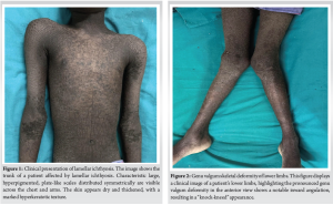
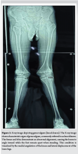
A 15-year-old boy came to the orthopedic outpatient department with progressive knee deformity and difficulty walking over the past 6 months, with a recent exacerbation in the last month. He is the fourth kid in his family and was delivered at term via a normal vaginal delivery to non-consanguineous parents. His eldest sister had similar symptoms and lived until the age of 5, but the exact cause of her death was not well understood by the parents. The patient’s skin condition was normal at birth but began to develop cracking within 48 h, followed by the appearance of fish-like, scaly skin lesions that persisted throughout his life. The scales would frequently shed, revealing underlying normal skin. Notably, he had negligible sweating from childhood and reported fluid-filled, raised lesions during summer that did not release fluid. He was immunized appropriately. The boy achieved normal developmental milestones in verbal, fine motor, gross motor, and social skills. For the first 5 years of life, he had no significant health complaints and no symptoms of photophobia, itching, or burning. From the age of 5 to 10 years, he performed well academically and enjoyed going to school. He was consistently strong in his scholastic achievements, showed interest in extracurricular activities, and actively participated in popular sports with his friends during school hours. His health was stable until the age of 10 years. Eventually, he began experiencing calf pain and knee pain after moderate activity. He also noted spasms in his fingers and jaw, suggestive of hypocalcemia, as indicated by Trousseau’s and Chvostek’s signs. During this period, he received Vitamin D and calcium supplements from a private practitioner, which temporarily alleviated his symptoms. At the age of 14, his leg pain became more severe over the course of the previous year, making it difficult for him to walk, climb stairs, and rise up from a squatting position, later causing his knees to acquire a valgus deformity. There was no past medical history of knee injuries, fractures, steatorrhea, chronic diarrhea, or renal problems. Even though he consumed enough calcium and Vitamin D through his diet, his symptoms continued. His treatment history includes 3 years of intermittent oral retinoid therapy and a variety of topical therapies, including urea cream and glycerin. Physical examination indicated 157 cm of height, 41 kg of weight, a 16.6 body mass index, an upper segment to lower segment ratio of 1.21, and a 156 cm arm spread. Numerous well-defined, hyperpigmented patches to plaques with large, gray scales were seen mostly on the trunk and limbs, with involvement of the face, palms, and soles (Fig. 1). Dandruff was present. The teeth and mucosal surfaces were normal. He also had bilateral lateral madarosis. A 12-cm intermalleolar gap was present in the bilateral genu valgum (Fig. 2). On radiographs, both knees displayed valgus deformity and a thinned-out cortex (Fig. 3).
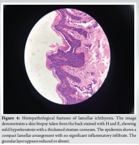
Skin biopsy revealed mild hyperkeratotic stratified squamous epithelium with elongated rete ridges and pigmented basal layer. The papillary dermis and dermis show perivascular lymphocytic infiltrate. Adnexal structures are also seen. Focal thickened stratum corneum with chronic inflammatory infiltrate in the dermis (Fig. 4).
No abnormalities were noted during ophthalmological and ears, nose, and throat examinations. He was started on topical emollients.
Investigations
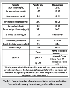
Lamellar ichthyosis with genu valgum caused by Vitamin D deficiency was diagnosed based on clinical and laboratory evaluation (Table 1). Insufficient cutaneous production of Vitamin D was considered the cause of Vitamin D insufficiency.
Treatment
For 8 weeks, he received oral cholecalciferol 60,000 IU once a week. After that, his serum 25(OH) Vitamin D level rose to 160.8 ng/mL. Because his skin condition was chronic, he was thereafter kept on an oral dose of 60,000 IU once a month. In addition, the orthopedics department was consulted for corrective osteotomy.
Outcome and follow up
Clinically, following corrective osteotomy, symptoms such as pain in the knees and thighs were much improved, and difficulty in climbing stairs, walking, and rising from a crouching position was resolved. His skin lesions appeared to be slightly improving at the follow-up, and no new skeletal deformities were discovered.
Vitamin D, a secosteroid hormone, significantly impacts human health, preventing and managing diseases such as cancer, cardiovascular, multiple sclerosis, Parkinson’s, obesity, aging, falls, and diabetes [5]. Its impact is due to its ability to regulate more than 200 human genes across various tissues [6]. Previtamin D3 is formed in the skin when UV rays convert 7-dehydrocholesterol. This is where Vitamin D production starts. The liver then transforms it into 25-hydroxyvitamin D3. It is further hydroxylated in the kidneys to 1,25-dihydroxyvitamin D3 (active form) (calcitriol), which acts through the Vitamin D receptor. The main functions of Vitamin D are to enhance the intestinal absorption of calcium and phosphorus, support the maturation of osteoclasts, and help in bone mineralization [2]. Childhood ichthyosis, a keratinization disorder, can hinder Vitamin D synthesis and bone mineralization through defects in Vitamin D synthesis, increased keratinocyte proliferation, calcium loss due to diseased skin, sun exposure avoidance, and systemic retinoid therapy, which can affect calcium absorption from the intestine [7]. Lamellar ichthyosis is a skin condition causing a distinctive appearance and texture. Infants with this condition are often born with a collodion membrane, a shiny, waxy layer that peels off within weeks of life, revealing skin with generalized scaling. It is caused by TGM1 gene mutations, which encode the transglutaminase 1 enzyme, causing impaired skin barrier function. Common features include ectropion, eclabion, superficial punctate keratitis, and loss of eyebrows and eyelashes. Genetic studies have linked the condition to chromosome 14q11 markers [8]. Rickets associated with ichthyosiform dermatoses are rare, but there are reports in the literature. Three of the 41 Sudanese children who had rickets as a result of Vitamin D insufficiency also had ichthyosis [9]. Five individuals with different keratinization disorders had low-to-normal 25-hydroxyvitamin D levels and high parathyroid hormone, according to .[7]. Nine patients with non-bullous congenital ichthyosis of European and North American heritage had a significant 25-(OH)D deficiency, according to Ingen-Housz-Oro et al. [10] Five children with lamellar ichthyosis and one with non-bullous ichthyosiform erythroderma/psoriasis with atopy were described by Sethuraman et al. [3]. Two of them had extremely severe skeletal abnormalities, and all exhibited biochemical and radiological signs of rickets. Two cases of severe bilateral rachitic genu valgum in patients with non-bullous congenital ichthyosiform erythroderma were reported by Bhagat et al. [11]. Our patient had lamellar ichthyosis, low serum Vitamin D levels, and bilateral genu valgum. Despite high serum parathyroid hormone levels, no rickets were present. The patient’s osteomalacia likely resulted from Vitamin D deficiency despite no radiological or clinical signs of rickets. Research indicates that Indian communities often have low serum levels of 25(OH) Vitamin D, mainly due to factors such as skin pigmentation and limited direct sunlight exposure [12]. Vitamin D is often not included in Indian dairy products or dietary fats, leading to widespread Vitamin D insufficiency. This, combined with a diet high in phytates and calcium deficiency, with the additional burden of ichthyosis could upset the already unstable equilibrium and could cause diseases such as osteomalacia and rickets [13].
The case report highlights the intricate relationship between lamellar ichthyosis, Vitamin D deficiency, and bone health. It emphasizes the need for monitoring and managing Vitamin D levels in patients with ichthyosis, particularly in India where dietary and environmental factors lead to Vitamin D deficiency. Early detection and intervention can prevent complications such as rickets and osteomalacia, thereby ensuring better overall management of patients with lamellar ichthyosis.
The case report highlights the link between lamellar ichthyosis, Vitamin D deficiency, and bone health, emphasizing the need for vigilant monitoring of Vitamin D levels in patients with ichthyosis, especially in India, and advocates for early diagnosis and intervention to improve patient management.
References
- 1.Deka N, Sarma D, Saikia UK. Lamellar ichthyosis with genu valgum: Unfolding the link. BMJ Case Rep 2012;2012:bcr1120115136. doi:10.1136/bcr.11.2011.5136 [Google Scholar]
- 2.Clinical Gate. Vitamin D: From Photosynthesis, Metabolism, and Action to Clinical Applications. Clinical Gate; 2015. Available from: https://clinicalgate.com/vitamin-d-from-photosynthesis-metabolism-and-action-to-clinical-applications [Last accessed on 2024 Aug 10]. [Google Scholar]
- 3.Sethuraman G, Khaitan BK, Dash SS, Chandramohan K, Sharma VK, Kabra M, et al. Ichthyosiform erythroderma with rickets: Report of five cases. Br J Dermatol 2008;158:603-6. [Google Scholar]
- 4.Richard G. Autosomal recessive congenital ichthyosis. In: Adam MP, Feldman J, Mirzaa GM, Pagon RA, Wallace SE, Bean LJ, et al., editors. GeneReviews®. Seattle, WA: University of Washington; 1993. Available from: https://www.ncbi.nlm.nih.gov/books/NBK1420 [Last accessed on 2024 Aug 11]. [Google Scholar]
- 5.5. Vitamin D and Diabetes. Diabetes Spectrum. American Diabetes Association. Available from: https://diabetesjournals.org/spectrum/article/24/2/113/32324/vitamin-d-and-diabetes [Last accessed on 2024 Aug 10]. [Google Scholar]
- 6.Diagnosis and Treatment of Vitamin D Deficiency: Expert Opinion on Pharmacotherapy. Vol 9. Available from: https://www.tandfonline.com/doi/full/10.1517/14656566.9.1.107 [Last accessed on 2024 Aug 10]. [Google Scholar]
- 7.Milstone, L. M., Ellison, A. F., & Insogna, K. L. (1992). Serum parathyroid hormone level is elevated in some patients with disorders of keratinization. Archives of Dermatology, 128(7), 926-930. Available from: https://doi.org/10.1001/archderm.1992.01680170058005 [Google Scholar]
- 8.Disorders of Keratinization - Rook’s Textbook of Dermatology. Wiley Online Library. Available from: https://onlinelibrary.wiley.com/doi/10.1002/9781444317633.ch19 [Last accessed on 2024 Aug 10]. [Google Scholar]
- 9.El Hag AI, Karrar ZA. Nutritional vitamin D deficiency rickets in Sudanese children. Ann Trop Paediatr 1995;15:69-76. [Google Scholar]
- 10.Ingen-Housz-Oro S, Boudou P, Bergot C, Ibrahim F, Souberbielle JC, Dubertret L, et al. Evidence of a marked 25-hydroxyvitamin D deficiency in patients with congenital ichthyosis. J Eur Acad Dermatol Venereol 2006;20:947-52. [Google Scholar]
- 11.Bhagat SB, Bhagat SS, Sharma HK, Naik M, Amin P, Pandit J. Severe bilateral rachitic genu valgum in patients with nonbullous congenital ichthyosiform erythroderma: A report of two cases and review of literature. J Pediatr Orthop B 2007;16:423-8. [Google Scholar]
- 12.Arya V, Bhambri R, Godbole MM, Mithal A. Vitamin D status and its relationship with bone mineral density in healthy Asian Indians. Osteoporos Int 2004;15:56-61. [Google Scholar]
- 13.Zargar AH, Ahmad S, Masoodi SR, Wani AI, Bashir MI, Laway BA, et al. Vitamin D status in apparently healthy adults in Kashmir Valley of Indian subcontinent. Postgrad Med J 2007;83:713-6. [Google Scholar]