
Osteochondroma, though typically a tumor of younger individuals, should not be excluded from the differential diagnosis of knee pain in elderly patients. Symptomatic lesions, even in uncommon locations such as the infrapatellar region, can be effectively managed with surgical excision, leading to significant improvement in function and quality of life.
Dr. Yogeshwar Agharkar, Department of Orthopaedics, Sree Balaji Medical College and Hospital, Chennai, Tamil Nadu, India. E-mail: yogeshwaragharkar1998@gmail.com
Introduction: Osteochondroma, a benign bone tumor commonly found in younger individuals, is seldom observed in older adults. This case report presents an atypical occurrence of an osteochondroma in a 65-year-old female, located in the infrapatellar region of the right knee. Clinical examination revealed a firm, non-tender mass in the infrapatellar area, accompanied by mild joint effusion and restricted range of motion. This case highlights the importance of including osteochondroma in the differential diagnosis for knee pain in elderly patients and demonstrates the effectiveness of surgical intervention in managing symptomatic lesions in uncommon anatomical locations.
Case Report: A 65-year-old female presented to the orthopedic outpatient department with a chief complaint of right knee pain and swelling that had progressively worsened over the last year. The pain was described as a dull ache, exacerbated by activity and partially relieved by rest. In addition, the patient experienced mechanical symptoms impacting her mobility and quality of life. Diagnostic imaging, including X-rays, magnetic resonance imaging, and computed tomography scans, confirmed an osteochondroma in the infrapatellar region, characterized by a bony outgrowth, with no signs suggesting malignant transformation. Due to the symptomatic nature and impact on the patient’s quality of life, surgical excision was performed achieving complete removal of the lesion.
Conclusion: This case illustrates a rare presentation of infrapatellar osteochondroma in an elderly patient, highlighting that such lesions, though uncommon in older adults, should remain a differential consideration in cases of unexplained knee pain with mechanical symptoms. Timely diagnosis through appropriate imaging and surgical excision can lead to excellent symptomatic relief and functional recovery, even in atypical anatomical locations.
Keywords: Osteochondroma, elderly, infrapatellar.
Osteochondromas are benign bony projections covered with a cartilaginous cap, often arising from the metaphysis of long bones. They account for 20–50% of all benign bone tumors and are usually discovered in patients under 30 years [1,2]. The occurrence in older adults is infrequent and poses diagnostic challenges, as symptoms can mimic more common degenerative joint diseases [3,4]. A rare case of Hoffa’s osteochondroma arising in the infrapatellar fat pad was reported previously, which emphasized that such atypical presentations can occur beyond the usual age group and mimic other joint pathologies [5]. This case report aims to highlight the clinical features, diagnostic approach, and successful management of a rare presentation of osteochondroma in the elderly, contributing valuable insights into its management.
A 65-year-old female presented to the orthopedic outpatient department with a chief complaint of right knee pain and swelling that had progressively worsened over the last year. The pain was described as a dull ache, exacerbated by activity and partially relieved by rest. In addition, the patient experienced mechanical symptoms impacting her mobility and quality of life. No significant past medical history. No history of trauma to the knee. No family history of bone tumors or hereditary multiple exostoses. Physical Examination revealed a visible swelling on the inferior aspect of the right knee, a firm, non-tender, immobile mass. Range of motion was limited, particularly in flexion. Neurovascular status of the limb was intact. Initial anteroposterior and lateral knee X-rays showed a well-defined, bony outgrowth from the inferior aspect of the patella (Fig. 1).
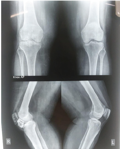
Figure 1: Pre-operative X-ray.
Magnetic resonance imaging (MRI) provided detailed visualization, with no signs of malignant transformation or soft tissue involvement with cartilage cap thickness of 1.3 mm (Fig. 2).
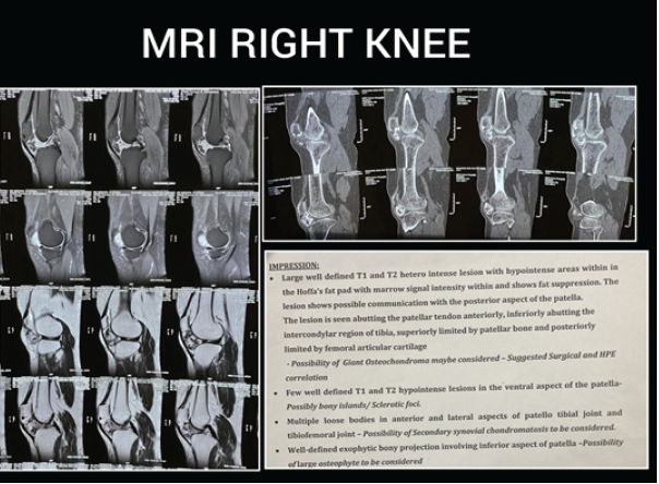
Figure 2: Magnetic resonance imaging right knee.
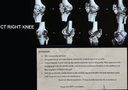
Figure 3: Computed tomography scan.
Computed tomography scan confirmed the continuity. Soft-tissue density lesion noted in the medial aspect of Hoffa’s fat pad and knee joint with multiple loose bodies in anterior and lateral aspects of patella-tibial joint and tibiofemoral joint (Fig. 3). Synovial chondromatosis/osteochondroma may be considered. Diagnosis was based on clinical and imaging findings: An osteochondroma of the posterior aspect of patella was confirmed. Given the patient’s significant symptoms and the impact on her daily activities, surgical excision was deemed necessary. Surgical procedure: A medial parapatellar approach was used to access the lesion [6,7]. Excision: The osteochondroma was excised en bloc, ensuring complete removal of the cartilaginous cap. Intraoperative findings were consistent with pre-operative imaging. The wound was irrigated and closed in layers. Hemostasis was achieved, and the extensor mechanism was carefully preserved (Fig. 4).
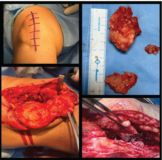
Figure 4: Intraoperative images.
Post-operative care: The patient was placed in a knee immobilizer post-surgery. Pain management was achieved with oral analgesics. Patient was encouraged early mobilization with gentle knee bending and quadriceps and hamstring exercises from pod 5. The patient was discharged with instructions for routine wound care and follow-up. At the 6-week follow-up, the patient reported marked reduction in pain and improved knee function. Physical examination showed no signs of recurrence, and the surgical site was healing well. Radiographs confirmed the absence of residual lesion. Histopathological diagnosis confirmed possibility of osteochondroma with chronic synovitis with thinned out overlying cartilage (Fig. 5).
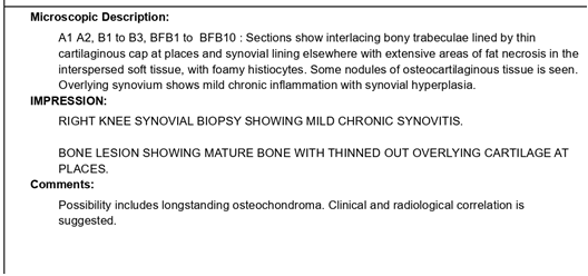
Figure 5: Histopathological report.
Outcome and follow-up
At the 6-week follow-up, the patient reported a marked reduction in pain and improved knee function. Physical examination showed no signs of recurrence, and the surgical site was healing well. Radiographs confirmed the absence of residual lesion (Fig. 6) [Table 1].
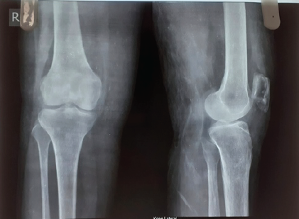
Figure 6: Post-operative X-ray.
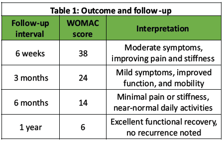
Table 1: Outcome and follow -up
This case highlights an unusual presentation of osteochondroma in an elderly patient, emphasizing the importance of considering benign bone tumors in differential diagnoses for knee pain and swelling in older adults. While osteochondromas commonly present in younger individuals, this case illustrates that they can remain asymptomatic until later in life or be discovered incidentally [ 7,2,8] . Surgical excision remains the treatment of choice, especially for symptomatic lesions, and offers excellent outcomes when malignancy is not a concern [9,10]. Extrasynovial osteochondromas in the infrapatellar area are rare but clinically significant, as they may interfere with the extensor mechanism of the knee and limit mobility. The mechanical symptoms can mimic meniscal injuries or osteoarthritic changes, delaying accurate diagnosis [1,3]. Imaging, particularly MRI, is essential not only for identifying the lesion but also for assessing cartilage cap thickness, which helps rule out malignant transformation. Differential diagnosis includes synovial chondromatosis, Hoffa’s disease, and loose bodies, which can be ruled out with MRI. Functional outcome assessment using the WOMAC scoring system demonstrated steady improvement in pain relief, stiffness reduction, and knee function over the follow-up period. This underscores the effectiveness of surgical excision and rehabilitation in restoring joint mechanics and quality of life, even in elderly patients [10,11]. This case highlights the importance of considering osteochondroma in the differential diagnosis of persistent knee pain in elderly patients and supports surgical excision as a safe and effective intervention for symptomatic lesions [6,11,12].
Osteochondromas, although rare in the elderly, should be considered in the differential diagnosis of knee pain and swelling. This case underscores the need for thorough clinical and radiological evaluation to ensure accurate diagnosis and effective treatment. Surgical excision of symptomatic osteochondromas provides significant relief and restores function, as demonstrated in this elderly patient. This report contributes to the limited literature on osteochondromas in older adults, offering insights into their management and outcomes. As this is a case report, a control group was not feasible. Nonetheless, we have compared our findings with previously published literature describing alternative management strategies (observation, excision).
- Single patient case report
- Histological grading or immunohistochemistry was not performed due to resource limitations. However, routine histopathological analysis was conclusive of the diagnosis
- Potential publication bias due to rare and successful cases is more likely to be reported, which can overestimate the ease of diagnosis or surgical success.
Osteochondroma is a common benign tumor in young patients but can rarely present in older adults, leading to diagnostic challenges. Infrapatellar localization is particularly uncommon and may mimic degenerative or inflammatory knee conditions. Careful imaging is essential to confirm the diagnosis and exclude malignant transformation. Surgical excision offers excellent outcomes in symptomatic cases, providing pain relief and functional recovery.


Related Articles in Journal of Orthopaedic Case Reports
 February 1, 2026 Isolated Osteochondromas of the Inner and Outer Tables of the Ilium: A Report of two Rare Cases
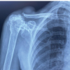 February 1, 2026 Minimally Invasive Double Loop Suture Technique for Recurrent Acromioclavicular Joint Ganglion Cyst in an Elderly Female: A Case Report
 February 1, 2026 A Rare Case of Calcaneal Osteochondroma: Case Report
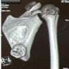 February 1, 2026 Symptomatic Ventromedial Scapular Osteochondroma Presenting with Restriction of Shoulder Movements: A Case Report