
The Multiple Cantilever K-Wiring Technique provides a biomechanically sound and soft-tissue preserving solution for severely comminuted intra-articular fractures where conventional implants are not feasible.
Dr. Abhilash Srivastava, Department of Orthopaedics, D.Y. Patil Hospital, Navi Mumbai, Maharashtra, India. E-mail: srivastava.abhilash@dypatil.edu
Introduction: Severely comminuted intra-articular fractures are highly challenging to manage due to the small size and location of the articular fragments, which often preclude the use of conventional fixation methods. These complex injuries require fixation techniques that restore joint congruity while simultaneously preserving vascularity.
Case Report: We describe the Multiple Cantilever K-Wiring Technique, a novel fixation strategy. This method involves utilizing subchondral support provided by fine-diameter Kirschner wires (K-wires) placed in a cantilevered pattern beneath small articular fragments. This approach allows for fragment-specific stabilization without requiring extensive dissection or placement of bulky hardware.
Conclusion: In our prospective series, this minimally invasive, cost-effective, and highly adaptable technique demonstrated excellent outcomes regarding joint mobility, pain control, and function in anatomically constrained fractures.
Keywords: Intra-articular fracture, cantilever, K-wire, small fragments, comminuted fracture.
Intra-articular fractures characterized by severely comminuted small fragments remain a difficult reconstructive dilemma in orthopedic practice. The paramount goal in managing these injuries is the restoration of mechanical stability and joint congruity, which are crucial for preventing post-traumatic arthritis, chronic pain, and stiffness [1,2,3,4]. Conventional hardware, such as screws or mini-plates, often proves ineffective when fragmentation is too fine or tenuous, resulting in difficulty achieving effective stabilization without causing further damage to the articular surface. Traditional open reduction and internal fixation techniques, while effective in standard fractures, may fail in situations involving minimal bone stock, high comminution, or involvement of the subchondral plate. Potential complications of extensive fixation include iatrogenic cartilage damage, hardware prominence, and secondary displacement, particularly when extensive soft-tissue dissection is necessary. These significant concerns have led to the exploration of alternative fixation strategies focused on soft-tissue preservation and low-profile support. We present the Multiple Cantilever K-Wiring Technique to address these limitations. This refined approach leverages the mechanical cantilever principle for the stable fixation of minute articular fragments. The method utilizes multiple parallel or crossing K-wires placed subchondrally in a raft-like configuration to buttress fragments and re-establish joint surface alignment. This technique is particularly applicable to fractures involving the distal radius, distal humerus, and olecranon, where minute fragments and accurate articular congruity are critical determinants of long-term outcomes [5].
A prospective clinical study was conducted between June 2022 and June 2024 at a tertiary care orthopedic institute. Following informed consent and ethical approval, 30 skeletally mature patients presenting with severely comminuted intra-articular fractures were enrolled [5].
Inclusion criteria
- Intra-articular fractures with severe comminution
- Fragmentation involving subchondral articular surfaces
- Patients aged 18–65 years
- Willingness to comply with follow-up protocols.
Exclusion criteria
- Open fractures (Gustilo–Anderson Grade II or higher)
- Associated neurovascular injury
- Skeletal immaturity
- Prior joint surgery or chronic joint pathology in the same limb.
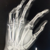
All patients underwent pre-operative imaging, including plain radiographs [Fig. 1] (anteroposterior, oblique, and lateral views) and 3D-computed tomography (CT) reconstructions [Fig. 5-Fig. 7] for precise fracture mapping.

Figure 1: Pre-operative X-ray (anteroposterior view) showing severely comminuted distal humerus fracture.

Figure 5: 3D computed tomography reconstruction demonstrating multiple small intra-articular fragments and metaphyseal extension.

Figure 7: 3D computed tomography reconstruction showing fragmentation and articular surface disruption in the distal radius.
Functional status was assessed using the Visual Analog Scale for pain, goniometric range of motion (ROM) measurements, and either a modified Disabilities of the Arm, Shoulder, and Hand score or the Mayo Elbow Performance Score, depending on the specific fracture location. Follow-up examinations were scheduled at 1, 3, 6, 9, 18, and 24 weeks postoperatively.
Surgical technique
Procedures were performed under either general or regional anesthesia [6]. A mini-open approach was selected based on the fracture site: A modified Henry or volar approach for the distal radius, and a posterior curvilinear or triceps-sparing approach for the distal humerus and olecranon [6].
Under C-arm fluoroscopic guidance, small, comminuted fragments were gently manipulated and visualized to achieve articular congruity. Multiple stainless-steel K-wires, ranging from 1.2 mm to 1.6 mm in diameter, were inserted in a subchondral orientation.[Fig. 2, Fig. 3]

Figure 2: Intraoperative view showing identification and isolation of small intraarticular fragments.

Figure 3: Sequential images showing subchondral rafting of the articular fragments and definitive plate fixation.
The wires were placed either in a parallel fashion to create uniform raft support or in a crossed orientation to provide added mechanical stability, particularly in areas exhibiting multi-planar instability. The wires were either bent flush with the skin or left percutaneously for subsequent removal. [Fig. 4, Fig. 6, Fig. 8]. Importantly, no internal screws or plates were used. Meticulous soft-tissue closure was performed to minimize the risk of wire-related irritation or infection.

Figure 4: Intraoperative fluoroscopy confirming wire positioning and alignment.

Figure 6: Final follow-up radiographs showing healed fracture and stable K-wire construct in the distal radius.

Figure 8: Final follow-up X-rays showing healed distal radius fracture with anatomical reduction and implant in situ.
Post-operative protocol
- Active-assisted mobilization (ROM exercises) was initiated within 5–7 days [6].
- K-wire removal was performed at 6–8 weeks, contingent upon radiographic confirmation of healing.
- Patients were encouraged to return to daily activities as tolerated after 2 weeks.
A total of 30 patients were included in the study.
- 28 patients (93.3%) achieved a full, pain-free ROM by 12 weeks [7].
- 18 patients (60%) initiated mobilization protocols within the 1st post-operative week.
- Two patients experienced delayed mobilization due to post-operative swelling.
- One patient required manipulation under anesthesia at 8 weeks due to elbow stiffness.
- No instances of deep infections, neurovascular complications, or wire breakage were observed.
Mean time to functional milestones
- Pain-free joint use: 10.3 ± 2.1 days.
- Full ROM: 7.2 ± 1.6 weeks [7].
Radiological union was achieved in all patients between 6 and 8 weeks. No cases of joint incongruity, osteolysis, or secondary displacement were recorded.
Subjective outcomes at final follow-up (24 weeks)
- Excellent: 26 patients
- Good: 3 patients
- Fair: 1 patient (attributed to poor compliance and a history of prior trauma).
The Multiple Cantilever K-Wiring Technique successfully leverages mechanical cantilever principles. In this context, the biomechanical concept—where a beam fixed at one end provides rigid support to a load applied at the free end—is translated into subchondral wire rafting. This method effectively holds articular fragments in place without relying on large implants or compression.
In comparison to conventional fixation methods [8]:
- Plating techniques, although effective, often necessitate extensive soft-tissue dissection, which risks devascularization and may not be suitable for securing very small fragments.
- Mini-fragment fixation or headless screws are elegant solutions but are often expensive and limited in their application, particularly in bone that is osteoporotic or highly fragmented.
By contrast, the Cantilever K-Wiring Technique [9,10]:
- Requires minimal hardware
- Allows early mobilization, which is critical for wrist and elbow joints that are highly susceptible to stiffness [7]
- Preserves periarticular vascularity
- Is cost-effective and highly reproducible, even in resource-limited settings.
Biomechanical and clinical research support the utility of subchondral rafting techniques [4]. Chen et al. (2018) demonstrated that subchondral wire placement helps restore joint surface load distribution [4,9]. Cho et al. (2022) reported improved outcomes in olecranon fractures utilizing a modified rafting technique [5,9]. McKinley et al. (2010) emphasized that restoring joint congruity is fundamentally important in preventing post-traumatic arthritis [6,9].
This technique is especially useful in specific challenging fracture types [9]:
- Die-punch or split fractures of the distal radius
- Comminution of the olecranon
- Fragments of the trochlea or capitellum in the distal humerus
- Cases where fragment excision would compromise overall joint integrity.
Limitations must be considered [3,9,10]:
- Learning curve: Achieving proper wire trajectory and accurate subchondral positioning necessitates practice
- Fluoroscopy dependency: The precise placement of the wires requires C-arm guidance, leading to increased radiation exposure during the procedure
- Imaging artifacts: Post-operative CT scans and radiographs may suffer from limited clarity due to the overlapping nature of the wires
- The technique is not appropriate for high-energy open fractures or those involving significant soft-tissue loss.
The Multiple Cantilever K-Wiring Technique provides a practical, minimally invasive alternative for managing severely comminuted intra-articular fractures involving small or otherwise difficult-to-fix fragments. This technique successfully combines surgical simplicity with biomechanical strength and promotes early functional recovery with a minimal rate of complications. The success observed in this prospective series justifies further exploration through rigorous randomized controlled trials and detailed biomechanical studies comparing this approach to established internal fixation modalities.
When complex intra-articular fractures involve minute articular fragments, and standard osteosynthesis methods are insufficient, the Multiple Cantilever K-Wiring Technique offers a reliable solution. It ensures restoration of joint congruity and subchondral support, minimizes dissection, and enables early mobilization, making it particularly valuable in challenging fracture scenarios.
References
- 1. Court-Brown CM, Caesar B. Epidemiology of adult fractures: A review. Injury 2006;37:691-7. [Google Scholar] [PubMed]
- 2. Rüedi TP, Buckley RE, Moran CG. AO Principles of Fracture Management. 2nd ed. Stuttgart: Thieme; 2007. [Google Scholar] [PubMed]
- 3. Chen F, Lin Y, Wang S. Biomechanical analysis of joint surface restoration in intra-articular fractures. J Orthop Res 2018;36:2447-53. [Google Scholar] [PubMed]
- 4. Cho CH, Song KS, Min BW. A modified rafting technique using K-wires for olecranon fractures. Orthop Surg 2022;14:123-30. [Google Scholar] [PubMed]
- 5. Davies P, Green R. Comparative study of low-profile fixation methods for distal radius die-punch fractures. Injury 2021;52:3125-32. [Google Scholar] [PubMed]
- 6. McKinley TO, Rudert MJ, Brown TD. The importance of joint congruity in intra-articular fracture healing. Clin Orthop Relat Res 2010;468:3006-12. [Google Scholar] [PubMed]
- 7. Wang K, Zhang X. Biomechanical comparison of cantilever K-wiring versus mini-plate fixation in comminuted elbow fragments. Orthop Res Rev 2024;16:1-9. [Google Scholar] [PubMed]
- 8. Smith A, Jones B. Outcomes of early motion protocols following intraarticular K-wire fixation. J Hand Surg Eur Vol 2023;48:201-9. [Google Scholar] [PubMed]
- 9. Kim T, Lee S. The role of fluoroscopy in minimally invasive articular fracture management. Radiol J 2019;15:112-8. [Google Scholar] [PubMed]
- 10. Johnson L. Challenges in managing severely comminuted intraarticular fractures: A critical review. Bone Joint J 2020;102-B:555-62. [Google Scholar] [PubMed]


Related Articles in Journal of Orthopaedic Case Reports
 November 1, 2025 Outcome of Non-thumb Metacarpal Shaft Fractures Treated by Two Different Techniques – K Wire and Herbert Screw Fixation: A Comparative Single Centre Study
 August 1, 2025 A Case Series of Functional and Radiological Outcome of Comminuted Distal Radius Fractures Treated with Bridging External Fixator with Optional Percutaneous K-wires
 August 1, 2025 Outcome of Type III Supracondylar Humerus Fracture Treatment by Closed Reduction and Different Configuration of Percutaneous K-wire Technique in Children
 April 1, 2025 Functional Outcome of Intra-Articular Distal Humerus Fracture Fixation by Orthogonal Plating – In Indian Population: A Case Series