
● Post-traumatic talar fractures complicated by avascular necrosis and non-union present significant treatment challenges, particularly following failure of initial internal fixation ● Total talus replacement may be considered a salvage option in selected patients to preserve ankle motion when the surrounding joint surfaces are relatively preserved ● Patient-specific 3D-printed talar implants, designed using contralateral ankle CT imaging and meticulous pre-operative planning, allow restoration of ankle alignment and joint congruency
Dr. Fatima Heba Habeeb, Department of Orthopedics, Yashoda Hospitals Somajiguda, Hyderabad, Telangana, India. E-mail: fatimaheba12@gmail.com
Abstract
Introduction: Avascular necrosis (AVN) of the talus is a known complication following trauma due to the bone’s unique anatomy and tenuous blood supply. A significant proportion of talar neck fractures may progress to AVN despite surgical fixation. Management of advanced talar AVN with collapse is challenging, with traditional salvage options including arthrodesis, which results in loss of ankle motion and may predispose to adjacent joint arthritis. Total talus replacement (TTR) has emerged as a motion-preserving alternative in selected cases. We report a case of post-traumatic talar AVN with non-union managed using a custom-made 3D-printed titanium talar implant.
Case Report: A 37-year-old male presented with persistent pain, deformity, and functional limitation of the right ankle following open reduction and internal fixation of a talar fracture. Over time, the patient developed progressive symptoms with restricted ankle motion. Physical examination revealed visible deformity of the ankle joint with limited ankle and subtalar joint range of motion. Radiological evaluation with radiographs and computed tomography demonstrated non-union of the talus with features of AVN. In view of progressive talar collapse and failure of initial fixation, the patient underwent TTR using a patient-specific 3D-printed titanium implant. Post-operative recovery was uneventful, with early improvement in pain and ankle mobility noted at 6-week follow-up.
Conclusion: TTR using a custom-made 3D-printed titanium implant may be considered an emerging salvage option in selected patients with post-traumatic AVN of the talus with collapse, with the potential to preserve ankle and hindfoot motion and provide early clinical improvement as indicated by improvements in Visual Analog Scale and American Orthopaedic Foot and Ankle Society scores at short-term follow-up.
Keywords: 3D-printed talus, talus avascular necrosis, titanium, total talus replacement, deformity.
Post-traumatic avascular necrosis (AVN) of the talus refers to temporary or permanent osteonecrosis resulting from a circulatory disturbance at the time of injury. The risk of post-traumatic AVN increases with high-energy trauma, fracture displacement, comminution, and associated soft tissue and vascular injury [1]. The talus is particularly susceptible due to its extensive articular cartilage coverage and tenuous blood supply, and disruption of this vascularity may result in osteonecrosis and collapse [2]. The majority of cases of talar AVN are post-traumatic in origin. Additional risk factors include fracture displacement, open injuries, dislocations, advanced age, high body mass index, and smoking [1]. Management options vary according to disease stage; however, in cases of complete AVN with collapse, treatment options are limited. Salvage procedures include necrectomy, bone grafting, and fusion of arthritic joints [1]. Arthrodesis is typically reserved for late-stage AVN with collapse and arthritis, and tibiotalocalcaneal fusion with bone grafting may be required in cases of significant bone loss [3]. However, fusion is associated with loss of ankle motion, altered gait mechanics, and the potential development of adjacent joint arthritis [4]. Total talus replacement (TTR) has emerged as a motion-preserving alternative in selected patients, with reports of symptomatic improvement and preservation of ankle motion in cases of end-stage talar AVN [5]. Advances in digital orthopedics and additive manufacturing have enabled the use of patient-specific 3D-printed talar prostheses for reconstruction [6]. The purpose of this case report is to describe the short-term outcome of TTR using a custom-made 3D-printed titanium implant in a patient with post-traumatic talar AVN and collapse.
We present the case of a 37-year-old male who sustained a right ankle injury following a road traffic accident. Initial imaging revealed fractures of the talus, medial malleolus, and lateral malleolus, along with a talonavicular joint dislocation. The patient underwent open reduction and internal fixation of the talus using two cannulated screws, along with application of an ankle-spanning external fixator. The external fixator was retained for 2 months during which the patient was kept non-weight bearing, following which the external fixator was removed, and weight-bearing was initiated. Over the subsequent months, the patient developed persistent ankle pain, difficulty with weight-bearing, and significant functional limitation (Fig. 1).
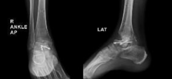
Figure 1: Pre-revision radiographs obtained 2 months after initial open reduction and fixation, demonstrating collapse of the talar body with screws in situ, along with loss of ankle congruency.
Physical examination revealed visible deformity of the ankle, including hindfoot varus and cavus with severely limited ankle joint range of motion and minimal subtalar joint motion compared to the contralateral limb. Pain and functional status were assessed using the Visual Analog Scale (VAS) and American Orthopaedic Foot and Ankle Society (AOFAS) scores pre and postoperatively. Radiographs obtained during follow-up showed collapse of the talus. A computed tomography scan performed subsequently demonstrated union of the medial and lateral malleoli, with non-union of the talus and sclerosis of the talar dome consistent with AVN (Fig. 2).
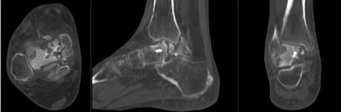
Figure 2: Pre-operative computed tomography images obtained immediately prior to revision surgery showing non-union of the talus with sclerosis of the talar dome consistent with avascular necrosis, while the medial and lateral malleoli demonstrate union.
In view of progressive talar collapse and failure of the initial fixation, the patient was admitted for implant removal and definitive surgical management. The patient underwent right talar replacement using a custom-made 3D-printed titanium implant manufactured by Bonetech Medysys, following detailed pre-operative planning. High-resolution computed tomography imaging of the contralateral healthy ankle was used to generate a three-dimensional model for the fabrication of patient-specific talar prosthesis. Magnetic resonance imaging was also performed to assess cartilage thickness, which aided implant size planning. Multiple sizes of the implant were prepared. Intraoperatively, the non-viable talus affected by AVN was excised, and the ankle joint was fully exposed. All the bony fragments of talus were removed. The posterior capsule was released to get an adequate range of movement, and the appropriately sized custom implant was positioned to restore joint alignment and function. The procedure was completed successfully, with stable intraoperative joint movement, and the patient was transferred to post-operative care in a hemodynamically stable condition. Postoperatively, a below-knee slab was applied, and the patient was kept on non-weight bearing for 1 month. At the 4-month follow-up, the patient reported an improvement in pain and function. Hindfoot varus and cavus were corrected, and gait improved significantly. Both VAS and AOFAS scores showed improvement at short-term follow-up. Post-operative X-rays taken at 4-week and 4-month follow-up showed that the implant was in a stable condition (Fig. 3 and 4).
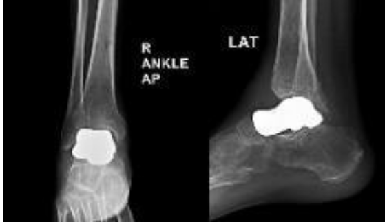
Figure 3: Post-operative anteroposterior and lateral views at 4-week follow-up showing a well-positioned custom 3D-printed talar implant with restored ankle alignment and no evidence of early subsidence.
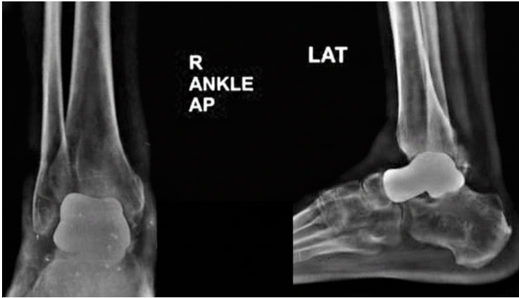
Figure 4: Post-operative anteroposterior and lateral radiographs of the right ankle at 4 months demonstrating maintained implant position and alignment following total talus replacement.
Post-traumatic AVN of the talus remains a challenging condition due to the high risk of progressive collapse and secondary arthritis. In advanced stages with collapse, arthrodesis is commonly employed as a salvage procedure, and tibiotalocalcaneal fusion with bone grafting may be required in cases with significant bone loss [3]. While fusion procedures provide pain relief and stability, they are associated with substantial functional limitations, including loss of ankle motion, altered gait mechanics, limb-length discrepancy, and the potential development of adjacent joint arthritis [4]. Traditional operative salvage techniques, such as talocrural arthrodesis utilizing the anterior talar segment or tibiocalcaneal shortening arthrodesis, have also been described; however, these procedures similarly result in loss of joint function and reduced mobility [7]. Due to these limitations, particularly in younger or active patients, alternative motion-preserving strategies have gained increasing attention. TTR has emerged as a motion-preserving alternative for selected patients with advanced talar AVN, with recent studies demonstrating superior preservation of ankle motion when compared to arthrodesis [8]. Reported advantages of talar replacement include preservation of ankle and hindfoot motion, maintenance of limb length, relatively shorter periods of restricted weight bearing, and rapid pain relief [7]. Recent reports have demonstrated encouraging short- to mid-term clinical outcomes following TTR in patients with post-traumatic talar AVN, with improvements in pain and functional ability [9]. Current advances in implant design and additive manufacturing have further enhanced the feasibility of TTR. Patient-specific talar implants manufactured using three-dimensional printing based on computed tomography data allow accurate anatomical reconstruction and improved implant fit. Titanium-based 3D-printed talar prostheses offer excellent biocompatibility, precise restoration of talar geometry, and the potential for osseointegration, contributing to favorable early clinical outcomes, although long-term durability remains under evaluation [10]. In the present case, the patient developed progressive talar collapse following failed internal fixation, rendering joint-preserving options unfeasible. Given the presence of talar AVN with collapse and minimal degenerative changes in the surrounding joints, TTR was considered an appropriate salvage option. Implantation of a custom-made 3D-printed titanium talus allowed restoration of ankle alignment and resulted in early improvement in pain and function at short-term follow-up. This case adds to the growing body of evidence supporting TTR as a motion-preserving alternative in carefully selected patients with post-traumatic talar AVN, while acknowledging the need for longer-term follow-up to evaluate implant longevity and functional outcomes.
TTR using a custom-made 3D-printed titanium implant represents an emerging surgical option for selected patients with advanced talar AVN and collapse. In this case, TTR resulted in early symptomatic improvement with preservation of ankle motion at short-term follow-up. While longer-term outcomes remain to be established, this technique may serve as a motion-preserving alternative to arthrodesis in appropriately selected patients.
Total talus replacement using a custom-made 3D-printed titanium implant represents an emerging motion-preserving treatment option for post-traumatic avascular necrosis of the talus, with encouraging short-term clinical outcomes in carefully selected patients.
References
- 1. Kopp L, Baba V, Marx C, Rammelt S. Post-traumatic osteonecrosis of the talus. Fuß Sprunggelenk 2025;23:41-59. [Google Scholar] [PubMed]
- 2. Gross CE, Sershon RA, Frank JM, Easley ME, Holmes GB Jr. Treatment of osteonecrosis of the Talus. JBJS Rev 2016;4:e2. [Google Scholar] [PubMed]
- 3. Dhillon MS, Rana B, Panda I, Patel S, Kumar P. Management options in avascular necrosis of talus. Indian J Orthop 2018;52:284-96. [Google Scholar] [PubMed]
- 4. Kubisa MJ, Kubisa MG, Pałka K, Sobczyk J, Bubieńczyk F, Łęgosz P. Avascular necrosis of the talus: Diagnosis, treatment, and modern reconstructive options. Medicina (Kaunas) 2024;60:1692. [Google Scholar] [PubMed]
- 5. Kadakia RJ, Akoh CC, Chen J, Sharma A, Parekh SG. 3D printed total talus replacement for avascular necrosis of the talus. Foot Ankle Int 2020;41:1529-36. [Google Scholar] [PubMed]
- 6. He X, Lu M, Zou C, Li Z, Gong T, Kenmegne GR, et al. Three-dimensional printed custom-made modular talus prosthesis in patients with talus malignant tumor resection. J Orthop Surg Res 2024;19:273. [Google Scholar] [PubMed]
- 7. Ando Y, Yasui T, Isawa K, Tanaka S, Tanaka Y, Takakura Y. Total talar replacement for idiopathic necrosis of the talus: A case report. J Foot Ankle Surg 2016;55:1292-96. [Google Scholar] [PubMed]
- 8. Harnroongroj T, Arunakul M, Reingrittha P, Chuckpaiwong B, Angthong C, Tharmviboonsri T, et al. Outcomes of tibiotalocalcaneal arthrodesis vs talar body prosthesis as treatment of collapsed avascular necrosis of the talus: A 10- to 13-year-follow-up retrospective comparative study. Foot Ankle Int 2024;45:435-43. [Google Scholar] [PubMed]
- 9. Siddiqi A, Fallat L. Use of 3-D printed total talus implant in a challenging case of post-traumatic arthritis and avascular necrosis of the talus. J Int Foot Ankle 2023;2:1. [Google Scholar] [PubMed]
- 10. Antounian F, Avagyan H, Ghaltaghchyan T, Holovenko Y, Khachatryan H, Aghayan M. Designing and additive manufacturing of talus implant for post-traumatic talus avascular necrosis: A case study. J Orthop Surg Res 2024;19:501. [Google Scholar] [PubMed]


Related Articles in Journal of Orthopaedic Case Reports
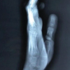 June 1, 2026 Hang Fire – Acute Crush Injury of Fingers – Till Appropriate Intercede – A Case Report
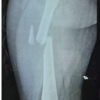 November 1, 2025 Outcome of Pediatric Femoral Shaft Fractures Treated with Titanium Elastic Nailing: A Prospective Study
 April 10, 2024 Utility of Asymmetric Multilevel Pontes Osteotomy in Ankylosing Spondylitis with Scoliosis using Ultrasonic Bone Scalpel: Case Report
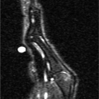 May 10, 2023 Swan Neck Deformity: An Unusual Complication Following Trigger Finger Release