

Large and painful soft-tissue masses with concerning malignant features may still represent benign pathology, highlighting the importance of appropriate imaging and histopathological evaluation for accurate diagnosis.
Dr. Khairul Arif Ahmad, Department of Family Medicine, Klinik Kesihatan Bandar Mentakab, Jalan Bunga Matahari, Mentakab Pahang, Malaysia. E-mail: khairularif_ahmad@yahoo.com
Abstract
Introduction: Soft-tissue sarcomas are uncommon malignant tumors that may present with enlarging, painful soft-tissue masses. However, certain benign lesions may occasionally demonstrate overlapping clinical features, creating diagnostic uncertainty. We report a case of a large lower limb lipoma that clinically mimicked soft-tissue sarcoma.
Case Report: A 30-year-old woman presented with a progressively enlarging right calf swelling associated with nocturnal pain for 3 years. Physical examination demonstrated a large tender mass with poorly defined margins measuring approximately 20 cm × 15 cm. Due to concerning clinical features suggestive of malignancy, further evaluation was performed. Plain radiography showed no bony involvement, while ultrasound did not demonstrate a definite soft-tissue mass or vascular malformation. Magnetic resonance imaging revealed diffuse thickening of subcutaneous fat with homogeneous signal intensity similar to adjacent fat tissue, favoring a benign lipomatous lesion. The patient subsequently underwent surgical excision. Histopathological examination demonstrated mature adipocytes without atypia or lipoblasts, confirming a benign lipoma.
Conclusion: This case highlights the diagnostic challenges in differentiating benign lipomatous lesions from soft-tissue sarcomas based solely on clinical presentation. Appropriate imaging evaluation and histopathological confirmation remain essential for accurate diagnosis and optimal management of suspicious soft-tissue masses.
Keywords: Lipoma, lower limb swelling, magnetic resonance imaging, soft-tissue sarcoma, soft-tissue tumor.

Soft-tissue sarcomas are malignant tumors arising from mesenchymal tissue and may present as enlarging soft-tissue masses with aggressive behavior and metastatic potential [1]. In contrast, lipomatous tumors are among the most common soft-tissue tumors, with lipomas representing the majority of benign adipocytic lesions [2]. Lipomas are the most common soft-tissue masses in adults and typically occur between 40 and 60 years of age [3,4]. Several clinical features may help differentiate benign from malignant soft-tissue lesions, including lesion size, rapid progression, pain, and anatomical location [1,3,5]. However, overlap in clinical presentation between benign and malignant lesions may create significant diagnostic uncertainty. This distinction is clinically important as both conditions differ considerably in prognosis and management. We report a case of a large lower limb lipoma presenting with multiple red flag features suggestive of soft-tissue sarcoma. Despite concerning clinical findings, further radiological and histopathological evaluation confirmed a benign lipoma. This case highlights the importance of a structured diagnostic approach when evaluating suspicious soft-tissue masses.
A 30-year-old woman presented with a 3-year history of progressively enlarging right calf swelling associated with nocturnal pain requiring analgesia. She reported a prior history of soft-tissue contusion following a motor vehicle accident before the onset of swelling. There were no constitutional symptoms or family history of malignancy.
Examination revealed a large, tender cystic mass over the proximal right calf measuring approximately 20 cm × 15 cm with poorly defined margins. The overlying skin was unremarkable without pulsation, bruising, or punctum (Fig. 1). Distal pulses and lower limb joint movements were preserved.
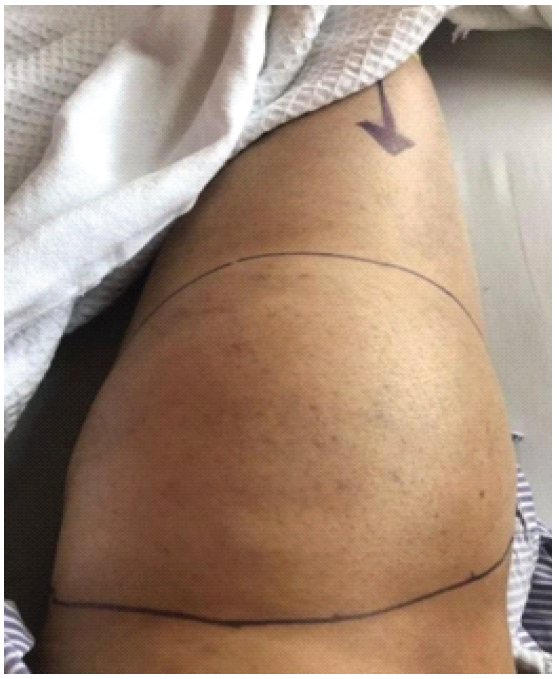
Figure 1: Pre-operative image showing a large, well-defined swelling over the proximal right calf (outlined), measuring approximately 20 cm × 15 cm. The mass appears smooth with no overlying skin changes, visible pulsation, or ulceration.
Plain radiography demonstrated no bony involvement or lytic lesions, while ultrasound showed no significant soft-tissue collection, vascular malformation, or definite mass. Due to concerning clinical features, including progressive enlargement, pain, large size, and poorly defined margins, contrast-enhanced magnetic resonance imaging (MRI) was performed. MRI revealed diffuse thickening of the subcutaneous fat at the anteromedial proximal right leg without a definite capsule. The lesion demonstrated homogeneous signal intensity similar to adjacent fat tissue without aggressive soft-tissue features, favoring a benign lipomatous lesion (Fig. 2).
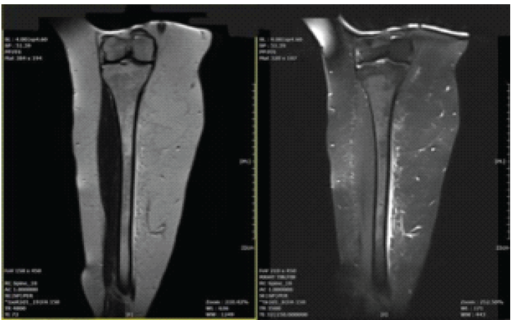
Figure 2: Magnetic resonance imaging of the right lower limb demonstrating diffuse thickening of the subcutaneous tissue at the anteromedial aspect of the proximal calf. The lesion shows homogeneous signal intensity similar to adjacent subcutaneous fat and lacks a well-defined capsule. There is no evidence of deep fascial invasion, bony involvement, or significant heterogeneous enhancement. These imaging features are suggestive of a benign lipomatous lesion.
The patient subsequently underwent complete surgical excision for diagnostic and therapeutic purposes. Intraoperatively, a poorly demarcated 25 cm × 15 cm mass with multiple internal seromas was identified within the subcutaneous tissue of the proximal right leg and excised completely. Histopathological examination demonstrated mature adipocytes without atypia, lipoblasts, necrosis, or increased mitotic activity, consistent with benign lipoma. The patient had an uneventful post-operative recovery with satisfactory wound healing (Fig. 3). No further oncological treatment was required following orthopedic oncology review.
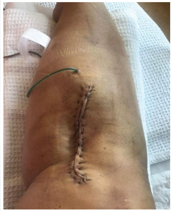
Figure 3: Demonstrating the surgical incision over the anteromedial aspect of the proximal right leg following excision of the mass. The wound is closed with sutures, and a drain is in situ.
Lumps in the body can range from benign to malignant conditions. Soft-tissue sarcomas are a group of malignant tumors that are relatively rare but important not to miss. These tumors can arise from muscles, fat, tendons, or blood vessels [6]. They tend to grow rapidly and may metastasize to other parts of the body. Lipomas, on the other hand, are benign growths and account for the majority of soft-tissue swellings [7]. They arise from mesenchymal tissue, consist of mature adipocytes, and are usually enclosed by a thin fibrous capsule [7]. Lipomas are typically slow-growing and localized. Although sarcomas can occur at any age, they are more common in children and young adults. A history of underlying conditions or genetic syndromes such as neurofibromatosis and Li-Fraumeni syndrome may also predispose individuals to soft-tissue sarcomas [1,5,8].
Differentiating benign from malignant soft-tissue masses may occasionally be challenging due to overlapping clinical features. Although lipomas are considerably more common than soft-tissue sarcomas, the presence of red flag features such as progressive enlargement, pain, large lesion size, and poorly defined margins may increase suspicion for malignancy [1,5]. In the present case, the lesion demonstrated several concerning clinical features commonly associated with soft-tissue sarcoma despite ultimately representing benign pathology. This overlap may create significant diagnostic uncertainty, particularly in the primary care setting.
MRI plays an important role in the evaluation of suspicious soft-tissue lesions due to its superior soft-tissue characterization and multiplanar capability [1,4,9]. MRI assists in determining lesion extent, relationship with surrounding structures, and features suggestive of benign or malignant pathology. In general, lesions with homogeneous signal intensity and smooth margins are more suggestive of benign lesions, whereas heterogeneous lesions with irregular borders may indicate malignancy [4]. In this case, MRI demonstrated homogeneous signal intensity similar to adjacent fat tissue without aggressive soft-tissue features, favoring a benign lipomatous lesion despite concerning clinical findings.
Histopathological examination remains the gold standard for definitive diagnosis. Although biopsy is commonly recommended before definitive management of suspicious soft-tissue tumors, combined diagnostic and therapeutic excision may be considered in selected cases when clinical and radiological findings are sufficiently informative [1]. Histopathological analysis in the present case demonstrated mature adipocytes without atypia or lipoblasts, confirming the diagnosis of benign lipoma.
Benign lipomas are usually adequately treated with complete surgical excision alone [10], whereas soft-tissue sarcomas often require multidisciplinary management involving surgery with or without adjuvant therapy [1]. In terms of prognosis, soft-tissue sarcomas can be life-threatening if not detected and treated early. Tumor type and the presence of metastasis are key determinants of prognosis [5].
This case highlights the importance of maintaining a high index of suspicion when evaluating enlarging soft-tissue masses. Early referral and appropriate imaging are essential to facilitate accurate diagnosis and timely management while minimizing delays in identifying malignant disease.
This case demonstrates that benign lipomatous lesions may occasionally present with clinical features concerning for soft-tissue sarcoma, creating significant diagnostic uncertainty. Careful clinical assessment combined with appropriate imaging and histopathological evaluation remains essential for accurate diagnosis of suspicious soft-tissue masses. Early referral is important to facilitate timely management while avoiding delayed diagnosis of malignancy and unnecessary overtreatment of benign conditions.
Benign lipomas may occasionally mimic soft-tissue sarcoma clinically; therefore, enlarging painful soft-tissue masses should undergo appropriate imaging and histopathological evaluation to establish an accurate diagnosis.
References
- 1. Ramu EM, Houdek MT, Isaac CE, Dickie CI, Ferguson PC, Wunder JS. Management of soft-tissue sarcomas; treatment strategies, staging, and outcomes. SICOT J 2017;3:20. [Google Scholar] [PubMed]
- 2. Bird JE, Morse LJ, Feng L, Wang WL, Lin PP, Moon BS, et al. Non-radiographic risk factors differentiating atypical lipomatous tumors from lipomas. Front Oncol 2016;6:197. [Google Scholar] [PubMed]
- 3. Johnson CN, Ha AS, Chen E, Davidson D. Lipomatous soft-tissue tumors. J Am Acad Orthop Surg 2018;26:779-88. [Google Scholar] [PubMed]
- 4. Walker EA, Song AJ, Murphey MD. Magnetic resonance imaging of soft-tissue masses. Semin Roentgenol 2010;45:277-97. [Google Scholar] [PubMed]
- 5. Grimer R, Judson I, Peake D, Seddon B. Guidelines for the management of soft tissue sarcomas. Sarcoma 2010;2010:506182. [Google Scholar] [PubMed]
- 6. Crombé A, Kind M, Fadli D, Miceli M, Linck PA, Bianchi G, et al. Soft-tissue sarcoma in adults: Imaging appearances, pitfalls and diagnostic algorithms. Diagn Interv Imaging 2023;104:207-20. [Google Scholar] [PubMed]
- 7. Yavari M, Afshar A, Shahraki SS, Tabrizi A, Doorandish N. Management of symptomatic lipoma of the hand: A case series and review of literature. Arch Bone Jt Surg 2022;10:530-5. [Google Scholar] [PubMed]
- 8. Penel N, Grosjean J, Robin YM, Vanseymortier L, Clisant S, Adenis A. Frequency of certain established risk factors in soft tissue sarcomas in adults: A prospective descriptive study of 658 cases. Sarcoma 2008;2008:459386. [Google Scholar] [PubMed]
- 9. Van Vliet M, Kliffen M, Krestin GP, Van Dijke CF. Soft tissue sarcomas at a glance: Clinical, histological, and MR imaging features of malignant extremity soft tissue tumors. Eur Radiol 2009;19:1499-511. [Google Scholar] [PubMed]
- 10. Bancroft LW, Kransdorf MJ, Peterson JJ, O’Connor MI. Benign fatty tumors: Classification, clinical course, imaging appearance, and treatment. Skeletal Radiol 2006;35:719-33. [Google Scholar] [PubMed]


Related Articles in Journal of Orthopaedic Case Reports
 April 1, 2025 Recurrent Intra-articular Lipoma of Ankle Joint; A Rare Case Report
 October 1, 2026 Magnetic Resonance Imaging-Arthroscopy Correlation in Traumatic Knee Injuries: A Retrospective Diagnostic Accuracy Study
 October 1, 2026 Incidence, Arthroscopic Detection, and Clinical Outcomes of Ramp Lesions in Patients Undergoing Anterior Cruciate Ligament Reconstruction: A Prospective Observational Study
 October 1, 2026 Acute Cauda Equina Syndrome in Rapid Succession: A Case Series of Eight Consecutive Patients Undergoing Emergency Surgical Decompression Over One Week