


Introduction
Osseous hemangiomas (OH) are benign lesions that account for only 1% of primary bony neoplasms [1]. They represent disorganized and expansive proliferation of blood vessels with blood contained in the thickened trabecular architecture. Predominantly found in the vertebrae and craniofacial bones, which account for about three-quarters of the incidence. Primary hemangioma of the long tubular bones is a rare entity [2]. Based on the anatomical location, they can be classified as medullary (55%), periosteal (33%), and intracortical (12%). In the long bones, they can be located in the diaphysis (78%), the metaphysis (12%), or the epiphysis (10%). Although OH in the axial skeleton are commonly asymptomatic, they usually present with pain, swelling, and pathological fracture in the appendicular skeleton, depending upon the size and site of the lesion [3,4]. We present a rare case of an asymptomatic OH located in the anteroinferior quadrant of the femoral head. To the best of our knowledge, this represents only the second reported solitary hemangioma in this juxta-articular location.
Case Report
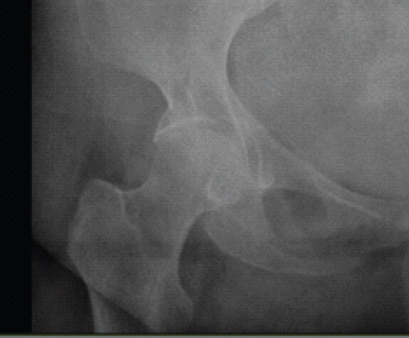
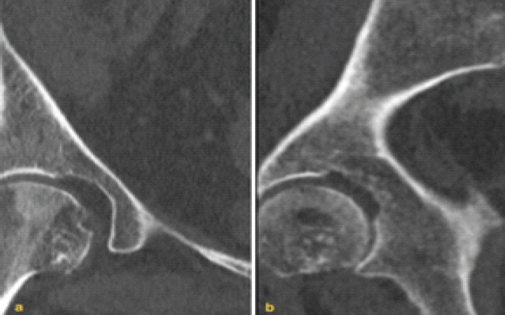
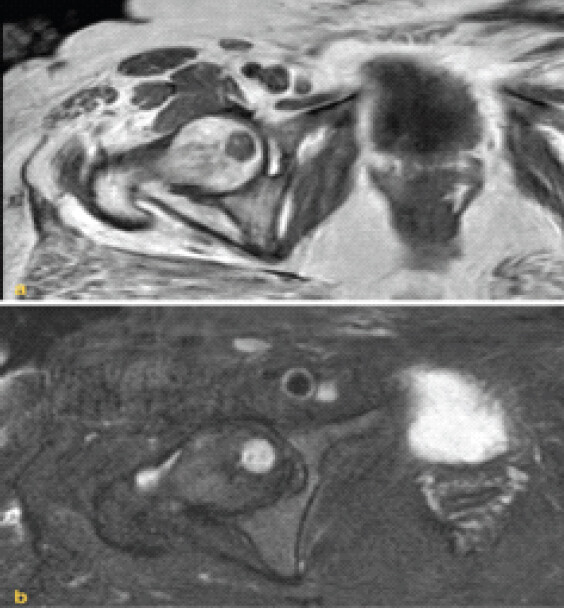
A 56-year-old lady presented with pain in the left hip and lower back, following a fall at home. On physical examination, the left hip movements were difficult to assess due to the severe pain. She had an antalgic gait, and the pain worsened with weight-bearing. Neurologic evaluation revealed no sensory or motor deficits. She reported no previous symptoms involving the right hip. Although initial radiographs could not exclude an occult neck of femur fracture, they incidentally demonstrated a well-circumscribed lytic lesion over the right head of the femur (Fig. 1). Computed tomography (CT) scan of the right hip revealed a lucent lesion adjacent to the fovea capitis showing typical “polka-dot” appearance of the trabeculae along the axis of the neck with a sclerotic rim (Fig. 2). Magnetic resonance imaging (MRI) was performed to further investigate both hips, which ruled out an occult neck of femur fracture in the left hip. In the right femoral head, MRI demonstrated a well-defined osteolytic lesion measuring 12 mm craniocaudally × 14 mm mediolaterally × 17 mm anteroposteriorly, with a peripheral sclerotic rim. The lesion extended into the articular surface infra-foveally on the non-weight-bearing part of the head, which may explain her absence of right hip pain. On axial images, hyperintense spots within a hypointense background representing thickened trabeculae could be seen within the lesion as honeycombed septations. It appeared to be hypointense on T1-weighted, hyperintense on T2-weighted with a sclerotic rim, regular margins with a narrow zone of transition (Fig. 3 and 4). Based on characteristic imaging findings, a radiological diagnosis of an infra-foveal intraosseous hemangioma was made. As the lesion was asymptomatic, demonstrated characteristic benign imaging features, and did not involve the weight-bearing dome, conservative management with clinical and radiological surveillance was agreed. The patient was counseled regarding the diagnosis and advised to return if she developed right-hip pain, impaired mobility, or other new symptoms.
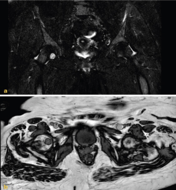
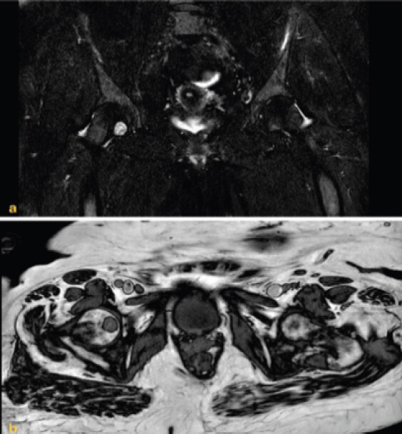
At 6-month follow-up, MRI demonstrated a stable appearance of the lesion without interval progression (Fig. 5). At 18 months, the patient remained asymptomatic, mobilized independently without walking aids, and reported no limitation in her activities of daily living.
Discussion
The first report of an OH in the appendicular skeleton was by Hitzrot in 1917, who described them as rare, benign, slow-growing vascular tumors of the bone. Embryologically, they are derived from hemangioblasts characterized by vascular hyperplasia with thin-walled sinusoids [5]. OHs of the axial skeleton are common and incidental, as opposed to OH of the appendicular skeleton, which account for only 15–20% and are more likely to be symptomatic. Interestingly, Kaleem et al. [6] reviewed all previously reported cases of solitary skeletal hemangiomas of the extremities in the English literature in 2000 and concluded that even experienced radiologists rarely encountered OH of the long tubular bones. In their series of 114 cases, none of the OH were mentioned to be located in the proximal femur [2, 6]. To the best of our knowledge, we report only the second case of solitary hemangioma located juxta-articularly in the femoral head. Chawla et al. [7] in 2006 presented a case of symptomatic OH in a middle-aged man, which involved the articular surfaces; it was managed with excision and uncemented total hip arthroplasty. Unlike other primary bony tumors, OHs typically have a female preponderance, with a female-to-male ratio of 3:2. They are most commonly encountered in middle age [3]. Our patient, a 56-year-old woman, was therefore within the commonly reported demographic group.
OHs in the appendicular skeleton can be classified anatomically as medullary, cortical, and periosteal. Medullary OHs can be further subcategorized as diaphyseal (48%), metadiaphyseal (30%), metaphyseal (12%), metaepiphyseal (4%), epimetadiaphyseal (3%), epiphyseal (1.5%), and involving the entire length of the tubular bone (1.5%) [8]. In this case, the lesion was medullary and epiphyseal in nature. The radiological appearance of osseous hemangiomas in long tubular bones is variable and may be non-specific. Plain radiographs may demonstrate a well-defined osteolytic lesion with a “honeycomb” or “soap-bubble’ appearance. On CT images, a characteristic “polka-dot” appearance corresponds to vertically thickened trabeculae. MRI provides further characteristic evidence, especially in diagnostic dilemmas, to distinguish it from other differentials, such as intrahemangioma cyst, fibrous dysplasia, and giant cell tumor. Signal intensity on T1-weighted images varies according to the proportion of fat and vascular tissue, whereas T2-weighted images generally demonstrate high signal intensity because of the lesion’s vascular or fluid-rich components.
The present case findings are consistent with a characteristic case of OH at an atypical location. The peculiarity is its asymptomatic presentation, albeit its location in the weight-bearing axis of the body. Pathological fracture has been reported in appendicular lesions and often requires prophylactic fixation [7,8,9]. In the present case, a radiological diagnosis of intraosseous hemangioma was made on the basis of the characteristic CT and MRI appearances. Histological confirmation was not pursued because the lesion was asymptomatic, well defined, surrounded by a sclerotic margin, and lacked aggressive imaging features. The potential risks of biopsy of a vascular lesion were considered to outweigh the likely diagnostic benefit. MRI provides excellent soft-tissue contrast and multiplanar assessment; however, histological confirmation should be considered when imaging findings are atypical, the lesion is symptomatic or progressive, or malignancy cannot be confidently excluded [10].
Conclusion
OH of the femoral head are exceptionally rare and may be discovered incidentally during imaging for unrelated conditions. Recognition of characteristic benign imaging features may prevent misdiagnosis and unnecessary invasive intervention. In carefully selected asymptomatic patients without aggressive radiological features, conservative management with clinical and radiological surveillance may be appropriate.
Clinical Message
Recognition of the characteristic CT and MRI features of intra-OH can facilitate a confident radiological diagnosis and avoid unnecessary biopsy or surgical intervention in carefully selected asymptomatic patients.
Conflict of Interest:
Nil
Source of Support:
Nil
Consent
The authors confirm that informed consent was obtained from the patient for publication of this article
How to Cite this Article
Sonkusale A, Ivanova S, Nikolaides AP. Incidentally Detected Osseous Hemangioma of the Femoral Head: Conservative Management of a Rare Case with Review of the Literature. J Orthop Case Rep 2026 November;16(11):27-30. doi: 10.13107/jocr.2026.v16.i11.8376.
References
- Chandra SP, Singh P, Kumar R, Agarwal D, Tandon V, Kale SS. Long-term outcome of treatment of vertebral body hemangiomas with direct ethanol injection and short-segment stabilization. Spine J 2019;19:131-43. [Google Scholar] | [PubMed]
- Rigopoulou A, Saifuddin A. Intraosseous hemangioma of the appendicular skeleton: Imaging features of 15 cases, and a review of the literature. Skeletal Radiol 2012;41:1525-36. [Google Scholar] | [PubMed]
- Reijnierse M, Kroon HM, Van Der Heul RO, Mulder JD. Mesenchymoma of bone. A case report. J Bone Joint Surg Am 1993;75:112-5. [Google Scholar] | [PubMed]
- Cha JG, Yoo JH, Kim HK, Park JM, Paik SH, Park SJ. PET/CT and MRI of intra-osseous haemangioma of the tibia. Br J Radiol 2012;85:e94-8. [Google Scholar] | [PubMed]
- Hitzrot JM. Haemangioma cavernosum of bone. Ann Surg 1917;65:476-82. [Google Scholar] | [PubMed]
- Kaleem Z, Kyriakos M, Totty WG. Solitary skeletal hemangioma of the extremities. Skeletal Radiol 2000;29:502-13. [Google Scholar] | [PubMed]
- Chawla A, Singrakhia M, Maheshwari M, Modi N, Parmar H. Intraosseous haemangioma of the proximal femur: Imaging findings. Br J Radiol 2006;79:e64-6. [Google Scholar] | [PubMed]
- Xia Z, Sittampalam K, Howe TS, Lo NN. Successful treatment of solitary intraosseous haemangioma of the femoral neck. Singapore Med J 2015;56:e65-70. [Google Scholar] | [PubMed]
- Ferrari L, Segura Fonseca JJ. Cavernous hemangioma of the proximal femur. Int J Surg Pathol 2011;19:617-9. [Google Scholar] | [PubMed]
- Singhal S, Saran S, Agarwal A, Dhingra M. Intramuscular hemangioma presenting as pathological femur fracture: A case report. Indian J Musculoskelet Radiol 2023;5:47-51. [Google Scholar] | [PubMed]
© 2026 Journal of Orthopaedic Case Reports - Published by Indian Orthopaedic Research Group
About the Authors
-

Aashay Sonkusale
-

Silviya Ivanova
-

A P Nikolaides1
Related Articles in Journal of Orthopaedic Case Reports
 October 1, 2026 Isolated Posterior Malleolus Fracture with Early Symptom Resolution: A Case Report
 November 1, 2026 Double-Layered Lateral Meniscus Mimicking Locking of Lateral Meniscus: A Case Report
 November 1, 2026 Second and Third Carpometacarpal Joint Dislocation in a Polytrauma Patient: Successful Closed Reduction and 2-Year Follow-Up
 November 1, 2026 Hallux Mystery: A Rare Case of Fibro-Osseous Pseudotumor