


Introduction
Reconstruction of the anterior cruciate ligament (ACL) with a quadrupled hamstring autograft is standard practice in many centres worldwide, supported by favourable long-term clinical outcomes and low donor-site morbidity relative to bone-patellar tendon-bone or quadriceps tendon autografts. However, the final diameter of a hamstring construct cannot be directly ascertained pre-operatively and depends heavily on individual tendon dimensions, which exhibit significant anatomical variability and cannot be reliably predicted by external anthropometric measurements alone.
A final quadrupled graft diameter below 8.0 mm has been strongly associated with elevated rates of graft failure and revision surgery in multiple clinical series, establishing 8.0 mm as a widely accepted benchmark for graft adequacy [1,2]. When the isolated semitendinosus (ST) tendon yields a graft diameter below this threshold, ipsilateral gracilis tendon harvesting and co-augmentation are routinely required. In conventional practice, this decision is made intraoperatively only after ST harvest and sizing, creating unforeseen operative delays, altered surgical plans, and additional morbidity.
Magnetic resonance imaging (MRI), routinely performed for diagnostic confirmation of ACL tears, provides high-resolution anatomical cross-sectional area (CSA) measurements of the hamstring tendons [1]. While previous studies have reported positive correlations (r = 0.70 to 0.95) between pre-operative CSA and harvested graft diameter [3,4,5,6], reported cut-off values vary widely (12 to 22 mm²) depending on the axial slice level, population demographics, and measurement protocols [7,8,9,10,11,12,13,14,15]. Furthermore, few studies present comprehensive diagnostic accuracy parameters – such as sensitivity, specificity, positive predictive value (PPV), negative predictive value (NPV), and receiver operating characteristic (ROC) curves – that directly guide surgical decision-making.
This study evaluates the diagnostic accuracy of an 18.5 mm² pre-operative ST CSA threshold derived from a prospective cohort in Southern India to predict whether a harvested quadrupled ST graft reaches the critical 8.0 mm diameter threshold.
Materials and Methods
Study design, setting, and ethics
This prospective observational diagnostic accuracy study was conducted in the Department of Orthopaedics and Traumatology at Pushpagiri Institute of Medical Sciences and Research Centre, Tiruvalla, Kerala, India, over an 18-month period following approval from the Institutional Ethics Committee (EC Reg. No. EC/NEW/INST/2020/1188; Approval ref: PIMSRC/E1/388A/52/2023, granted on June 29, 2023). Written informed consent was obtained from all patients prior to enrolment.
Participant eligibility and flow
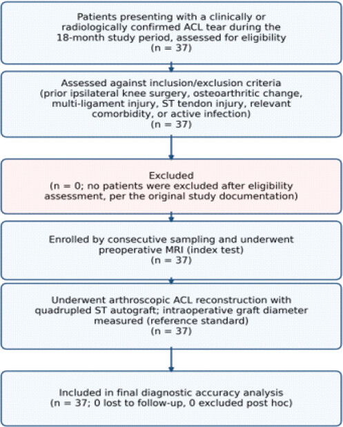
Consecutive patients aged 18–50 years presenting with a clinically and radiologically verified complete ACL rupture scheduled for arthroscopic primary ACL reconstruction using a quadrupled ST autograft were screened. Exclusion criteria comprised previous ipsilateral knee surgery, established osteoarthritic changes (Kellgren-Lawrence grade ≥2), multi-ligamentous knee injuries, previous ST injury, active local/systemic infection, and systemic inflammatory diseases. A total of 37 consecutive patients met all eligibility criteria and completed both the index test (preoperative MRI) and reference standard (intraoperative graft measurement), with zero losses to follow-up (Fig. 1).
Sample size calculation
The sample size was determined using Fisher’s z-transformation formula for detecting a significant correlation coefficient (Equation 1):
N = [ (Z1−α/2 + Z1−β) / (0.5 × ln[(1+r) / (1−r)])]2 +3
where N is the required sample size, Z1−α/2 is the standard normal deviate for a two-tailed test at the 5% significance level (1.96), Z1β is the standard normal deviate corresponding to 80% power (0.84), and r is the anticipated correlation coefficient between CSA and graft diameter (0.45, based on prior literature). Substituting these values yielded a minimum required sample size of approximately 37 participants, which determined the target enrolment for this study.
Index test: Pre-operative MRI protocol
All enrolled participants underwent standard knee MRI prior to surgery. Axial sequences were evaluated by a senior musculoskeletal radiologist blinded to clinical and intraoperative findings. ST CSA was measured at a standardized joint-line level corresponding to the axial slice where the anteroposterior diameter of the medial femoral condyle was maximal, utilizing continuous free-hand region-of-interest tracing.
Reference standard: Intraoperative graft sizing
Arthroscopic ACL reconstruction was performed by a single experienced orthopedic knee surgeon blinded to the pre-operative MRI CSA values. The ST tendon was harvested using an open tendon stripper, prepared, cleaned of muscular tissue, and quadrupled. The quadrupled graft diameter was measured using a calibrated cylindrical graft-sizing block in 0.5 mm increments before any gracilis tendon harvest or augmentation. An adequate graft was defined as diameter ≥8.0 mm; an undersized graft (<8.0 mm) was augmented with the ipsilateral gracilis tendon per institutional protocol.
Statistical analysis
Statistical evaluations were performed using Jamovi (v2.3.28). Normality of continuous data was evaluated using the Shapiro–Wilk test. Continuous variables are reported as mean ± standard deviation (SD), or median with interquartile range (IQR); categorical variables are expressed as frequencies and percentages. The correlation between pre-operative ST CSA and intraoperative graft diameter was assessed through Spearman’s rank correlation. ROC analysis was applied to derive the optimal CSA cut-off maximizing the Youden index (J = Sensitivity + Specificity − 1). Diagnostic performance metrics (sensitivity, specificity, PPV, NPV, positive likelihood ratio [LR+], negative likelihood ratio [LR−], and accuracy) were calculated with 95% confidence intervals (CI). Significance was set at P < 0.05.
Results
Cohort characteristics
The prospective cohort included 37 patients: 31 males (83.8%) and 6 females (16.2%). The median age was 31 years (IQR: 24–38; range: 18–56). The mean body weight was 74.0 ± 14.0 kg (range: 45–102 kg) and mean height was 171.0 ± 8.4 cm (range: 152–186 cm). Right-sided knee involvement was noted in 20 patients (54.1%) and left-sided in 17 patients (45.9%) (Table 1).
Demographic and anthropometric characteristics of the study cohort (n=37)
| Variable | Value (n =37) |
|---|---|
| Age, years, median (IQR; range) | 31 (24–38; 18–56) |
| Sex, n (%) | Men: 31 (83.8%); Women: 6 (16.2%) |
| Weight, kg, mean±SD (range) | 74.0±14.0 (45–102) |
| Height, cm, mean±SD (range) | 171.0±8.4 (152–186) |
| Side of injury, n (%) | Right: 20 (54.1%); Left: 17 (45.9%) |
IQR: Interquartile range, SD: standard deviation, n: Number of participants
Graft dimensions and correlation analysis
The median pre-operative ST CSA on MRI was 20.7 mm² (IQR: 18.1–22.4 mm²; range: 14.5–23.6 mm²). Intraoperatively, quadrupled ST graft diameter averaged 8.56 ± 1.14 mm (range: 6.7–10.5 mm) and graft length averaged 30.90 ± 5.05 mm (range: 22–40 mm). Overall, 24 patients (64.9%) had an adequate graft (≥8.0 mm), while 13 patients (35.1%) had an inadequate graft (<8.0 mm) requiring gracilis augmentation. A very strong positive correlation was identified between pre-operative ST CSA and intraoperative quadrupled graft diameter (Spearman’s ρ = 0.948, p² = 0.90, P < 0.001).
Diagnostic accuracy of the 18.5 mm² cut-off
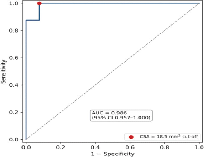
ROC curve analysis identified 18.5 mm² as the optimal cut-off for predicting an intraoperative graft diameter ≥8.0 mm, yielding an area under the curve (AUC) of 0.986 (95% CI: 0.957–1.000, P < 0.001) (Fig. 2). The 2 × 2 contingency table (Table 2) showed 24 true positives, 11 true negatives, 2 false positives, and 0 false negatives. At the 18.5 mm² threshold (adequate graft as the target condition), diagnostic sensitivity was 100.0% (95% CI: 85.75–100.0%), specificity was 84.62% (95% CI: 54.55–98.08%), PPV was 92.31% (95% CI: 77.03–97.72%), NPV was 100.0% (95% CI: 71.51–100.0%), LR+ was 6.50 (95% CI: 1.82–23.26), LR− was 0.00 (95% CI: 0.00–0.38), and overall diagnostic accuracy was 94.59% (95% CI: 81.81–99.34%) (Table 3). When the threshold is used to detect an inadequate graft (CSA <18.5 mm² as test-positive: 11 true positives, 2 false negatives, 0 false positives, 24 true negatives), sensitivity was 84.62% (95% CI: 54.55–98.08%), specificity 100.0% (95% CI: 85.75–100.0%), PPV 100.0% (95% CI: 71.51–100.0%), NPV 92.31% (95% CI: 74.87–99.05%), and accuracy 94.59%.
2×2 Contingency table for the 18.5 mm² CSA threshold against the 8.0 mm graft adequacy standard (n=37)
| Preoperative MRI ST CSA | Graft > 8.0 mm (Adequate) | Graft < 8.0 mm (Inadequate) | Total |
|---|---|---|---|
| CSA > 18.5 mm2 (Test Positive) | 24 (True Positive) | 2 (False Positive) | 26 |
| CSA < 18.5 mm2 (Test Negative) | 0 (False Negative) | 11 (True Negative) | 11 |
| Total | 24 | 13 | 37 |
CSA: Cross-sectional area; ST: Semitendinosus.
Diagnostic accuracy parameters of the 18.5 mm² CSA threshold for predicting graft adequacy (≥8.0 mm)
| Diagnostic Parameter | Value (%) | 95% confidence interval (CI) |
|---|---|---|
| Sensitivity | 100 | 85.75%–100.0% |
| Specificity | 84.62 | 54.55%–98.08% |
| Positive predictive value (PPV) | 92.31 | 77.03%–97.72% |
| Negative predictive value (NPV) | 100 | 71.51%–100.0% |
| Overall diagnostic accuracy | 94.59 | 81.81%–99.34% |
| Positive likelihood ratio (LR+) | 6.5 | 1.82–23.26 |
| Negative likelihood ratio (LR–) | 0 | 0.00–0.38 |
| Area Under the ROC curve (AUC) | 0.986 | 0.957–1.000 |
ROC: Receiver operating characteristic, AUC: Area under the curve, CI confidence interval.
Values are for an adequate graft (≥8.0 mm) as the target condition; the LR− CI was calculated with a 0.5 continuity correction.
Discussion
The primary objective of this study was to evaluate the diagnostic capability of pre-operative MRI-derived ST CSA to predict quadrupled graft adequacy in ACL reconstruction. The most notable finding was that an 18.5 mm² threshold provided 100% sensitivity and 100% NPV for an adequate graft: all 24 patients with an adequate graft had CSA ≥18.5 mm², and all 11 patients with CSA <18.5 mm² had an inadequate graft (PPV 100% for an undersized graft). However, the threshold missed 2 of the 13 patients whose quadrupled ST graft failed to achieve the 8.0 mm benchmark (sensitivity 84.62% for detecting an undersized graft). This strong predictive fidelity is supported by a correlation of ρ = 0.948 and an AUC of 0.986.
A quadrupled hamstring graft diameter <8.0 mm has been repeatedly highlighted in the literature as a prominent risk factor for construct failure, with Magnussen et al. demonstrating substantially increased revision rates in younger, active cohorts receiving undersized grafts [16]. Hence, precise pre-operative knowledge of graft thickness is critical for surgical decision-making. If an ST autograft is predicted to be undersized (<18.5 mm²), surgeons can pre-operatively plan gracilis tendon co-harvesting, prepare allograft augmentation, or select an alternative primary autograft (e.g., bone-patellar tendon-bone or quadriceps tendon).
Several prior studies have explored pre-operative MRI and ultrasound measurements for graft diameter estimation [3,4,5,6,7,8,9,10,11,12,13,14,15]. Çeliktaş et al. and Manni et al. observed moderate associations between anthropometric markers and graft dimensions, though anthropometry alone lacked sufficient precision for clinical decision-making [2,3]. Cobanoglu et al. and Movahedinia et al. demonstrated that MRI CSA is a superior predictor compared to height or weight [4,5]. Liau et al. reported an AUC of 0.856 using geometric tendon modeling on MRI [12], while Mayr et al. emphasized that axial slice selection significantly influences cross-sectional thresholds [13]. In the present study, measuring ST CSA at the maximal medial femoral condyle diameter at the joint line offered high diagnostic yield; however, inter- and intra-observer reliability of the CSA measurement was not assessed.
Limitations of the study include the single-center design and a predominantly male cohort (83.8%), reflecting regional athletic presentation patterns. Importantly, the 18.5 mm² cut-off was derived and evaluated within the same small cohort (n = 37) without internal or external validation, which likely overestimates its diagnostic accuracy and yields wide confidence intervals; the findings should therefore be considered preliminary. Inter- and intra-observer reliability of the CSA measurement was also not assessed, and external validation across multicentric, diverse cohorts is recommended.
Conclusion
A pre-operative MRI ST tendon CSA threshold of 18.5 mm² provides high sensitivity (100%) for an adequate graft and overall accuracy (94.59%) for predicting quadrupled ST graft adequacy (≥8.0 mm). All patients below the threshold had an inadequate graft, although 2 of 13 undersized grafts were missed. Incorporation of this measurement into pre-operative ACL planning may help identify patients requiring gracilis augmentation or alternative graft constructs and reduce unanticipated intraoperative changes, pending external validation.
Clinical Message
Measuring the ST tendon cross-sectional area on routine pre-operative axial MRI at the joint line is a simple method that may help predict quadrupled graft diameter. An ST CSA <18.5 mm² alerts the orthopedic surgeon to prepare for gracilis tendon harvest and co-augmentation, optimizing operative readiness and surgical outcomes..
Conflict of Interest:
Nil
Source of Support:
Nil
Consent
The authors confirm that informed consent was obtained from the patient for publication of this article
How to Cite this Article
Mathew T, Mathew A, Faris S, Johnson F, Krishna A. Diagnostic Accuracy of an 18.5 mm² Magnetic Resonance Imaging Cross-Sectional Area Threshold for Predicting Graft Adequacy (≥8.0 mm) in Quadrupled Hamstring Anterior Cruciate Ligament Reconstruction. J Orthop Case Rep. 2026 November;16(11):405-410. doi: 10.13107/jocr.2026.v16.i11.8510.
References
- amarda L, Grassedonio E, Albano D, Galia M, Midiri M, D'Arienzo M. MRI evaluation to predict tendon size for knee ligament reconstruction. Muscles Ligaments Tendons J 2017;7:478-84. [Google Scholar] | [PubMed]
- Çeliktaş M, Gölpinar A, Köse Ö, Sütoluk Z, Çelebi K, Sarpel Y. Prediction of the quadruple hamstring autograft thickness in ACL reconstruction using anthropometric measures. Acta Orthop Traumatol Turc 2013;47:14-8. [Google Scholar] | [PubMed]
- Manni D, Kalyan HK, Hegde SP, Arya S, Ajay Kumar SP, Reddy RR. Prediction of autologous hamstring graft size in ACL reconstruction using gender, activity level, anthropometric parameters and MRI - a prospective and retrospective study. Int J Orthop Sci 2019;5:1058-62. [Google Scholar] | [PubMed]
- Cobanoglu M, Ozgezmez FT, Omurlu IK, Ozkan I, Savk SO, Cullu E. Preoperative magnetic resonance imaging evaluation of semitendinosus tendon in anterior cruciate ligament reconstruction: Does this have an effect on graft choice?. Indian J Orthop 2016;50:499-504. [Google Scholar] | [PubMed]
- Movahedinia M, Movahedinia S, Hosseini S, Motevallizadeh A, Salehi B, Shekarchi B. Prediction of hamstring tendon autograft diameter using preoperative measurements with different cut-offs between genders. J Exp Orthop 2023;10:4. [Google Scholar] | [PubMed]
- Goto K, Hara M, Yamazaki Y, Urata T, Shimizu Y, Shimizu N. Smaller cross-sectional areas of the hamstring tendon measured from preoperative ultrasonography are likely to need additional Gracilis harvesting for double-bundle anterior cruciate ligament reconstructions. Knee Surg Relat Res 2020;32:34. [Google Scholar] | [PubMed]
- Erquicia JI, Gelber PE, Doreste JL, Pelfort X, Abat F, Monllau JC. How to improve the prediction of quadrupled semitendinosus and gracilis autograft sizes with magnetic resonance imaging and ultrasonography. Am J Sports Med 2013;41:1857-63. [Google Scholar] | [PubMed]
- Corey S, Mueller T, Hartness C, Prasad BM. Correlation of intra-operative hamstring autograft size with pre-operative anthropometric and MRI measurements. J Orthop 2018;15:988-91. [Google Scholar] | [PubMed]
- Ro DH, Lee S, Cho Y, Lee YM, Lee MC, Kim SH. Factors that predicts the size of autologous hamstring tendon graft for double-bundle ACL reconstruction. Indian J Orthop 2020;54:444-53. [Google Scholar] | [PubMed]
- Alkhalaf FN, Hanna S, Alkhaldi MS, Alenezi F, Khaja A. Autograft diameter in ACL reconstruction: Size does matter. SICOT J 2021;7:16. [Google Scholar] | [PubMed]
- Sherman B, Kwan K, Schlechter J. Magnetic resonance imaging predictive model determines hamstring autograft size for anterior cruciate ligament reconstruction in patients under 18 years old. Arthrosc Sports Med Rehabil 2021;3:e715-20. [Google Scholar] | [PubMed]
- Liau ZQ, Ng MS, Low SS, Chin BZ, Hui JH, Kagda FH. A novel practical method to predict anterior cruciate ligament hamstring graft size using preoperative MRI. Knee Surg Relat Res 2024;36:17. [Google Scholar] | [PubMed]
- Mayr MF, Siegel M, Taghizadeh E, Obid P, Schmal H, Izadpanah K. Prediction of the hamstring graft size for ACL reconstruction using different axial Layers in preoperative MRI. J Pers Med 2024;14:582. [Google Scholar] | [PubMed]
- Bhamare DS, Sirasala S, Jivrajani P, Nair A, Taori S. Preoperative MRI assessment of hamstring tendons to predict the quadruple hamstring graft diameter in anterior cruciate ligament reconstruction. Cureus 2022;14:e21753. [Google Scholar] | [PubMed]
- Astur DD, Novaretti JV, Liggieri AC, Janovsky C, Nicolini AP, Cohen M. Ultrasonography for evaluation of hamstring tendon diameter: Is it possible to predict the size of the graft?. Rev Bras Ortop 2018;53:404-9. [Google Scholar] | [PubMed]
- Magnussen RA, Lawrence JT, West RL, Toth AP, Taylor DC, Garrett WE. Graft size and patient age are predictors of early revision after anterior cruciate ligament reconstruction with hamstring autograft. Arthroscopy 2012;28:526-31. [Google Scholar] | [PubMed]
© 2026 Journal of Orthopaedic Case Reports - Published by Indian Orthopaedic Research Group
About the Authors
-

Thomas Mathew
-

Ashish Mathew
-

Salman Faris
-

Francis Johnson
-

Athul Krishna
Related Articles in Journal of Orthopaedic Case Reports
 October 1, 2026 Magnetic Resonance Imaging-Arthroscopy Correlation in Traumatic Knee Injuries: A Retrospective Diagnostic Accuracy Study
 September 1, 2026 Diagnostic Accuracy of Magnetic Resonance Imaging Signs for Detecting Meniscal Root Tears: A Prospective Comparison with Arthroscopic Findings
 August 1, 2026 Comparative Outcomes of Hamstring Tendon versus Peroneus Longus Tendon Autograft in Arthroscopic Single-Bundle Anterior Cruciate Ligament Reconstruction: A Prospective Comparative Study
 May 1, 2026 Clinical and Radiological Outcomes of All-inside Versus Complete Tibial Tunnel Techniques in Anterior Cruciate Ligament Reconstruction using Hamstring Tendon Autograft: A Prospective Comparative Study