

Posterior paratricipital approach should be considered a reliable option in the armamentarium for pediatric complex Gartland type III supracondylar humeral fractures.
Dr. Rishab Kafley, Department of Orthopaedics, Kalinga Institute of Medical Sciences, Bhubaneswar - 751024, Odisha, India. E-mail: rishabkafley15@gmail.com
Abstract
Introduction: Gartland type III supracondylar fractures of the humerus in children are completely displaced injuries often posing challenges to closed reduction, particularly in cases with severe swelling, instability, or neurovascular compromise. Open reduction and internal fixation remains a reliable option in such cases. Mini open methods from the lateral aspect are frequently used to avoid injury to the blood supply and major scars in children. However, the posterior paratricipital (triceps-sparing) approach allows direct fracture visualization to achieve better reduction and avoid growing deformities.
Materials and Methods: This prospective case series included patients aged 5–10 years with Gartland type III supracondylar fractures treated at Kalinga Institute of Medical Sciences, Bhubaneswar, between November 2023 and January 2025 after approval from the ethical committee. All patients underwent open reduction through a posterior paratricipital approach and Kirschner wire fixation. Demographic data, injury characteristics, operative details, and complications were recorded. Patients were followed for a minimum of 1 year. Outcomes were assessed using Flynn’s criteria, range of motion, carrying angle, and radiographic alignment.
Results: A total of 38 patients (mean age ~7.4 years) were included. According to Flynn’s criteria, excellent to good outcomes were observed in 92.1% (35) of the patients. The mean loss of elbow motion was minimal, and no patient had clinically significant cubitus varus deformity. There were no cases of deep infection, triceps weakness, or iatrogenic neurovascular injury.
Conclusion: Open reduction and pinning via the posterior paratricipital approach is a safe, effective, and reproducible technique for managing Gartland type III supracondylar humeral fractures, providing stable fixation and satisfactory functional outcomes.
Keywords: Supracondylar, fracture, open reduction, posterior, paediatrics.

Supracondylar fractures of the humerus are the most common elbow fractures in the pediatric population, accounting for approximately 50–70% of all elbow injuries in children and nearly 15% of all pediatric fractures [1,2]. These injuries typically occur in the first decade of life, with a peak incidence between 5 and 8 years, and are most frequently caused by a fall on an outstretched hand. Despite being common, supracondylar fractures continue to pose significant challenges to the treating surgeon because of their proximity to vital neurovascular structures, potential for instability, and risk of long-term deformities if inadequately managed. The Gartland classification remains the most widely used system to describe extension-type supracondylar humerus fractures. Gartland type III fractures are completely displaced with no cortical contact and are inherently unstable [3]. These fractures are associated with a higher incidence of complications, including neurovascular injury, compartment syndrome, malunion resulting in cubitus varus deformity, elbow stiffness, and iatrogenic nerve injury during treatment [4,5,6]. As a result, Gartland type III fractures usually require operative management to restore anatomy and allow early functional recovery.
Closed reduction and percutaneous pinning (CRPP) is the widely accepted treatment for displaced supracondylar fractures [7,8]. When an acceptable closed reduction can be achieved, CRPP offers the advantages of minimal soft tissue dissection, shorter operative time, and reduced risk of infection. However, closed reduction may not always be feasible or successful in Gartland type III fractures due to factors such as severe displacement, rotational instability, swelling, soft tissue interposition (including brachialis muscle, periosteum, or neurovascular structures), delayed presentation, or failed attempts at closed reduction [9,10]. In such scenarios, open reduction and internal fixation (ORIF) becomes necessary to achieve anatomic reduction and stable fixation.
Several surgical approaches for open reduction of supracondylar humerus fractures have been described in the literature, including anterior, medial, lateral, and posterior approaches [11,12]. Each approach has its own advantages and limitations. The anterior approach allows direct visualization of the neurovascular structures and is particularly useful in fractures associated with vascular compromise; however, it requires extensive dissection and carries a risk of scar-related morbidity. The medial approach provides excellent access for addressing medial column comminution but poses a risk to the ulnar nerve. The lateral approach is familiar to many surgeons and avoids ulnar nerve dissection but may provide limited visualization of the medial column, especially in severely displaced fractures. The posterior approach, although less commonly used, offers the advantage of wide exposure of both the medial and lateral columns of the distal humerus, facilitating accurate reduction in complex and severely displaced fractures [13]. Traditional posterior approaches involving triceps splitting or olecranon osteotomy have been criticized for their potential to cause postoperative elbow stiffness, extensor mechanism weakness, and delayed rehabilitation [14]. These concerns have led to reluctance in adopting posterior approaches for pediatric supracondylar fractures. To address these limitations, the posterior paratricipital (triceps-sparing) approach has been described as a modification that preserves the integrity of the triceps mechanism while still providing excellent visualization of the distal humerus [15]. In this approach, the triceps muscle is mobilized by developing medial and lateral paratricipital windows without splitting or detaching the triceps tendon. This technique allows circumferential exposure of the supracondylar region, facilitates precise fracture reduction including rotational alignment, enables stable pin fixation including medial pinning while minimizing ulnar nerve damage risk due to safe identification and protection.
Despite these potential benefits, the posterior paratricipital approach has not been widely adopted for pediatric supracondylar fractures, and literature on its outcomes remains limited compared to more conventional approaches. Concerns persist regarding operative time, learning curve, risk of elbow stiffness, cosmetic scar outcomes, and possible risk of injury to distal humeral blood supply. However, case series suggest that when performed meticulously, the posterior paratricipital approach can yield satisfactory radiological alignment, stable fixation, good functional outcomes, even in severely displaced fractures or in cases where closed reduction has failed [13,15,16].
In resource-limited settings or in late-presenting cases, where swelling, callus formation, or soft tissue interposition makes closed reduction difficult, it can also be a reliable and extensile approach that allows controlled reduction. Furthermore, its utility in revision surgeries or fractures with associated neurovascular complications warrants further exploration. Given the ongoing debate regarding the optimal surgical approach for open reduction of Gartland type III supracondylar fractures, there is a need for additional clinical data evaluating the safety, efficacy, and functional outcomes of the posterior paratricipital approach.
The present study aims to evaluate the clinical and radiological outcomes of Gartland type III supracondylar humerus fractures treated with ORIF using the posterior paratricipital approach. By analyzing fracture union, alignment, range of motion (ROM), functional recovery, and complication rates, this case series seeks to contribute to the existing literature and provide further insight into the role of the posterior paratricipital approach as a viable and effective option in the management of complex pediatric supracondylar fractures.
Study design
This was a prospective observational case series conducted at the Department of Orthopaedics, Kalinga Institute of Medical Sciences (KIMS), Bhubaneswar. The study evaluated the clinical and radiological outcomes of pediatric Gartland type III supracondylar humerus fractures managed with open reduction and percutaneous pinning using the posterior paratricipital (triceps-sparing) approach operated within the period from November 2023 to January 2025. All Gartland type III extension-type fractures were initially attempted with closed reduction under general anesthesia under fluoroscopic guidance. Standard reduction maneuvers including longitudinal traction, correction of translation and rotational displacement, and flexion maneuver were performed. In difficult reductions, assisted reduction techniques including the milking maneuver were additionally attempted. Open reduction was undertaken only when all standard and assisted closed reduction attempts failed to achieve acceptable alignment or stable reduction intraoperatively as it is ethically inappropriate to open-reduce fractures that can be achieved with closed reduction. During the study period, approximately 12% of all operated Gartland type III fractures required open reduction and formed the present study cohort. This study which uses a posterior paratricipital approach for reduction of pediatric supracondylar fractures is taken as a pilot study; furthermore, studies with more cases and comparison may add to the evidence. At present, no studies suggest this, even though our study with this approach proved to have good results in follow-ups of at least 1 year.
Delayed presentation was defined as presentation more than 48 h after injury; two patients in the present cohort met this criterion. Open and complex injuries were not taken into account to have a more uniform type of injury to comment upon the outcome of the chosen approach and taking those into account would have added confounding factors. No cases with associated vascular compromise (pink pulseless hand or white pulseless hand) were included, and no patient had associated motor nerve injury at presentation. All surgical procedures were performed by a single orthopedic surgeon at a single institution, as it is a pilot study to ensure uniformity of surgical technique and minimize inter-surgeon variability. Institutional ethical committee approval was obtained before the commencement of the study (IEC Reg No.-KIIT/KIMS/IEC/1102/2023), and informed consent was obtained from the parents or legal guardians of all patients.
The population included children aged 5–10 years with extension-type Gartland type III supracondylar humerus fractures (Fig. 1), as supracondylar fractures are most common in this age group. It provides a homogeneous cohort with similar skeletal maturity and treatment characteristics, improving the validity of our case series.
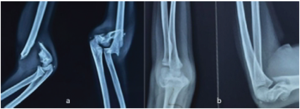
Figure 1: Radiographs of Gartland type III supracondylar humerus fracture in anteroposterior and lateral projection (a and b).
Patients <5 years appear too young for operative management, whereas those above 10 years have a relatively more mature skeleton, frequently needing internal fixation. All fractures were attempted with CRPP. Fractures requiring open reduction due to failed closed reduction, irreducible fractures, severe displacement or rotational instability, or delayed presentation were included in the study. These patients were managed with ORIF through posterior paratricipital approach and 3-pin fixation. All patients were followed up for a minimum of 1 year, as considering the volatile growth at the age of 5–10 years, a growth disturbance would be evident in even 1 year, which was not seen in any of the cases which goes in favor of the approach. Gartland type I and II fractures, open fractures, associated ipsilateral fractures of the upper limb, pathological fractures, and patients lost to follow-up before 1 year were excluded from the concerned study.
Preoperative assessment
All patients underwent a thorough clinical evaluation at presentation, which included mechanism of injury, limb alignment, swelling, skin condition, and neurovascular examination, with documentation of median, radial, and ulnar nerve function and distal pulses. Standard anteroposterior (AP) and lateral radiographs of the elbow were taken. Temporary immobilization using an above-elbow posterior slab was provided before surgery. Patient-reported outcomes were not separately taken into consideration as ROM, deformity, and cosmesis are the primary tasks to be followed; hence, they were the ones taken into account.
Surgical technique
All surgeries were performed under general anesthesia with the patient positioned in the lateral decubitus position with the affected limb supported over a padded bolster. Adequate aseptic painting and draping were done from the upper arm. A midline posterior longitudinal incision from approximately 5–6 cm proximal to the olecranon was made over the distal arm, curving laterally around the olecranon and ending just distal to it. The ulnar nerve was identified along the medial border of the triceps, carefully dissected up to 5–6 cm proximal to the cubital tunnel and protected throughout the procedure using a soft vessel loop. Medial and lateral paratricipital intervals were developed by elevating the triceps muscle off the posterior surface of the distal humerus without splitting or detaching the triceps tendon. Hematoma and interposed soft tissues were cleared. Fracture fragments were visualized directly. Anatomical reduction was achieved under direct vision by correcting coronal, sagittal, and rotational displacement. Definitive fixation was performed using percutaneous Kirschner wires, 2 from lateral and 1 from medial. The pins were left exposed over the skin for easy removal (Fig. 2). Pin placement and fracture alignment were confirmed using an image intensifier in AP and lateral views. Hemostasis was achieved. The triceps muscle was allowed to fall back into its anatomical position. The ulnar nerve was ensured to be free and not under tension. Wound closure was done in layers using poliglecaprone sutures. Adequately padded dressing was done.
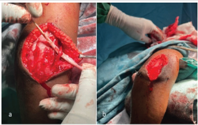
Figure 2: Intraoperative clinical pictures showing ulnar nerve isolation (a) and percutaneous pin placement (b).
Post-operative management
An above-elbow posterior slab was applied with the elbow in 90° flexion and forearm supination. This slab was kept for 6 weeks with 2 dressings on post-operative day 2 and day 14 for skin evaluation. Intravenous antibiotics were administered for 48 h, followed by oral antibiotics for 5 days. Neurovascular status was reassessed postoperatively. Patients were discharged once pain control and limb status were satisfactory.
Follow-up protocol
Patients were followed up at 2 weeks for Pin tract inspection and wound healing assessment and at 6 weeks for removal of K-wires. Range-of-motion exercises of elbow joint was started gradually as per patient tolerance. Follow-up visits at 2 weeks, 6 weeks, 3 months, 6 months, and 1 year. Flynn’s criteria were calculated at 1-year follow-up. Triceps strength in all cases was 5/5, as seen while evaluating ROM of the elbow in follow-ups.
Outcome assessment
Clinical and radiological outcomes were assessed using Flynn’s criteria, evaluating loss of carrying angle (cosmetic outcome), loss of elbow motion (functional outcome), Baumann’s angle (radiological outcome), radiographic evidence of union and alignment. Complications such as pin tract infection, neurovascular injury, elbow stiffness, malunion (cubitus varus), and reoperation were looked for.
Statistical analysis
Data were recorded using a structured pro forma and analyzed using descriptive statistics. Continuous variables were expressed as mean and range, while categorical variables were expressed as frequencies and percentages. Outcomes were categorized according to Flynn’s grading system
A total of 38 pediatric patients with Gartland type III supracondylar fractures of the humerus were included in the study (Table 1) and followed up for a minimum duration of 1 year.
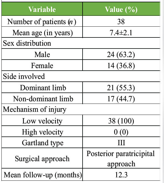
Table 1: Demographic and injury characteristics of study population
All fractures were managed with ORIF through a posterior paratricipital approach, followed by cross-pinning using three Kirschner wires. The study population comprised 24 males (63.2%) and 14 females (36.8%). The mean age at presentation was 7.4 ± 2.1 years (range: 5–10 years). The dominant limb was involved in 21 patients (55.3%), while the non-dominant limb was affected in 17 patients (44.7%). All injuries resulted from low-energy falls, most commonly falls while playing.
At final follow-up, 35 patients (92.1%) achieved near-full ROM of the elbow, with loss of flexion or extension of <10° compared to the contralateral side. Mild elbow stiffness was noted in 3 patients (7.9%), all of whom had a terminal restriction of motion ranging from 15° to 20°, without any functional limitation in daily activities. No patient had moderate or severe stiffness. The mean elbow flexion achieved at final follow-up was 132.6° ± 6.4°, and the mean extension loss was 3.2° ± 2.1°. No cases had any residual cubitus varus deformity by the last follow-up. Mean difference in carrying angle of the operated side with respect to the normal side, measured clinically, was about 3.7 ± 1.8°. However, 4 cases had nearly 10° of hyperextension with no loss of normal arc of elbow motion (Table 2).
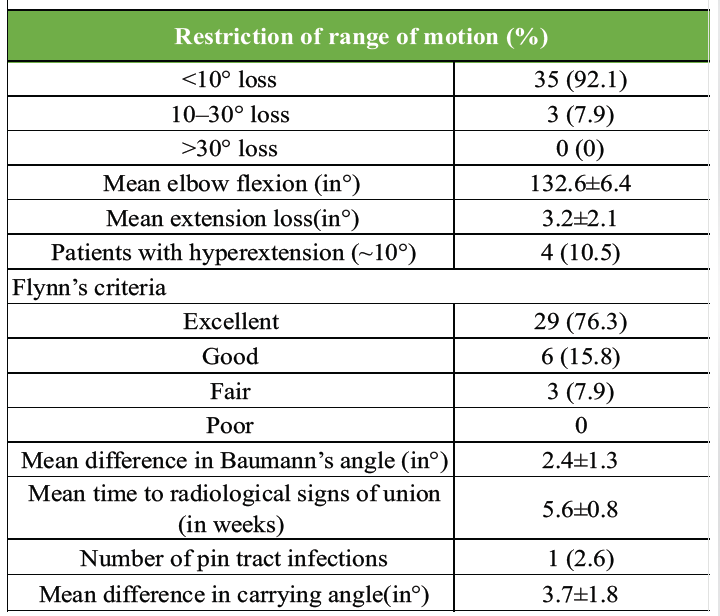
Table 2: Functional and radiological outcomes at final follow-up
Radiological assessment demonstrated adequate fracture reduction and maintenance of alignment in all cases throughout the follow-up period (Fig. 3).
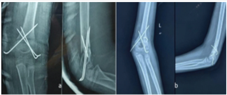
Figure 3: Post-operative and follow-up radiographs after open reduction and pinning (a and b).
The mean Baumann’s angle on the operated side at final follow-up was 72.4° ± 4.1°, which was comparable to the contralateral normal elbow (70.4° ± 3.9°), with no statistically or clinically significant difference. No case showed a change in Baumann’s angle exceeding 5°, indicating stable fixation and absence of secondary displacement. All cases had neutral reduction in lateral view with no extension deformity from normal anatomy. In AP views, no cases had any coronal plane malalignment of more than 5° from the contralateral side. All fractures achieved radiological union by a mean duration of 5.6 ± 0.8 weeks. No cases of malunion, non-union, or loss of reduction were observed. No rotational malalignment was observed.
Functional and cosmetic outcomes were assessed using Flynn’s criteria at 1-year follow-up (Fig. 4). Excellent results were observed in 29 patients (76.3%), good results in 6 patients (15.8%), and fair results in 3 patients (7.9%). Poor results were not observed. Thus, 92.1% of patients achieved excellent or good outcomes, indicating a high rate of satisfactory functional recovery.
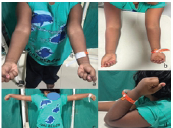
Figure 4: Post-operative carrying angle (a) and range of motion of the elbow (b, c, d).
Pin tract infection was noted in one patient (2.6%), which was superficial and resolved completely with oral antibiotics and local pin site care. No cases of deep infection, iatrogenic ulnar nerve injury, vascular compromise, compartment syndrome, or cubitus varus deformity were observed. None of the patients had any form of scar tenderness or wound dehiscence.
The optimal surgical management and approach of Gartland type III supracondylar humerus fractures in children remains an area of ongoing debate. While CRPP is widely accepted as the primary treatment modality for displaced fractures, ORIF becomes necessary when acceptable closed alignment fails or when the injury pattern precludes safe manipulation under fluoroscopy. In this series of 38 patients managed via a posterior paratricipital approach with three-pin fixation, we observed good to excellent results in 92.1% of cases and only mild to moderate restriction of elbow ROM in the remaining three, highlighting the viability of this approach in selected cases.
The outcomes reported in this series align with the broader body of evidence that supports satisfactory functional and cosmetic results following ORIF irrespective of surgical approach, provided an adequate reduction and stable fixation are achieved. In a multicenter comparative study of open surgical exposures used for Gartland type III fractures, posterior, medial, lateral, and anterior approaches demonstrated comparable functional and cosmetic outcomes when assessed by Flynn’s criteria, with no significant differences in major complications across groups [17]. This suggests that surgeon experience and precise execution of the chosen approach may be more pivotal than the approach itself. However, the posterior approach has historically been viewed with caution in some literature due to concerns regarding soft tissue disruption and triceps injury when the extensor mechanism is violated. It also considers the posterior approach as a potential threat to the condylar blood supply of the distal humerus. Conventionally, anterior or lateral approaches have been most commonly used considering the above problems along with avoidance of injury to the ulnar nerve and direct visualization of anterior neurovascular structures [11,12,18]. In contrast, the posterior paratricipital approach, as used in our series, has increasingly been shown to mitigate these drawbacks by preserving the extensor mechanism while providing excellent visualization of both medial and lateral columns and protecting the ulnar nerve, even when a medial pin is given. In our study, the majority of patients achieved near-normal elbow motion with minimal stiffness at final follow-up. This aligns with conclusions from recent cohorts reporting that posterior approaches can be safe and effective, allowing direct reduction and secure fixation while reducing the risk of iatrogenic ulnar nerve injury due to better visualization [15,16,19].
The Flynn criteria remain a cornerstone for assessing pediatric supracondylar fracture outcomes, incorporating both functional ROM and cosmetic carrying angle. The high rate of good to excellent outcomes (92.1%) in our series demonstrates that when posterior paratricipital exposure is combined with careful triceps preservation and stable three-pin configuration, the functional results can be comparable to those reported with other surgical approaches. Multicenter data also support the finding that posterior approaches yield satisfactory Flynn scores similar to medial and lateral exposures when performed by experienced surgeons [17,19]. Some systematic reviews suggest a trend toward better functional outcomes with lateral approaches compared to posterior ones, though these differences were not statistically significant [20], whereas some study suggests there was no clinically significant difference in elbow arc of motion and adverse events between the anterior approach and the posterior approach [21]. Such analyses noted a higher likelihood of excellent and good functional outcomes with lateral exposures, and fewer fair/poor results, although with overlapping confidence intervals. Even so, these comparisons underscore that posterior approaches remain within the spectrum of acceptable techniques in experienced hands.
Cosmetically, posterior incisions are often considered less favorable than lateral or anterior scars due to visibility; however, none of our patients reported dissatisfaction significant enough to affect overall outcome scores. No incident of significant cubitus varus deformity was noted in any of the cases, emphasizing the reliability of the intervention in preventing pediatric elbow deformities which are rather common in supracondylar fracture sequelae. Contemporary literature indicates that while cosmetic outcomes may vary with incision location, acceptable cosmesis can be achieved with all major approaches when appropriate soft-tissue handling is employed.
Debate persists regarding optimal pin configuration, especially regarding cross pinning versus lateral pins alone. Biomechanical analyses generally favor crossed pins for rotational stability in highly unstable fractures, but traditional concerns focus on iatrogenic ulnar nerve injury during medial pin insertion [22,23,24]. While some studies advocate lateral pinning to reduce nerve risk, clinical outcomes with cross pins have nevertheless been satisfactory when careful medial nerve protection is employed, a principle adhered to in our surgical protocol. Although our study did not directly compare pin configuration, our outcomes reinforce that three-pin crossed constructs, when placed under direct vision via open exposure, provide reliable fixation with low incidence of loss of reduction later in rehabilitation and follow-up. This is consistent with broader clinical evidence that direct visualization during medial pin placement can mitigate iatrogenic nerve risks while delivering robust biomechanical stability. Moreover, visualization of both the condyles and their fracture reduction reduces the chances of a rotational malalignment being present without additional amounts of fluoroscopic exposure [25].
The decision to proceed with ORIF for Gartland III fractures is well supported when anatomical reduction cannot be reliably achieved with CRPP, especially in the context of severe swelling, rotational deformity, soft-tissue interposition, or neurovascular compromise [9,10,26]. Repeated attempts at closed reduction not only risk further soft-tissue injury but may also prolong surgery and anesthesia time, potentially impacting outcomes. Increasing age is associated with increased elbow stiffness after percutaneous fixation of Gartland Type III and Type IV SCH fractures [27]. Hence, early recognition of “difficult” fractures and timely conversion to ORIF, as practiced in our series, can optimize alignment and eventual function.
This series is limited by its observational design and absence of a randomized comparison group, which restricts interpretation of approach-specific advantages. While complications such as ulnar nerve palsies or significant stiffness were rare in our case series, the sample size limits broad generalization. Moreover, while Flynn’s criteria are widely accepted, they might not capture subtleties in strength or patient-reported outcomes. The learning curve is not relevant as it is a common approach in adults and only physis is the consideration.
Nevertheless, the study’s strengths include a uniform surgical approach, consistent pin configuration, and medium-term follow-up allowing evaluation of functional restoration. The strong clinical outcomes observed reinforce that, when indicated, posterior paratricipital ORIF with three-pin fixation is a valid option that compares well with other published approaches in the management of complex supracondylar fractures. Surgeons managing pediatric supracondylar fractures should maintain familiarity with multiple exposure techniques; however, the choice of approach should be guided by fracture characteristics, soft-tissue conditions, and surgeon expertise rather than dogmatic preference. Our results support the concept that posterior paratricipital exposure can achieve outcomes comparable to lateral or medial approaches, especially when preservation of the triceps, ulnar nerve isolation and protection, anatomic fracture reduction, and careful pin placement are prioritized.
The posterior paratricipital approach is particularly valuable in completely displaced fractures where closed reduction is unsuccessful or contraindicated due to swelling, instability, or neurovascular concerns. In our case series, the majority of patients achieved good to excellent outcomes as assessed by Flynn’s criteria and ROM while producing minimal complications. When performed meticulously, it offers outcomes comparable to, and in selected cases, superior to other open approaches, without a significant increase in complications. Based on our findings, this technique should be considered a reliable option in the armamentarium for pediatric complex Gartland type III supracondylar humeral fractures. Further multicentric randomized studies with larger samples may help to strengthen the evidence and define its precise role relative to other surgical approaches in the scenario.
The posterior paratricipital approach is a safe, triceps-sparing technique for open reduction of irreducible Gartland type III pediatric supracondylar humerus fractures. It provides excellent fracture visualization while preserving the extensor mechanism, facilitates accurate reduction and stable K-wire fixation. It achieves favorable functional and radiological outcomes with a low risk of complications.
References
- 1. Wilkins KE. Supracondylar fractures: What’s new? J Pediatr Orthop 1997;17:193-8. [Google Scholar] [PubMed]
- 2. Cheng JC, Lam TP, Maffulli N. Epidemiological features of supracondylar fractures of the humerus in Chinese children. J Pediatr Orthop B 2001;10:63-7. [Google Scholar] [PubMed]
- 3. Gartland JJ. Management of supracondylar fractures of the humerus in children. Surg Gynecol Obstet 1959;109:145-54. [Google Scholar] [PubMed]
- 4. Otsuka NY, Kasser JR. Supracondylar fractures of the humerus in children. J Am Acad Orthop Surg 1997;5:19-26. [Google Scholar] [PubMed]
- 5. Dormans JP, Squillante R, Sharf H. Acute neurovascular complications with supracondylar humerus fractures in children. J Hand Surg Am 1995;20:1-4. [Google Scholar] [PubMed]
- 6. Skaggs DL, Cluck MW, Mostofi A, Flynn JM, Kay RM. Lateral-entry pin fixation in the management of supracondylar fractures in children. J Bone Joint Surg Am 2004;86:702-7. [Google Scholar] [PubMed]
- 7. Flynn JC, Matthews JG, Benoit RL. Blind pinning of displaced supracondylar fractures of the humerus in children. Sixteen years’ experience with long-term follow-up. J Bone Joint Surg Am 1974;56:263-72. [Google Scholar] [PubMed]
- 8. Mangwani J, Nadarajah R, Paterson JM. Supracondylar humeral fractures in children: Ten years’ experience in a teaching hospital. J Bone Joint Surg Br 2006;88:362-5. [Google Scholar] [PubMed]
- 9. Abzug JM, Herman MJ. Management of supracondylar humerus fractures in children. Orthop Clin North Am 2012;43:567-77. [Google Scholar] [PubMed]
- 10. Choi PD, Melikian R, Skaggs DL. Risk factors for failure of closed reduction of supracondylar humerus fractures. J Pediatr Orthop 2008;28:535-9. [Google Scholar] [PubMed]
- 11. Boyd HB, Altenberg AR. Fractures about the elbow in children. Arch Surg 1944;49:213-24. [Google Scholar] [PubMed]
- 12. Pretell-Mazzini J, Rodriguez-Martin J, Andres-Esteban EM. Does open reduction and pinning affect outcome in displaced supracondylar humerus fractures? Clin Orthop Relat Res 2010;468:1698-705. [Google Scholar] [PubMed]
- 13. Ersan O, Gonen E, Ilhan R, Boysan E, Ateş Y. Comparison of anterior and lateral approaches in the treatment of extension-type supracondylar hume rus fractures in children. J Pediatri Orthop B 2012;21:121-12. [Google Scholar] [PubMed]
- 14. Campbell CC. Posterior exposure of the elbow joint. J Bone Joint Surg Am 1932;14:63-73. [Google Scholar] [PubMed]
- 15. Bryan RS, Morrey BF. Extensive posterior exposure of the elbow. A triceps-sparing approach. Clin Orthop Relat Res 1982;166:188-92. [Google Scholar] [PubMed]
- 16. Kumar R, Malhotra R. Medial paratricipital approach for displaced supracondylar fractures. Indian J Orthop 2015;49:203-9. [Google Scholar] [PubMed]
- 17. Shannon FJ, Mohan P, Chacko J, D’Souza LG. “Dorgan’s” percutaneous lateral cross-wiring of supracondylar fractures of the humerus in children. J Pediatr Orthop 2004;24:376-9. [Google Scholar] [PubMed]
- 18. Lyons JP, Ashley E, Hoffer MM. Ulnar nerve palsies after percutaneous cross-pinning of supracondylar fractures in children’s elbows. J Pediatr Orthop 1998;18:43-5. [Google Scholar] [PubMed]
- 19. Ponce BA, Hedequist DJ, Zurakowski D, Atkinson CC, Waters PM. Complications and timing of follow-up after closed reduction and percutaneous pinning of supracondylar humerus fractures: Follow-up after percutaneous pinning of supracondylar humerus fractures. J Pediatr Orthop 2004;24:610-4. [Google Scholar] [PubMed]
- 20. Brauer CA, Lee BM, Bae DS, Waters PM, Kocher MS. A systematic review of medial and lateral entry pinning versus lateral entry pinning for supracondylar fractures of the humerus. J Pediatr Orthop 2007;27:181-6. [Google Scholar] [PubMed]
- 21. Gerami MH, Naderian R, Nemati A, Abdoos P, Saeedi F. Anterior approach versus posterior approach for the open reduction of displaced pediatric supracondylar humerus fracture. J Orthop 2023;42:70-3. [Google Scholar] [PubMed]
- 22. Zionts LE, McKellop HA, Hathaway R. Torsional strength of pin configurations used to fix supracondylar fractures of the humerus in children. J Bone Joint Surg Am 1994;76:253-6. [Google Scholar] [PubMed]
- 23. Lee SS, Mahar AT, Miesen D, Newton PO. Displaced pediatric supracondylar humerus fractures: Biomechanical analysis of percutaneous pinning techniques. J Pediatr Orthop 2002;22:440-3. [Google Scholar] [PubMed]
- 24. Skaggs DL, Hale JM, Bassett J, Kaminsky C, Kay RM, Tolo VT. Operative treatment of supracondylar fractures of the humerus in children. The consequences of pin placement. J Bone Joint Surg Am 2001;83:735-40. [Google Scholar] [PubMed]
- 25. Sankar WN, Hebela NM, Skaggs DL, Flynn JM. Loss of pin fixation. J Pediatr Orthop 2007;27:763-9. [Google Scholar] [PubMed]
- 26. Ramachandran M, Birch R, Eastwood DM. Clinical outcome of nerve injuries associated with supracondylar fractures of the humerus in children: The experience of a specialist referral centre. J Bone Joint Surg Br 2006;88:90-4. [Google Scholar] [PubMed]
- 27. Gupta K, Erdman MK, Siddiqui A, Schur M, Meisel E, Goldstein RY. Age is a predictor of elbow stiffness after type III or IV supracondylar humerus fractures. Eur J Orthop Surg Traumatol 2024;34:3067-71. [Google Scholar] [PubMed]






Related Articles in Journal of Orthopaedic Case Reports
 May 1, 2025 Surgical Decision-Making in Thoracolumbar Fractures: A Systematic Review of Anterior and Posterior Approach
 March 1, 2025 Prospective Study of Functional and Radiological Outcome after Operative Management of Supracondylar Fracture Humerus in Children
 July 10, 2024 Management of Bilateral Posterior Sternoclavicular Joint Dislocation: A Case Report
 December 10, 2022 Analysis of Functional Outcomes Following Surgical Management of Neglected Multiple Carpometacarpal Dislocations: A Series of 4 four Cases and Review of Literature