

A commissural webspace approach offers improved anatomical visualization for surgical release of interosseous-lumbrical adhesions in saddle syndrome, and repetitive use should be considered a causative mechanism.
Dr. Edward J Wu, Department of Orthopedic Surgery, University of Minnesota Medical School, Minneapolis - 55454, Minnesota. E-mail: ewu@umn.edu
Abstract
Introduction: Saddle syndrome is caused by post-traumatic adhesions between the interosseous and lumbrical tendons and the intermetacarpal ligament in the webspaces of the hand. Diagnosis can be challenging, requiring a high index of suspicion with inadequately defined diagnostic criteria.
Case Report: We present a case of saddle syndrome secondary to repetitive motorcycle use in a 56-year-old female to highlight the pathoanatomy, workup, diagnostic magnetic resonance imaging findings, and a new surgical approach for this disorder. Given persistent symptoms, surgical intervention was pursued using a commissural approach that provided superior en face visualization of the pathological anatomy. The adhesions between the intrinsic tendons and the intermetacarpal ligament were released with partial ligament resection.
Conclusion: Immediate post-operative mobilization led to complete pain resolution and return to full function by 3 months. This report provides the first high-quality in vivo photographs correlating with advanced imaging findings and demonstrates how a commissural surgical approach offers improved anatomical visualization for treating this condition.
Keywords: Saddle syndrome, saddle deformity, intrinsics, interosseous-lumbrical, adhesions.

Saddle syndrome represents a distinct post-traumatic hand pathology first described by Watson and colleagues in 1974 [1]typically occurring after trauma and characterized by adhesion formation between the intrinsic muscles of the hand [2]. Saddle syndrome affects approximately 0.5–1% of hand trauma patients, though the true prevalence may be underestimated due to delayed presentation and diagnostic challenges [2,3]. The intrinsic hand muscles demonstrate a unique anatomical arrangement where lumbrical muscles course palmarly and interosseous (IO) muscles travel dorsally relative to the deep transverse metacarpal ligament (DTML) [4]. These muscular structures unite distally before their common insertion into the extensor mechanism. During intrinsic contraction, this musculotendinous junction travels proximally, with the two muscle bellies positioned on either side of the DTML, creating the characteristic “saddle formation.”[2,5]. Adhesions can occur either between the lumbrical and IO muscles distal to the DTML or between the intrinsic muscles and the DTML within the webspace at the level of the metacarpal head [2].
The first pattern involves adhesions between the lumbrical and IO muscles distal to the DTML, creating impingement during intrinsic muscle contraction. The second pattern consists of direct adhesions between the intrinsic muscles and the DTML itself or the metacarpophalangeal (MCP) joint capsule [2]. This can produce a positive Bunnell test where proximal interphalangeal joint (PIP) flexion with MCP joint extension elicits tightness and pain due to restricted intrinsic muscle excursion. Clinically, patients present with pain in the affected webspace, decreased grip strength, and diminished range of motion with finger flexion. Conservative management includes anti-inflammatory medication, splinting, occupational therapy, activity modification, and steroid injections. Surgical management is reserved for refractory cases and involves adhesiolysis for both patterns, with the second type specifically necessitating partial DTML resection after adhesion release to prevent recurrent impingement.
We present a case of saddle syndrome secondary to repetitive motorcycle use that ultimately required surgical intervention. Saddle syndrome can be challenging to diagnose and treat. The pathological adhesions can be difficult to visualize via traditional volar or dorsal approaches. Previous cadaveric studies and case series do not provide adequate intraoperative photographs to guide surgical decision-making. Our report is the first to provide in vivo photographs of the pathology that correlate with advanced imaging findings associated with this condition. We also present a novel surgical approach that offers improved access and visualization of the affected structures.
A 56-year-old right-hand dominant female presented to the clinic with 4 months of pain and swelling in the second webspace of her right hand. She denied any acute trauma or injury. Radiographs did not demonstrate any significant abnormality. Her symptoms were aggravated by carrying heavy objects, prolonged typing, and riding her motorcycle. While riding, she repeatedly flexed her right index and middle fingers for the throttle and brake controls. Physical examination revealed tenderness in the second webspace between the index and middle metacarpal heads and a positive Bunnell intrinsic tightness, with pain elicited during passive PIP flexion of the index and middle fingers while maintaining MCP extension.
Her symptoms persisted despite conservative treatment, including anti-inflammatory medications, topical treatments, splinting, occupational therapy, and a series of steroid injections. A magnetic resonance imaging (MRI) demonstrated adhesions of the DTML in the second webspace, with palmar adherence to the lumbrical myotendinous junction and dorsal adherence to the IO myotendinous junction (Fig. 1a). Notably, the normal fat plane between the dorsally located IO musculature and the DTML was absent. In addition, the border between the lumbrical and IO musculature was blurred along the ulnar aspect of the DTML. Along the ulnar aspect of the DTML, there was blurring of the border between lumbrical and IO musculature. There was also thickening of the IO tendon when compared to analogous levels on subsequent images. Comparison with the third webspace proximally showed a normal fat plane between the IO and lumbrical musculature (Fig. 1b).
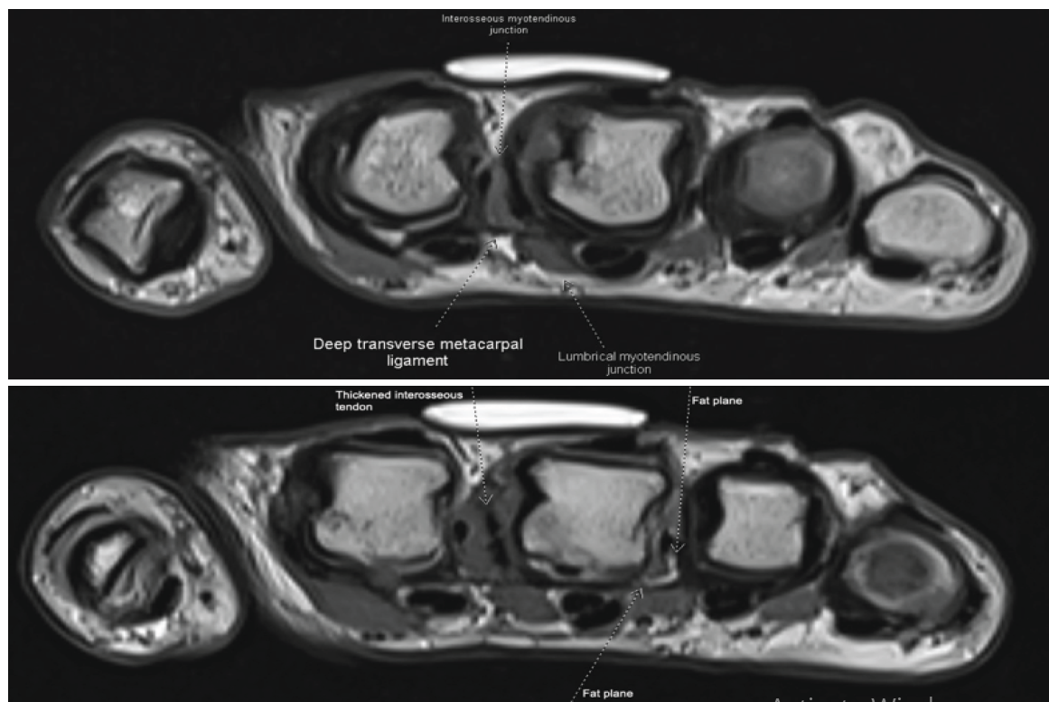
Figure 1: (a) Axial proton density magnetic resonance imaging images at the level of the second metacarpal head. Notable is the absence of fat plane between the interosseous musculature (dorsal second interspace) and the volar deep transverse metacarpal ligament (DTML), with blurring of the lumbrical-interosseous border along the ulnar aspect of the DTML. Thickening of the interosseous tendon is evident compared to more proximal levels. (b) Axial images 3 mm proximal to the previous image in Figure 1a. In comparison, the third interspace shows a fat plane between the interosseous and lumbrical musculature. The second interspace demonstrates a thickened interosseous tendon, particularly on the ulnar aspect.
Given that her symptoms remained refractory to conservative interventions, the patient ultimately elected to proceed with surgery. This was performed under sedation with a regional block to minimize local anesthetic in the surgical field. A novel commissural approach was utilized at the apex of the second webspace to allow for direct en face visualization of the intrinsic anatomy around the DTML (Fig. 2). Intraoperative findings revealed adhesions between the lumbrical and IO tendons and the intermetacarpal ligament adjacent to the radial aspect of the third MCP joint in the second webspace (Fig. 3). Notably, there were no apparent adhesions on the ulnar aspect of the index MCP joint as there was only a single palmar IO tendon volar to the intermetacarpal ligament. Release of the intrinsic tendon adhesions was performed along with partial excision of the distal half of the intermetacarpal ligament in the second webspace (Fig. 4).
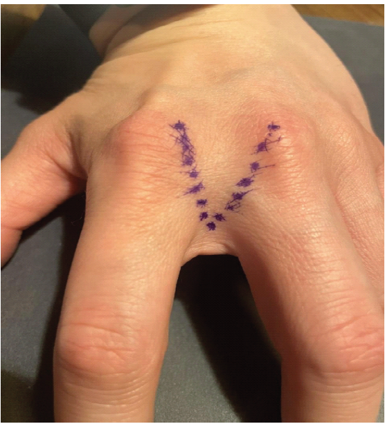
Figure 2: Surgical incision for commissural approach to the second webspace.
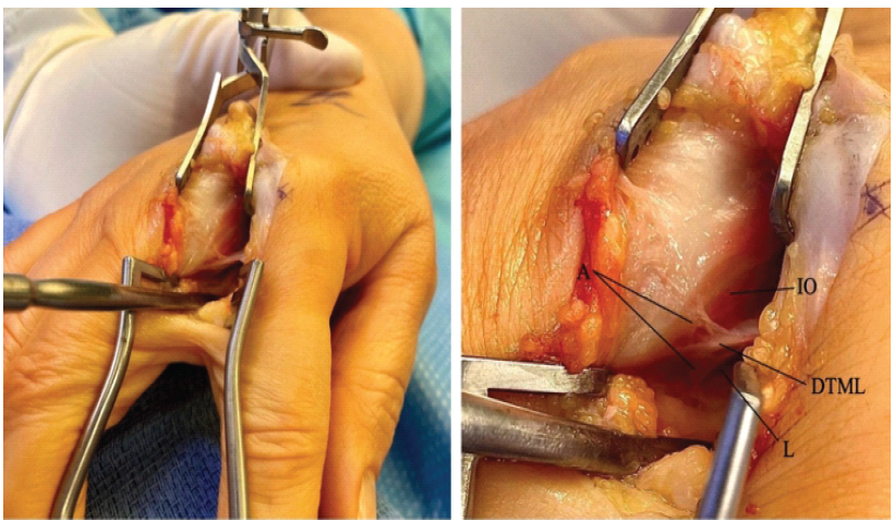
Figure 3: Intraoperative photographs demonstrating adhesions between the deep transverse metacarpal ligament and the lumbrical and interosseous tendons at the second webspace. (Right) Labeled structures demonstrating adhesions (A) between the intrinsic tendons and transmetacarpal ligament. L: Lumbrical muscle, DTML: Deep transverse metacarpal ligament, IO: Interossei muscle, A: Adhesions.
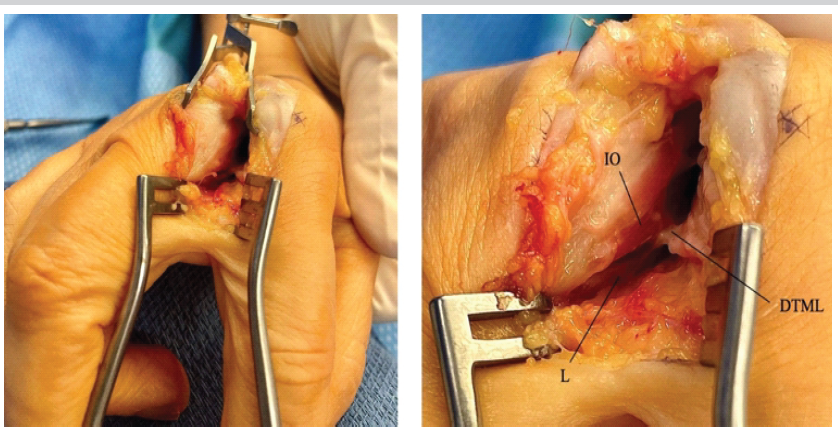
Figure 4: Intraoperative photographs following intrinsic tendon release, adhesiolysis, and partial excision of the distal half of the intermetacarpal ligament. L: Lumbrical muscle, DTML: Deep transverse metacarpal ligament, IO: Interosseous muscle.
The patient was instructed to begin immediate active and passive finger range of motion with a one-pound weight-bearing restriction. Occupational therapy was initiated 4 days postoperatively, focusing on range of motion, edema reduction, and functional activities. Sutures were removed 7 days after surgery, with minimal edema noted. However, 6 days postoperatively, the patient experienced a temporary setback with increased pain and swelling in the webspace secondary to overuse. This was managed with compression glove therapy, ice, and activity modification. One month postoperatively, she reported continued stiffness in the long finger MCP joint despite ongoing occupational therapy, though swelling was gradually improving. Two months after surgery, she was able to make a full fist and successfully returned to motorcycle riding without pain. Three months postoperatively, the pre-operative pain had completely resolved, including with computer work and motorcycle operation. The compression glove was discontinued after 3 months, and occupational therapy was completed 4 months postoperatively with satisfactory functional recovery.
In previous reported cases of saddle deformity, the majority of causes resulted from acute trauma, such as crush injuries and contusions [2]. Non-traumatic causes of saddle deformity are uncommon, with only a few case reports in the literature describing infection-related adhesions. Repetitive use as an etiology represents a unique mechanism. The repetitive gripping and releasing motions required for motorcycle throttle and brake control may create microtrauma and inflammatory responses leading to adhesion formation, similar to mechanisms seen in other overuse syndromes. In addition, there is typically a delay in presentation between injury and symptoms, which can further confound the diagnosis. In prior series, surgery occurred on average 16–19 months after injury [2,6].
Our surgical approach differed from the traditional palmar Brunner incision technique previously described for saddle deformity release. The standard approach, as outlined in the original Watson series, involves palmar incisions directly over the metacarpal heads to access the intermetacarpal spaces and visualize the pathologic adhesions between the lumbrical and IO muscles [1]. However, the commissural webspace approach provided an unparalleled view of the pathology, offering visualization of the DTML and the intrinsic muscle adhesions from a more direct anatomical angle. This approach allowed for exposure of both the palmar lumbrical and the dorsal IO structures without risking or disrupting the palmar digital neurovascular structures, which can be challenging to protect with traditional palmar incisions. However, a potential limitation of this approach is limited access to more proximal adhesions.
Current treatment recommendations for saddle syndrome suggest surgical intervention for definitive management when conservative measures prove ineffective for established adhesions. The literature consistently demonstrates that surgical release of IO-lumbrical adhesions combined with partial resection of the DTML yields favorable outcomes, with significant improvement in 87% of patients who underwent early mobilization following adhesiolysis [2,7]. Post-operative rehabilitation protocols emphasize immediate active range of motion within the 1st week to prevent recurrent scar formation, as delayed mobilization has been associated with suboptimal outcomes [2]. Advanced imaging techniques, particularly MRI, can aid in pre-operative planning and diagnosis, as described in our study as well as by Tan et al. in 2002 [8,9]. Therefore, a hand MRI, preferably with a 3 Tesla magnet, should be considered when there is suspicion for saddle syndrome [8,10]. Ultrasound may represent a promising diagnostic tool, but its efficacy remains unexplored in the literature. Return to full activity can be prolonged, with swelling and stiffness typical through 4–6 weeks postoperatively and full recovery often takes 3 months.
To the best of our knowledge, this is the first report to provide high-quality in vivo intraoperative photographs correlating with MRI findings in saddle syndrome. A commissural webspace approach allowed direct visualization of the IO-lumbrical adhesions. It facilitated safe release without disrupting the palmar neurovascular structures, resulting in complete pain resolution and return to full function at 3 months. These findings may serve as a practical reference for hand surgeons managing this uncommon condition.
This case contributes several important clinical insights to the management of saddle syndrome. First, repetitive use should be considered a potential etiology in patients presenting with webspace pain without an acute trauma history. Second, we highlight pathological findings to evaluate for on MRI. Third, the commissural approach may offer better visualization of adhesions and make surgical treatment easier. Finally, the only other published intraoperative images are from 40 years ago and are of limited definition in black and white. We present photographs that clearly and accurately depict the delicate pathological adhesions to look for intraoperatively.
References
- 1. Watson HK, Ritland GD, Chung EK. Post-traumatic interosseus-lumbrical adhesions. A cause of pain and disability in the hand. J Bone Joint Surg Am 1974;56:79-84. [Google Scholar] [PubMed]
- 2. Chicarilli ZN, Watson HK, Linberg R, Sasaki G. Saddle deformity. Posttraumatic interosseous-lumbrical adhesions: Review of eighty-seven cases. J Hand Surg Am 1986;11:210-8. [Google Scholar] [PubMed]
- 3. Murphy RX Jr., Chernofsky MA. Tear of an anomalous lumbrical muscle as a source of pain in the hand. J Hand Surg Am 1999;24:1078-82. [Google Scholar] [PubMed]
- 4. Campbell BR, Wu M, Kistler JM, Hozack BA, Rivlin M, Jones CM. Anatomic relationship of hand intrinsic tendons at the metacarpal head as it relates to the diagnosis of saddle syndrome: A cadaveric study. J Hand Surg 2025;50:232.e1-6. [Google Scholar] [PubMed]
- 5. Muder D, Vedung T. Interosseous-lumbrical adhesions secondary to an infection: A case report. J Med Case Reports 2014;8:301. [Google Scholar] [PubMed]
- 6. Jann D, Vedung T, Giesen T, Muder D. Interosseous-lumbrical adhesions – a rare condition? A series of five cases. J Plast Surg Hand Surg 2021;55:368-72. [Google Scholar] [PubMed]
- 7. Topper SM. Symptomatic adhesions of the index interosseous-lumbrical tendons: A case report. J Hand Surg Am 1997;22:1025-6. [Google Scholar] [PubMed]
- 8. Tan V, Rothenfluh DA, Beredjiklian PK, Potter HG, Weiland AJ. Interosseous-lumbrical adhesions of the hand: Contribution of magnetic resonance imaging to diagnosis and treatment planning. J Hand Surg Am 2002;27:639-43. [Google Scholar] [PubMed]
- 9. Van der Veen FJ, Karim R, van der Woude HJ. The additional value of magnetic resonance imaging in suspected saddle deformity. Eur J Radiol Extra 2009;69:77-9. [Google Scholar] [PubMed]
- 10. Pakeerappa P, Wichmann A, Hedrick M, Vargas-Ortiz O, Hedrick M. Ultrasound-guided, percutaneous injection of steroids as a treatment for symptomatic adhesions between the lumbrical and interosseous muscles of the hand: A case report. Hand Surg Rehabil 2019;38:328-31. [Google Scholar] [PubMed]



Related Articles in Journal of Orthopaedic Case Reports
 January 10, 2016 Training by Publication: Can Journal be mentors
 October 10, 2022 Arthroscopic Treatment of a Ganglion Cyst Originating from the Transverse Acetabular Ligament in the Hip Joint A Case Report
 June 1, 2026 Acute Capitellum Fracture with Distal Radio-ulnar Joint Disruption: An Atypical Variant of an Essex Lopresti Injury – Case Report
 May 1, 2025 Comparative Outcomes of Arthroscopic versus Open Surgical Approaches in the Management of Septic Arthritis of the Knee: A Retrospective Study