

The combination of the impaction bone grafting technique and a collared fully hydroxyapatite-coated cementless stem provides excellent early stability and reliable biological fixation in revision total hip arthroplasty with femoral bone defects.
Dr. Nakamura Junichi, Department of Orthopedic Surgery, Graduate School of Medicine, Chiba University, Chiba - 260-0856, Japan. E-mail: njonedr@chiba-u.jp
Abstract
Introduction: Impaction bone grafting (IBG) with cemented stems in revision total hip arthroplasty (THA) is a well-established method, but reports using cementless stems are scarce. The purpose of this study is to report short-term outcomes of six revision THAs using the IBG technique with a collared fully hydroxyapatite (HA)-coated stem.
Materials and Methods: From 2014 to 2021, six joints (median age 64.3 years; median follow-up 4.5 years) were revised for aseptic loosening (n = 5) or septic loosening (n = 1). All cases used the IBG technique with a collared fully HA-coated stem (CORAIL or UNIVERSIA). Full weight-bearing was allowed post-operative day 1. Outcomes included the Japanese Orthopedic Association Score, stem subsidence, and radiographic remodeling.
Results: Median surgical time was 127 min; blood loss was 855 mL. No complications (infection, polyethylene wear, loosening, or dislocation) occurred. The JOA score improved from 63 (preoperatively) to 95 (final follow-up). Median stem subsidence was 1.4 mm (1–3 mm), with no case >5 mm. Radiographs showed no radiolucent lines and remodeling in all cases. Computed tomography (CT) images of one case confirmed bone ingrowth between the stem and the compressed allograft.
Conclusion: Short-term outcomes of revision THA with IBG using a collared fully HA-coated stem are favorable. This technique, supported by radiographic and CT evidence of bone ingrowth, is a useful and effective treatment option for femoral component revision.Short-term outcomes of revision THA with IBG using a collared fully HA-coated stem are favorable. This technique, supported by radiographic and CT evidence of bone ingrowth, is a useful and effective treatment option for femoral component revision.
Keywords: Impaction bone grafting, collared fully hydroxyapatite-coated stem, revision total hip arthroplasty

Since the development in 1987 of the impaction bone grafting (IBG) technique using allograft bone to fill bone defects with the Exeter stem [1], many short-term and long-term outcomes of revision total hip arthroplasty (THA) using the IBG technique with a cemented stem have been reported [2,3,4,5]. Gie et al. in 1993 first reported good outcomes in 56 cases over 18–49 months [1]. Subsequently, Lamberton et al. performed revision THA using IBG in 540 joints, reporting a loosening-related re-revision rate of 2% at an average follow-up of 6.7 years [5]. Wilson et al. performed revision THA on 705 joints, reporting a loosening-related re-revision rate of 1.2% at an average post-operative follow-up of 14.7 years [3]. Thus, revision THA using IBG with cemented systems has been reported to have good long-term outcomes in many cases. Long-term outcomes of IBG with a cementless cup on acetabular revision have also been reported [6,7]. However, few reports exist on revision THA using the IBG technique with a cementless stem. We have used a surgical technique using the combination of IBG and a collared fully hydroxyapatite (HA)-coated stem for revision THA [8]. The purpose of this study is to report the good short-term outcomes of six cases of revision THA using IBG with the cementless stem (collared fully HA-coated stem).
This study was approved by the Institutional Review Board of Funabashi Orthopedic Hospital (Approval Number: 2025007; Date of Approval: March 27, 2025). All procedures were performed in accordance with the ethical standards of the 1964 Declaration of Helsinki and its later amendments. Informed consent was obtained from all individual participants included in the study. From December 2014 to March 2021, six cases (three males and three females) involving six joints were revised using IBG with a collared fully HA-coated stem. The median age was 64.3 years (47–78 years), and the median follow-up period was 4.5 years (3.1–9.5 years). The reasons for revision surgery were aseptic loosening in 5 joints and delayed septic loosening in 1 joint. The bone defect level at the time of revision was classified according to the Paprosky classification [9] as Type I in 1 joint, Type II in 4 joints, and Type IIIa in 1 joint. The surgeries were all performed using the direct anterior approach (DAA). The stems used were collared fully HA-coated CORAIL stems (CORAIL, Depuy Synthes, Warsaw, Indiana) in 3 joints and UNIVERSIA stems (UNIVERSIA, Teijin Healthforce, Okayama, Japan) in 3 joints. Surgical data includes operation time, blood loss, length of hospital stay, and complications. Complications included intraoperative fractures, symptomatic pulmonary embolism, aseptic loosening, septic loosening, dislocation, and post-operative fractures. As a patient-reported outcome measure, the Japanese Orthopedic Association Hip Score (JOA Score) was used to evaluate hip condition preoperatively and at the final follow-up. The JOA hip score is a clinician-assessed, joint-specific outcome measure in which pain (40 points), range of motion (20 points), gait and walking ability (20 points), and activities of daily living (20 points) are summed to a maximum of 100 points per hip, with higher scores indicating better hip function. Radiographic review examined the length of stem subsidence and the presence of bone graft remodeling. Stem subsidence of 5 mm or more was evaluated as loosening. Remodeling was determined by the presence of spot welds on X-ray images.
Surgical technique:
All revision surgeries were performed by a single senior author (KO) using a DAA to the hip. In December 2014, KO invented a surgical technique using the combination of IBG and a cementless collared fully HA-coated stem (Fig. 1).
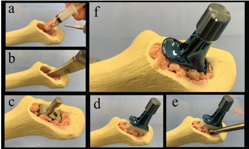
Figure 1: Demonstration of surgical technique using the combination of impaction bone grafting and a collared fully hydroxyapatite-coated stem. The bone grafting procedure involves the following steps: After removing the old stem, morselized allograft bone is packed into the intramedullary canal using a syringe (a), compressed through broaching (b), and the stem size is determined after confirming sufficient fixation (c). The new stem is manually inserted, and the grafted bone around the stem is impacted (d). Additional bone is packed around the stem (e), and the stem is driven in with bulk bone grafting at the calcar to complete the procedure (f).
Two types of stems were utilized: CORAIL and UNIVERSIA. The IBG technique involves filling the medullary cavity from which the stem was removed with morselized allograft bone and compressing it with a broach. This process is repeated until a sufficiently compressed cancellous bone layer is formed to allow insertion of an undersized stem, one size smaller than the original stem. The collared, fully HA-coated stem is then manually inserted, and additional bone graft is added to the side of the stem as needed before the stem is hammered to seat the collar properly. A block bone graft is placed between the calcar and collar to reinforce the calcar area as needed. Post-operative therapy allowed full weight-bearing walking from the day after surgery. All activity restrictions were canceled 3 months postoperatively.
The median follow-up period was 4.5 years (range, 3.1–9.5 years). The median operation time was 127 min, the median intraoperative blood loss was 855 mL, and the median length of hospital stay was 8.0 (7–41) days. There were no complications such as intraoperative fractures, symptomatic pulmonary embolism, aseptic loosening, septic loosening, dislocation, or postoperative fractures. The JOA score improved from a median of 63 points preoperatively to a median of 95 points at the final follow-up. The median stem subsidence was 1.4 mm (1–3 mm), with no subsidence exceeding the loosening threshold of 5 mm. There was no circumferential bone radiolucent line around the stem, and spot welds were observed in all cases, indicating remodeling (Table 1). At the latest follow-up, all six stems were retained, with no re-revision or reoperation for any reason.
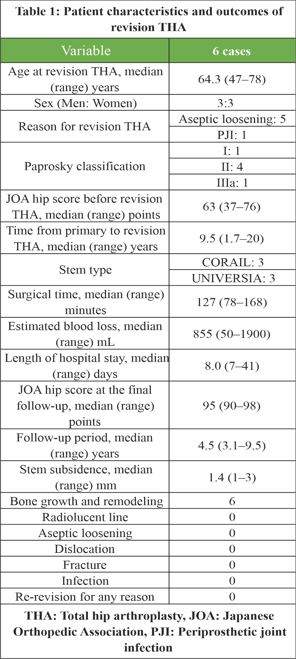
Table 1: Patient characteristics and outcomes of revision THA.
Case 1:
A 48-year-old male underwent bipolar hip prosthesis surgery for secondary right hip osteoarthritis following synovectomy for pigmented villonodular synovitis at another hospital. Ten years post-surgery, he experienced pain in the right hip joint during walking. X-rays revealed stem subsidence, central migration of the femoral head, and leg length discrepancy, leading to a diagnosis of aseptic loosening of the stem (Fig. 2a). The bone defect was classified as Paprosky Type II. The femoral prosthesis was revised using IBG with CORAIL, and a cementless cup was placed (Fig. 2b). The operation lasted 168 min with a blood loss of 910 mL. Autologous blood transfusion was used, with transfusion consisting only of recovered blood. Eight and a half years later, X-rays showed stem subsidence of 1 mm (Fig. 2c). In addition, comparing computed tomography (CT) images taken 3 months, 2 years, and 5 years post-surgery, remodeling of the bony mass was observed in the calcar region. Remodeling and bone ingrowth were noted in the allograft cancellous bone layer around the stem (Fig. 2d, e, f). The JOA score improved from 62 points preoperatively to 96 points at 8.5 years post-surgery.
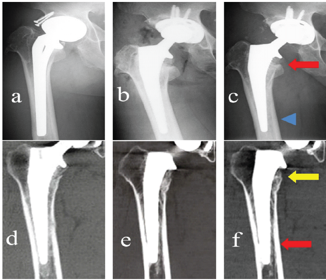
Figure 2: X-ray and computed tomography (CT) images in Case 1. (a) 10 years post-operative X-ray. (b) X-ray after revision surgery. (c) X-ray eight and a half years postoperatively. The allograft bone at the greater trochanter had integrated, making the boundary with the host bone indistinct (red arrow). Cortical hypertrophy at the distal stem had not progressed (blue arrowhead). (d) CT images taken 3 months post-surgery. (e) CT images taken 2 years post-surgery. (f) CT images taken 5 years post-surgery revealed remodeling of the bony mass in the calcar region (yellow arrow) and remodeling and bone growth in the allograft cancellous bone layer around the stem (red arrow).
Case 2:
A 78-year-old female underwent THA at our hospital for right hip osteoarthritis (Fig. 3a). Although the post-operative course was uneventful, X-rays taken 3 years after surgery revealed osteolysis (Fig. 3b). The magnetic resonance imaging of the hip joint showed an abscess around the right hip joint, leading to a diagnosis of pyogenic hip arthritis (Fig. 3c).
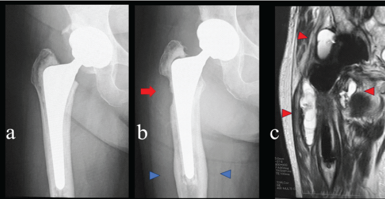
Figure 3: Reason for revision of total hip arthroplasty in Case 2. (a) X-ray after primary cemented total hip arthroplasty. (b) Three years post-operative X-ray showed bone resorption in the proximal lateral femur (red arrow) and cortical hypertrophy at the distal stem (blue arrowheads). (c) Magnetic resonance imaging T2-weighted coronal imaging revealed abscess formation around the prosthesis (red arrowhead).
The bone defect was classified as Paprosky Type IIIa. A cementless cup was replaced on the acetabulum. Strut bone was grafted to the area of the metaphyseal bone defect, and wiring was performed. Femoral prostheses were revised using IBG with CORAIL (Fig. 4a). The operation lasted 167 min with a blood loss of 1400 mL. This case involved an infection, and transfusion of recovered blood was not possible, requiring 4 units of red blood cell transfusion. This case was the only one where the stem subsided more than 2 mm, with a 3 mm subsidence seen on X-ray images a year post-surgery (Fig. 4b). Three years after, remodeling of the strut bone was observed (Fig. 4c). Remodeling of the transplanted bone around the stem was also noted. The JOA score improved from 67 points preoperatively to 97 points at 3 years post-surgery.
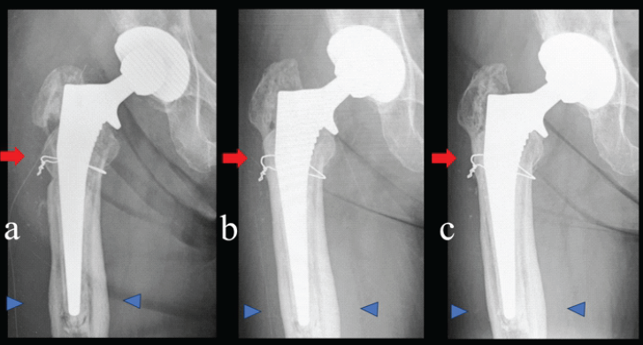
Figure 4: Bone growth and remodeling on X-ray images in Case 2. (a) X-ray after revision total hip arthroplasty. A stem revision surgery was performed using a combination of IBG and a collared fully hydroxyapatite-coated stem. The morselized allograft bone and bulk bone grafting were performed at the lateral defect of the greater trochanter (red arrow). (b) X-ray 1 year postoperatively. The stem had subsided by 3 mm, but the grafted bulk bone showed signs of remodeling (red arrow). (c) X-ray 3 years postoperatively. The allograft bone at the greater trochanter had integrated, making the boundary with the host bone indistinct (red arrow). Cortical hypertrophy at the distal stem had not progressed (blue arrowhead).
Good short-term outcomes were demonstrated in these six cases of revision THA using IBG with a collared fully HA-coated stem. The use of IBG with a collared, fully HA-coated stem is suggested as a potentially effective means for revision THA of the femoral component. Revision THA using IBG is often reported with the use of cement systems. Starting from the report by Gie et al. [1] in 1993, good outcomes were reported in 56 cases over 18–49 months. Subsequently, Lamberton et al. [5] performed revision THA using IBG in 540 joints, reporting a loosening-related re-revision rate of 2% at an average follow-up of 6.7 years. Furthermore, Wilson et al. [3] performed revision THA on 705 joints, reporting a loosening-related re-revision rate of 1.2% at an average post-operative follow-up of 14.7 years. Thus, revision THA using the IBG with cement systems has been reported to have good long-term outcomes in many cases. A recent systematic review and meta-analysis of 4102 hips confirmed a 10-year survivorship of 95% for femoral IBG with cemented stems [10], and a Japanese series using the Exeter stem reported a 15-year stem survival of 99% for aseptic loosening [11]. However, there are few reports on revision THA using the IBG with cementless stems. Ishiguro et al. [8] reported a case (our case 1). Nesse et al. [12] randomized Paprosky Grade II 14 patients into two groups intraoperatively, and compared cementless impaction grafting using CORAIL stems with cemented IBG using polished femoral stems. Functional scores and bone density did not differ between the two groups during the 5-year follow-up period. However, radiological data have not been discussed. Masterson et al. [13] reported the long-term results of revision THA using IBG with both cementless and cemented Freeman stems. In the cementless group, stem subsidence of more than 4 mm occurred in half of the cases, intraoperative fractures were observed in 10% of cases, and revision surgery due to loosening was required in another 10%. These results indicate a high incidence of complications. Masterson et al. [13] speculated that the poor outcomes in the cementless group were due to the need for more robust bone grafting and the necessity to restrict weight-bearing until bone fusion was achieved. Nickelsen et al. [14] combined IBG and a cementless, proximally titanium-coated long stem for 100 cases of revision THA, grading bone loss and aseptic loosening according to Engh et al. [15]. Their complication rates showed that 14% of cases had intraoperative fractures, and 17 patients required revision during the average 112-month follow-up period. Nickelsen et al. [14] attributed the poor outcomes in the cementless group to two main factors: First, predominantly early failures associated with early subsidence, indicating a lack of sufficient initial stability, and second, a higher incidence of failures in extra-long stems (250 or 300 mm). A recent systematic review comparing cemented and uncemented stems in femoral IBG found that the uncemented technique was associated with a lower rate of femoral component loosening (1.7% vs. 8.0%), while emphasizing that adequate initial stability is essential to avoid early subsidence [16]. In this study, good post-operative functional scores were obtained in all cases. From the X-ray image, bone fixation was observed in all cases, with a median stem subsidence of only 1.4 mm. There were no cases of intraoperative fractures or cases that led to re-replacement. Collared fully HA-coated stems (CORAIL and UNIVERSIA) used in this study are stems that follow the shape of cement systems and are designed to exert compressive forces circumferentially on the stem. The stem stability and bone ingrowth using IBG with a cement system have been elucidated in previous studies [1,2,3,4,5]. It is believed that similar compressive forces were applied with the CORAIL-type stem, forming a good compressive cancellous bone layer. Furthermore, the use of a collared stem and transplantation of bulk allogenic bone graft into the calcar region contributed to the good fixation by suppressing subsidence. The cementless CORAIL stem has an established long-term track record, with favorable 20-year survivorship reported in primary THA [17]. Although experimental work has shown that early loading influences the incorporation of impacted morselized allograft [18], full weight-bearing from the 1st post-operative day did not compromise graft incorporation or stem stability in the present series. Most significantly, the CT image of Case 1 provided critical evidence that bone ingrowth occurs between a collared fully HA-coated stem and the compressive allograft cancellous bone layer. The paper presenting this radiographic image is only the case report of Ishiguro [8] and this study, demonstrating the usefulness of using IBG with a collared fully HA-coated stem. This study had several limitations. First, the sample size was small, and the follow-up period was short due to the decreasing number of revision surgeries as a result of improved outcomes of primary THA in recent years. Second, as cases classified as Paprosky IIIB and C with large bone defects were not included, the outcomes of more complex revision surgeries remain unclear.
IBG combined with a collared fully HA-coated cementless stem may be a useful and effective option for femoral component reconstruction in revision THA, offering both early mechanical stability and biological fixation. Larger series with longer follow-up are warranted to confirm the durability of this technique.
The combination of the impaction bone grafting technique and a collared fully hydroxyapatite (HA)-coated cementless stem provides an effective biological solution for femoral component revision in total hip arthroplasty. This technique achieves early mechanical stability through the collar and promotes reliable biological fixation via bone ingrowth into the HA-coated surface from the impacted allograft. Clinicians can consider this method as a viable alternative to cemented systems, particularly when biological reconstruction of femoral bone stock is required.
References
- 1. Gie GA, Linder L, Ling RS, Simon JP, Slooff TJ, Timperley AJ. Impacted cancellous allografts and cement for revision total hip arthroplasty. J Bone Joint Surg Br 1993;75:14-21. [Google Scholar] [PubMed]
- 2. Ling RS. Femoral component revision using impacted morselized cancellous graft. J Bone Joint Surg Br 1997;79:874-5. [Google Scholar] [PubMed]
- 3. Wilson MJ, Hook S, Whitehouse SL, Timperley AJ, Gie GA. Femoral impaction bone grafting in revision hip arthroplasty: 705 Cases from the originating centre. Bone Joint J 2016;98-B:1611-9. [Google Scholar] [PubMed]
- 4. Ornstein E, Atroshi I, Franzén H, Johnsson R, Sandquist P, Sundberg M. Results of hip revision using the Exeter stem, impacted allograft bone, and cement. Clin Orthop Relat Res 2001;389:126-33. [Google Scholar] [PubMed]
- 5. Lamberton TD, Kenny PJ, Whitehouse SL, Timperley AJ, Gie GA. Femoral impaction grafting in revision total hip arthroplasty: A follow-up of 540 hips. J Arthroplasty 2011;26:1154-60. [Google Scholar] [PubMed]
- 6. Palm L, Jacobsson SA, Kvist J, Lindholm A, Ojersjö A, Ivarsson I. Acetabular revision with extensive allograft impaction and uncemented hydroxyapatite-coated implants. Results after 9 (7-11) years follow-up. J Arthroplasty 2007;22:1083-91. [Google Scholar] [PubMed]
- 7. Etienne G, Bezwada HP, Hungerford DS, Mont MA. The incorporation of morselized bone grafts in cementless acetabular revisions. Clin Orthop Relat Res 2004;428:241-6. [Google Scholar] [PubMed]
- 8. Ishiguro S, Asanuma K, Tamaki T, Oinuma K, Sudo A. A case of cementless impaction bone graft in a revision total hip arthroplasty requiring calcar reconstruction. Case Rep Orthop 2021;2021:8811593. [Google Scholar] [PubMed]
- 9. Paprosky WG, Lawrence JM, Cameron HU. Femoral defect classification in revision arthroplasty. Orthop Rev 1990;19 Suppl: 9-14. [Google Scholar] [PubMed]
- 10. Klimko A, Andronic O, Lu VY, Dimitriou D, Hoch A, Zingg PO. Survivorship and patient-reported outcomes in femoral impaction bone grafting for revision hip arthroplasty: A systematic review and meta-analysis of 4,102 hips. J Arthroplasty 2026;41:283-97.e2. [Google Scholar] [PubMed]
- 11. Iwase T, Otsuka H, Katayama N, Fujita H. Impaction bone grafting for femoral revision hip arthroplasty with Exeter stem in Japan: An extended 10- to 15-year stem survival analysis of the previously reported series. J Orthop Sci 2024;29:151-6. [Google Scholar] [PubMed]
- 12. Nesse E, Nielsen EW, Bastian D. Zementierte versus unzementierte revisions- hüftarthroplastik mit verdichtetem knochenallograft–prospektiv randomisierte vergleichsstudie mit jahres-follow-up [Cemented versus cementless revision femoral stems using morselized allograft–a prospective, randomized study with 5 years follow-up]. Z Orthop Ihre Grenzgeb 2003;141:678-83. [Google Scholar] [PubMed]
- 13. Masterson S, Lidder S, Scott G. Impaction femoral allografting at revision hip arthroplasty: Uncemented versus cemented technique using a Freeman femoral component. J Bone Joint Surg Br 2012;94:51-5. [Google Scholar] [PubMed]
- 14. Nickelsen TN, Erenbjerg M, Retpen JB, Solgaard S. Femoral revision with impaction allografting and an uncemented femoral component. Hip Int 2008;18:278-85. [Google Scholar] [PubMed]
- 15. Engh CA, Glassman AH, Griffin WL, Mayer JG. Results of cementless revision for failed cemented total hip arthroplasty. Clin Orthop Relat Res 1988;235:91-110. [Google Scholar] [PubMed]
- 16. Zhong Yi K, Selvaratnam V. A systematic review comparing the outcomes of cemented versus uncemented stems in femoral impaction bone grafting for revision hip arthroplasty. Cureus 2024;16:e71560. [Google Scholar] [PubMed]
- 17. Vidalain JP. Twenty-year results of the cementless Corail stem. Int Orthop 2011;35:189-94. [Google Scholar] [PubMed]
- 18. Van Der Donk S, Buma P, Verdonschot N, Schreurs BW. Effect of load on the early incorporation of impacted morsellized allografts. Biomaterials 2002;23:297-303. [Google Scholar] [PubMed]






Related Articles in Journal of Orthopaedic Case Reports
 August 1, 2026 Outcome of Large-Diameter Jumbo Cups or Acetabular Mesh with Impaction Bone Grafting for Acetabular Defects in Primary and Revision Total Hip Arthroplasty: A Case Series
 September 1, 2026 Use of a Robotic Platform Compared with Manual Preparation for Revision Total Hip Arthroplasty: A Cadaver-Based Feasibility Study
 August 1, 2026 Metallosis Following Total Hip Arthroplasty: A Case Report
 July 1, 2026 Custom-Made Monoflange Acetabular Component for Revision Hip Arthroplasty in Paprosky Type IIIA Defect: A Case Report