


Introduction
Distal femur fractures represent <1% of all fractures [1,2]. These fractures are frequently comminuted and have intra-articular involvement [3]. Principles of management of distal articular femur fractures are similar to those of any other articular fracture, i.e., to achieve anatomical reduction of the fracture fragments while providing rigid fixation to allow early functional mobilization [3]. Management of periarticular fractures requires a careful understanding of fracture characteristics and proper pre-operative planning [4]. Management of combined distal femoral intercondylar fractures along with coronal plane fractures has received very little attention in the past [5]. Proper pre-operative imaging of these injuries may assist in the planning of surgical approaches and the selection of implants [6].
The present AO-OTA classification is a radiograph-based classification system. The presence of multiple fragments and the orientation of the fragments are now simplified with the use of computed tomography [6]. The presence of combined sagittal and coronal plane fractures of either or both condyles necessitates that it be classified as AO-OTA type 33 C3 or multifragmentary articular type [5]. A single category 33 C 3 does not provide adequate information about the degree of articular comminution and cannot predict the functional outcome. Similarly, B3 represents all types of coronal plane or Hoffa’s fractures. However, a combined unicondylar sagittal fracture associated with a coronal plane fracture of the same condyle cannot be categorized according to the AO-OTA classification. Thus, we feel that there is a necessity for a new CT-based classification system for distal articular femur fractures, which can predict the severity, assist in the planning of surgical approaches, aid in implant selection, and suggest the functional outcome. The proposed CT-based classification is described (See Table 1) and its implications for the surgical management of biplanar articular fractures have been discussed in this article (Table 2 and Fig. 1).
Describing the CT-based proposed classification system
| Fracture | Type | Subtype | AO-OTA Equivalent |
|---|---|---|---|
| Uniplanar | A. Sagittal | 1. Medial condyle | B1 |
| 2. Lateral condyle | B2 | ||
| 3. Intercondylar | C1 | ||
| B. Coronal | 1. Medial Hoffa | B3.2 | |
| 2. Lateral Hoffa | B3.2 | ||
| 3. Bicondylar Hoffa | B3.3 | ||
| Biplanar | C. Two part | 1. Medial | – |
| 2. Lateral | – | ||
| D. Three part | 1. Medial | C3 | |
| 2. Lateral | |||
| 3. Central | |||
| E. Four part | 4 parts in the distal femur | C3 | |
| F. Multifragment | 1. Unicondylar | C3 | |
| 2. Bicondylar | C3 |
CT: Computed tomography
Patient demographic details and post-operative follow-up findings
| Mean age | 35 years |
| M: F ratio | 05:01 |
| Classification | Number of patients |
| Type C | 24 |
| Type D | 18 |
| Type E | 5 |
| Type F | 3 |
| Classification | Mean post-operative ROM |
| Type C | 0–115° |
| Type D | 0–102° |
| Type E | 10–70° |
| Type F | 10–50° |
| Complications | |
| Infection | 1 patient (Type E) |
| Non-union | 1 |
| Delayed union | 1 patient (Type D2) |
| Deformity | 2 patients Varus >15° |
| 1 patient Valgus >15° | |
| Shortening | 1 patient type (D3 M3) |
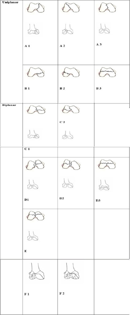
In this study, we have included only the biplanar distal femoral articular fractures and evaluated the fixation techniques and functional outcomes in these patients. The degree of metaphyseal comminution should be considered separately, as the restoration of the articular surface is essential, irrespective of the degree of metaphyseal comminution. Accordingly, we have graded the degree of metaphyseal comminution separately.
Materials and Methods
This study was conducted at the Department of Orthopedics between May 2018 and June 2023. A total of 50 patients with distal articular biplanar femur fractures were included in our study. Pre-operative CT evaluation was done in all patients. Demographic details and the mechanism of injury were documented for all the patients. Surgical planning, including the approach and implant selection, was made based upon the proposed classification (Fig. 1). The suggested classification is as follows. Type A and B are uniplanar fractures but, for the sake of completeness, have been included in the picture. Types C, D, and E are biplanar fractures with increasing degree of comminution. Type C represents a 2-part fracture with combined coronal and sagittal fractures in one condyle. C-L and C-M have been used to represent the medial and lateral condyles. In type C fractures, there is metaphyseal continuity in the other condyle. Type D represents a 3-part fracture with combined sagittal and coronal fractures in one condyle, whereas the other condyle has a uniplanar fracture. Terminologies D-L and D-M are used to suggest the biplanar involvement of the lateral or medial condyle, whereas D and C suggest that there is a distal femur fracture associated with a bicondylar Hoffa’s fracture. Type E represents a 4-part fracture, wherein there is a combined coronal and sagittal fracture in both condyles. Type F suggests a comminuted fracture.
The metaphyseal comminution was graded as M0 if there was no comminution. M1 and M2 represent moderate and severe comminution, respectively, and M3 suggests comminution with bone loss.
Intraoperative photographs were also taken to confirm the CT findings. Postoperatively, patients were reviewed once every 15 days, and radiographs were repeated every 30 days. All the patients were followed up regularly for 6 months. Fracture healing time, post-operative complications, and Knee Society Scores were recorded in all patients.
Interobserver and intraobserver reliability for the classification was also assessed. To assess the interobserver reliability, five orthopedic consultants with more than 6 years of clinical experience and one radiologist were contacted. Each consultant received five randomly selected radiographs, CT images, and intra-operative photographs of different patients and was asked to classify them according to the above classification. The same exercise was repeated after an interval of 4 weeks to assess the intraobserver reliability.
General principles
-
Approach: We prefer an anterior approach for biplane articular fractures. The medial or lateral parapatellar approach is decided based upon the coronal fracture.
-
Reduction: An accurate anatomical articular reduction is essential before proceeding to metaphyseal fixation. Reduction of coronal fracture fragments is achieved first before the sagittal fracture fixation.
-
Articular fixation: Headless compression screws and countersunk cancellous screws were used for coronal plane fractures. The sagittal fractures were fixed with one or two cancellous screws. This converts the multifragment articular fracture into one articular block, which can be fixed with the metaphyseal segment. In 2-part biplane fractures (Type C), only screw fixation is sufficient, with or without a buttress plate. In 3-part and 4-part biplane fractures, we need to identify the condyle that has no associated coronal plane fracture. This condyle was labeled as “the stable fragment.” In such fractures, a locking plate should be applied between the stable fragment and the metaphysis. If both condyles have coronal fractures (Type E), then the decision for bicolumnar plate fixation should be taken intra-operatively, depending upon the stability of the fixation.
-
Metaphyseal fixation: In M1 cases, we fix the articular block and the metaphyseal fragments with a locking plate using the minimally invasive method. In M2 fractures, the plate is fixed in bridging mode. M3 cases are mostly associated with open wounds, and bone cement was used to fill the metaphyseal defect. The bone cement is later replaced by bone grafting using the Masquelet technique.
Results
Fifty patients with biplanar distal articular fractures were included in our study. Road traffic accidents with a head-on collision were the most common mode of injury. Eight patients had a compound wound. Four patients had other associated fractures. Anterior cruciate ligament and posterior cruciate ligament avulsions were present in three patients.
The mean age was 35 years. The male-to-female ratio in our study was 5:1. The most common fracture type was D, followed by C, E, and F, respectively. ROM in each group of fractures was recorded at every follow-up. Patients with C- and D-type fractures had a good range of motion of more than 100°. While the patients who suffered type E and type F fractures had an average range of motion of <80°. One patient who suffered from a type E fracture and had a grade IIIB compound injury developed wound infection, which subsided with secondary debridement and cement bead placement. Two patients who had M3-type metaphyseal fractures required secondary bone grafting after 6 weeks. One patient of with a type D3M3 had a 1.5 cm shortening, which was managed with a shoe raise. Two patients had a significant post-operative varus deformity, and one had a valgus deformity. One patient had non-union and was lost to follow-up. The clinical and radiological representative pictures are shown in Fig. 2.
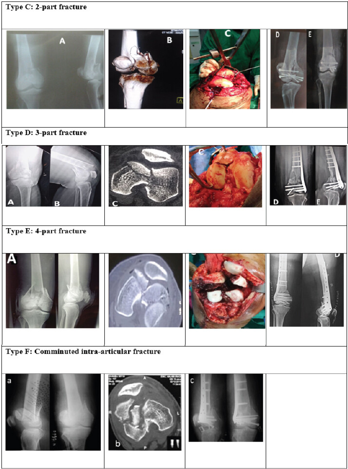
The interobserver agreement was 87.5%, and the intraobserver agreement was 100%. The chance-corrected and weighted kappa statistics for observer agreement, both for inter-observer and intra-observer variability, demonstrated satisfactory repeatability of the classification system.
Discussion
Various classification systems have been described in the literature [7,8]. The most commonly used classification system for distal femur fractures is the AO OTA classification [9]. In this classification, the distal femur fractures are broadly classified into type A, B, and C, corresponding to extra-articular, partially intra-articular, and fully intra-articular [9]. Each group is further subclassified into three types based on the pattern of the fracture and degree of comminution. Type B1 involves sagittal split of the Lateral condyle; B2 involves sagittal split of the medial condyle, and type B3 represents a coronal plane fracture known as a Hoffa’s fracture. Type C is subclassified into C1 (simple articular, simple metaphyseal), C2 (simple articular, multifragmentary metaphyseal), and C3 (multifragmentary articular and metaphyseal).
The above classification is simple, valid, and provides a good picture of the severity of the distal femur fractures. However, it is an X-ray-based classification. The inability of the AO system in classifying biplane fractures has been well explained by Nork et al. [5]. In their study, they found that associated coronal plane fractures were found in 38% of the distal femoral intra-articular fractures. All these combined fractures have to be invariably labeled as C3 type or multifragmentary articular fractures. The term multifragmentary is very subjective. What actually accounts for multifragmentary is not understood by the classification. The C3 type is further subclassified into C3.1, C3.2, and C3.3. However, all these subtypes focus on metaphyseal comminution and not articular fragments. A detailed CT-based evaluation irrespective of the degree of metaphyseal comminution is essential in all intra-articular fractures. CT will not only help in the proper identification of fracture fragments, but it will also help in planning the surgery and implant selection. Moreover, we found it difficult to classify a few fractures using the AO OTA system. One example is a combined sagittal and coronal plane fracture of an isolated medial or lateral condyle.
Although there is a consensus in the literature regarding the validity of AO OTA classification for distal femur fractures, there are a few studies that highlight the problems with it. Elsoe et al. conducted a population-based epidemiological study of distal femur fractures among Danish patients [10]. They found that approximately 10 % of the total distal femur fractures could not be classified using the AO Classification. They have subclassified this group as the Missing AO group. Similarly, Agrawal and Kiyawat conducted a study on the management of complex distal femur fractures [11]. The inability of the AO classification system to classify these fractures is very evident by the fact that they have used two sub-classifications for extra-articular and intra-articular parts of the same fracture. Bagaria et al. proposed a new classification system for the coronal plane fractures of the distal femur [12]. They have suggested a separate subclass for combined coronal and sagittal fracture of the femoral condyle, which could not be classified by the AO system. These studies highlight the necessity of a CT-based classification system.
Based upon these needs, we have described a classification system which is CT-based and provides a complete description of the articular fragments. This classification is based upon the planes of the fracture lines. Type A is a uniplanar sagittal fracture, and type B is a uniplanar coronal fracture. Type C, D, E, and F represent biplanar fractures with increasing grades of comminution. This classification system helps us in deciding the approach to the fracture and also helps in prognostication of fractures as well. Fractures with increasing degrees of articular comminution have less post-operative ROM, and there are increased chances of arthritis; e.g., type C has a better prognosis than type F. A metaphyseal fracture classification system is also added to the articular fracture classification. M1 is simple, M2 is comminuted, and M3 is associated with bone loss. A description of the applicability of the classification in terms of approach, surgical planning, implant selection, and prognosis is provided in Table 3.
Describing the fracture classification, approach, implant selection, and prognosis following fracture fixation
| Classification | Description | Approach | Implant | Average Knee Society scores |
|---|---|---|---|---|
| CM | Two-part fracture with medial condyle coronal fracture | Medial parapatellar | Headless screws with or without a buttress plate | 84 |
| CL | Two-part fracture with medial condyle coronal fracture | Lateral parapatellar | Headless screws with or without a buttress plate | 86 |
| DM | Three-part articular fracture with coronal fracture involving the medial condyle | Medial parapatellar | Headless screw and standard distal femur LCP | 78 |
| DL | Three-part articular fracture with coronal fracture involving the lateral condyle | Lateral parapatellar | Headless screw and standard distal femur LCP and additional medial condyle plate | 76 |
| DC | Central articular fracture with bicondylar coronal fracture | Medial or lateral parapatellar | Headless screw and standard Distal femur LCP with or without medial condyle plate | 74 |
| E | Four-part articular fracture | Medial or lateral parapatellar | Headless screw and standard distal femur LCP | 68 |
| F | Comminuted unicondylar/bicondylar fracture | Medial or lateral parapatellar | Multiple screws and bridge plating | 35 |
| Percutaneous pin fixation for external fixator | Knee spanning external fixator | 22 |
CM: Medial condyle, CL: Lateral condyle, DM: biplanar involvement of medial condyle, DL: biplanar involvement of lateral condyle, DC: distal femur fracture associated with a bicondylar Hoffa fracture.
Limitations
The sample size of this study is very small. This classification system requires large group studies to assess its validity and reliability.
Conclusion
Thus, we conclude that this CT-based classification system is a focused tool to assess the degree of severity of the articular comminution, plan our approach to the fracture, decide the implants necessary, and grade the prognosis. Simultaneously, this classification provides an addition to the metaphyseal comminution, which can be written together. In spite of being very elaborate, it is easy to reciprocate and communicate.
Clinical Message
This classification helps young surgeons who have little experience to understand the pattern of fracture, plan for fixation of the fragments, determine the types of implants needed, anticipate intraoperative difficulties, and be ready with armamentarium.
Conflict of Interest:
Nil
Source of Support:
Nil
Consent:
The authors confirm that informed consent was obtained from the patient for publication of this article
How to Cite this Article
Sadiq M, Neerudi SR, Kasam RR, Kumar KU, Pradeep KS, Manne A. Biplane Fractures of the Distal Femur: A Proposed Computed Tomography-Based Classification and Surgical Management. Journal of Orthopaedic Case Reports 2026 October;16(10): 635-641.
References
- Court-Brown CM, Caesar B. Epidemiology of adult fractures: A review. Injury 2006;37:691-7. [Google Scholar] | [PubMed]
- Martinet O, Cordey J, Harder Y, Maier A, Bühler M, Barraud GE. The epidemiology of fractures of the distal femur. Injury 2000;31 Suppl 3:C62-3. [Google Scholar] | [PubMed]
- Imam MA, Torieh A, Matthana A. Double plating of intra-articular multifragmentary C3-type distal femoral fractures through the anterior approach. Eur J Orthop Surg Traumatol 2018;28:121-30. [Google Scholar] | [PubMed]
- Gangavalli AK, Nwachuku CO. Management of Distal femur fractures in adults. Orthop Clin North Am 2016;47:85-96. [Google Scholar] | [PubMed]
- Nork SE, Segina DN, Aflatoon K, Barei DP, Henley MB, Holt S. The association between supracondylar-intercondylar distal femoral fractures and coronal plane fractures. J Bone Joint Surg 2005;87:564-9. [Google Scholar] | [PubMed]
- Allmann KH, Altehoefer C, Wildanger G, Gufler H, Uhl M, Seif El Nasr M. Hoffa fracture--a radiologic diagnostic approach. J Belge Radiol 1996;79:201-2. [Google Scholar] | [PubMed]
- Neer CS 2nd, Grantham SA, Shelton ML. Supracondylar fracture of the adult femur. A study of one hundred and ten cases. J Bone Joint Surg Am 1967;49:591-613. [Google Scholar] | [PubMed]
- Seinsheimer F. Fractures of the distal femur. Clin Orthop 1980;153:169-79. [Google Scholar] | [PubMed]
- Marsh JL, Slongo TF, Agel J, Broderick JS, Creevey W, DeCoster TA. Fracture and dislocation classification compendium -2007: Orthopaedic trauma association classification, database and outcomes committee. J Orthop Trauma 2007;21 Suppl 10:S1-133. [Google Scholar] | [PubMed]
- Elsoe R, Ceccotti AA, Larsen P. Population-based epidemiology and incidence of distal femur fractures. Int Orthop 2018;42:191-6. [Google Scholar] | [PubMed]
- Agrawal A, Kiyawat V. Complex AO type C3 distal femur fractures: Results after fixation with a lateral locked plate using modified swashbuckler approach. Indian J Orthop 2017;51:18-27. [Google Scholar] | [PubMed]
- Bagaria V, Sharma G, Waghchoure C, Chandak RM, Nemade A, Tadepelli K. A proposed radiological classification system of Hoffa's fracture based on fracture configuration and consequent optimal treatment strategy along with the review of literature. SICOT J 2019;5:18. [Google Scholar] | [PubMed]
© 2026 Journal of Orthopaedic Case Reports - Published by Indian Orthopaedic Research Group
About the Authors
-

Mohammed Sadiq
-

Sharon Rose Neerudi
-

Rohith Reddy Kasam
-

Kothalanka Udaya Kumar
-

Koribilli Sai Pradeep
-

Anudeep Manne
Related Articles in Journal of Orthopaedic Case Reports
 May 1, 2026 Outcomes of Open Reduction and Internal Fixation in Intra-articular Distal Femur Fractures with Incarcerated Patella: A Case Series of a Rare Injury Pattern
 February 1, 2025 Outcomes of Combined Distal Femur Plating and Retrograde Femur Nailing in Comminuted Distal Femur Fractures: Case Series of Seven Cases with 6 Months Follow-up
 December 10, 2023 Bilateral Hoffa Fractures of the Medial Femoral Condyles: A Case Report and Review of the Literature
 July 10, 2020 Neglected Idiopathic Clubfoot Successfully Treated by the Ponseti Method: A Case Report of an Adult Patient who Started Treatment at 26 Years of Age