


Introduction
Low back pain (LBP) is the leading cause of years lived with disability worldwide and represents a major public health challenge. It affects approximately 619 million people globally, with the burden projected to increase to over 840 million cases by 2050 due to population growth and aging [1]. Persistent pain often results in compensatory postural adaptations, including pelvic asymmetry, altered lumbar lordosis, trunk imbalance, coronal deviation, vertebral rotation, and abnormal muscle activation patterns. These biomechanical alterations contribute to functional disability, impaired mobility, and diminished quality of life [2, 3].
Current evidence-based guidelines recommend a multimodal management approach comprising patient education, exercise therapy, pharmacological treatment, and rehabilitation as first-line interventions for chronic LBP (CLBP). However, a considerable proportion of patients continue to experience persistent symptoms despite adequate conservative treatment. In such cases, fluoroscopy-guided transforaminal epidural steroid injection (TFESI) is widely employed as an intermediate intervention before surgical consideration. By delivering corticosteroid and local anesthetic directly into the ventral epidural space adjacent to the affected nerve root, TFESI reduces local inflammation, neural edema, and ectopic nerve discharge, thereby providing significant improvements in pain and functional disability in appropriately selected patients [4, 5].
Although numerous studies have demonstrated improvements in pain and disability following TFESI, relatively few have objectively evaluated the associated changes in spinal posture and biomechanics. Conventional radiographic assessment of spinal alignment exposes patients to ionizing radiation and is therefore unsuitable for repeated follow-up evaluations. Recent advances in optical surface topography have enabled non-invasive, radiation-free assessment of spinal posture.
The DIERS formetric 4D spine posture analysis system is a validated raster stereography-based optical imaging technology that reconstructs a three-dimensional model of the spine and trunk surface using the principle of structured light projection. The system automatically identifies anatomical landmarks, including the vertebra prominens and posterior superior iliac spines (PSIS), and computes clinically relevant parameters such as trunk inclination, trunk imbalance, pelvic tilt, pelvic torsion, pelvic rotation, thoracic kyphosis, lumbar lordosis, surface rotation, lateral deviation, sagittal balance, and coronal alignment. The four-dimensional (4D) analysis incorporates repeated image acquisition over several seconds, thereby reducing motion artifacts caused by physiological sway and improving measurement precision. Multiple validation studies have demonstrated excellent intra- and inter-observer reliability, high reproducibility, and good agreement with radiographic assessment for evaluating spinal alignment while eliminating radiation exposure. Consequently, the DIERS formetric 4D system has become an increasingly valuable tool for longitudinal monitoring of spinal posture in patients undergoing rehabilitation and musculoskeletal interventions [6, 7, 8, 9].
Therefore, the present study aims to evaluate the effects of fluoroscopy-guided TFESI on pain, disability, quality of life, neural mobility, and spinal posture in patients with CLBP. Spinal posture will be objectively assessed using the DIERS formetric 4D spine posture analysis system, enabling quantitative evaluation of three-dimensional spinal alignment and biomechanical changes.
Materials and Methods
Study design and setting
This prospective, single-center, longitudinal observational study included a total of 43 consecutive patients with CLBP who met the eligibility criteria after approval from the Institutional Ethics Committee (IEC No-AIIMS/MG/IEC/ 2024-25/132 dated November 23, 2024).
Inclusion criteria
-
Aged between 18 and 60 years
-
CLBP of more than 3 months duration
-
Magnetic resonance imaging-confirmed lumbar disc degeneration or contained lumbar disc prolapse correlating with clinical findings
-
Failed to respond to at least 6 weeks of conservative treatment.
Exclusion criteria
-
Previous lumbar spine surgery
-
Lumbar spinal instability or grade II or higher spondylolisthesis
-
Non-discogenic back pain.
Intervention
All participants who were planned for fluoroscopy-guided TFESI (Triamcinolone acetonide 80 mg/2 mL) were included in the study. Spine posture analysis was done before the intervention at baseline (0 weeks) and after the intervention at 8 weeks. Following the procedure, all patients were advised for standardized rehabilitation program which includes strengthening exercises and activity modification.
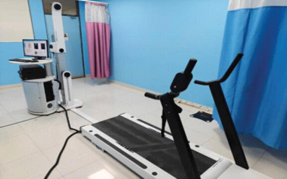
Three-dimensional spinal posture analysis was performed using the DIERS formetric 4D surface topography system (DIERS International GmbH, Schlangenbad, Germany). The assessment was conducted in a dedicated examination room under standardized lighting conditions by the same trained examiner throughout the study to minimize inter-observer variability.
Participants were instructed to remove clothing covering the back, footwear, and accessories that could interfere with image acquisition. They stood barefoot on the designated platform in a relaxed, habitual posture with feet positioned shoulder-width apart, knees fully extended without locking, arms hanging naturally by the sides, and eyes directed toward a fixed point at eye level. Participants were instructed to remain still and breathe normally during image acquisition.
The DIERS formetric 4D system utilizes raster stereography, a radiation-free optical technique that projects parallel light stripes onto the dorsal surface of the trunk. Surface asymmetries are captured by a high-resolution camera, and specialized software reconstructs a three-dimensional model of the back to estimate the underlying spinal alignment using validated anatomical landmarks, including the vertebra prominens (C7) and bilateral PSIS (Fig. 1 and 2).
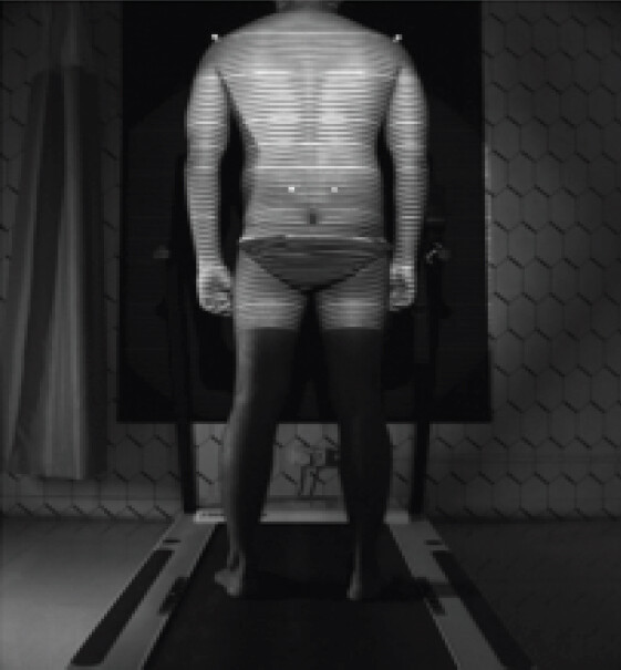
For each assessment, a sequence of images was acquired over approximately 6 s (4D dynamic acquisition), and the software automatically averaged the measurements to reduce the influence of postural sway. Three consecutive recordings were obtained, and the mean values were used for statistical analysis.
Outcome measures
Clinical and surface topography assessments were performed at baseline and at 8 weeks following the intervention.
Clinical outcome measures included:
-
Visual analog scale (VAS) for pain intensity
-
Modified Oswestry disability index (MODI)
-
Short form-12 (SF-12) physical component score (PCS)
-
SF-12 mental component score (MCS)
-
Straight leg raise test (SLRT).
Surface topography parameters assessed included:
-
Sagittal imbalance
-
Cervical sagittal arrow
-
Lumbar sagittal arrow
-
Kyphotic angle.
-
Lordotic angle
-
Pelvic obliquity
-
Pelvic torsion
-
Coronal imbalance
-
Vertebral rotation.
Statistical analysis
Statistical analysis was performed using IBM Statistical Package for the Social Sciences Statistics version 25.0. Continuous variables were expressed as mean ± standard deviation for normally distributed data or median with interquartile range for non-normally distributed data. Categorical variables were summarized as frequencies and percentages.
Comparisons between baseline and 8-week follow-up measurements were performed using the Wilcoxon signed-rank test for paired continuous variables and the McNemar test for paired categorical variables. A two-tailed P < 0.05 was considered statistically significant.
Results
A total of 43 participants were included in the study. The mean age of the participants was 41.91 ± 5.83 years. Females constituted 58.14% (n = 25) of the study population, while males accounted for 41.86% (n = 18), indicating a predominantly middle-aged cohort with a slight female predominance.
Significant improvements were observed in all clinical outcome measures at the 8-week follow-up compared with baseline. The median VAS score decreased significantly from 7 (6–8) at baseline to 4 (4–5) at 8 weeks (P < 0.0001). Similarly, the MODI improved from 56 (53–66) to 40 (37–48) (P < 0.0001), demonstrating a substantial reduction in disability. Health-related quality of life also improved significantly, with the median SF-12 PCS increasing from 32 (30–35) to 42 (39–45) and the SF-12 MCS increasing from 38 (35–41) to 46 (43–48) (both P < 0.0001) (Table 1).
Comparison of clinical outcome measures between 0 and 8 weeks
| Clinical outcome measures | At 0 week (n=43) | At 8 weeks (n=43) | P-value |
|---|---|---|---|
| Visual analog scale | 7 (6–8) | 4 (4–5) | <0.0001¶ |
| Modified Oswestry disability index | 56 (53–66) | 40 (37–48) | <0.0001¶ |
| SF-12 physical component score | 32 (30–35) | 42 (39–45) | <0.0001¶ |
| SF-12 mental component score | 38 (35–41) | 46 (43–48) | <0.0001¶ |
¶
Wilcoxon signed-ranks test. SF-12: Short Form-12
There was no significant change in the proportion of participants with positive SLRT findings between baseline and 8 weeks, with 23 participants (53.49%) remaining SLRT positive at both assessments (McNemar test, P = 1.000). Likewise, the affected side remained unchanged, with left-sided involvement in 52.17% and right-sided involvement in 47.83% of positive cases. However, the median SLRT angle improved significantly from 50° (42.5–60°) at baseline to 60° (55–65°) at 8 weeks (P = 0.0001), indicating improved neural mobility following treatment (Table 2).
Comparison of SLRT between 0 and 8 weeks
| SLRT | At 0 week | At 8 weeks | P-value |
|---|---|---|---|
| SLRT (%) | |||
| Negative | 20 (46.51) | 20 (46.51) | 1†† |
| Positive | 23 (53.49) | 23 (53.49) | |
| SLRT side (%) | |||
| Left | 12 (52.17) | 12 (52.17) | |
| Right | 11 (47.83) | 11 (47.83) | |
| SLRT angle (degrees) | 50 (42.5–60) | 60 (55–65) | 0.0001¶ |
¶
Wilcoxon Signed-Ranks Test,
††
McNemar test. SLRT: Straight leg raise test
Assessment of sagittal alignment parameters revealed no statistically significant changes after treatment. Median sagittal imbalance remained comparable (2° [1–3] vs. 2° [2–3]; P = 0.929), as did the cervical sagittal arrow (40 mm [30–59] vs. 40 mm [34–45]; P = 0.430] and lumbar sagittal arrow (36 mm [33–57.5] vs. 38 mm [35–47.5]; P = 0.104] (Table 3).
Comparison of sagittal alignment between 0 and 8 weeks
| Sagittal alignment | At 0 week (n=43) | At 8 weeks (n=43) | P-value |
|---|---|---|---|
| Sagittal imbalance (°) | 2(1–3) | 2 (2–3) | 0.929¶ |
| Cervical sagittal arrow (Fleche cervicale) (mm) | 40 (30–59) | 40 (34–45) | 0.430¶ |
| Lumbar sagittal arrow (Fleche lombaire) (mm) | 36 (33–57.5) | 38 (35–47.5) | 0.104¶ |
¶
Wilcoxon Signed-Ranks Test
Similarly, spinal curvature parameters showed no significant changes over the study period. The kyphotic angle changed from 46° (45–55) at baseline to 45° (45–50) at 8 weeks (P = 0.258), while the lordotic angle remained stable at 40° (34–49 vs. 35–42.5; P = 0.451), suggesting that TFESI did not significantly influence sagittal spinal curvature within the 8-week follow-up (Table 4).
Comparison of spinal curvature angles between 0 and 8 weeks
| Spinal curvature angles | At 0 week (n=43) | At 8 weeks (n=43) | P-value |
|---|---|---|---|
| Kyphotic angle (ICT-ITL, maximum) (°) | 46 (45–55) | 45 (45–50) | 0.258¶ |
| Lordotic angle (ITL-ILS, maximum) (°) | 40 (34–49) | 40 (35–42.5) | 0.451¶ |
¶
Wilcoxon Signed-Ranks Test
Regarding pelvic parameters, the side of pelvic obliquity and pelvic torsion remained unchanged throughout the study, with the left-sided pelvic obliquity observed in 69.77% of participants and left-sided pelvic torsion in 69.05% at both assessments. Although pelvic torsion magnitude did not change significantly (2° [2–3] vs. 2° [2–3]; P = 0.564), pelvic obliquity demonstrated a small but statistically significant improvement from 3 mm (2.5–4) to 3 mm (2–4) (P = 0.011), indicating modest improvement in frontal plane pelvic alignment (Table 5).
Comparison of pelvic parameters between 0 and 8 weeks
| Pelvic parameters | At 0 week | At 8 weeks | P-value |
|---|---|---|---|
| Pelvic obliquity (mm) (%) | |||
| Left | 30 (69.77) | 30 (69.77) | – |
| Right | 13 (30.23) | 13 (30.23) | |
| Pelvic torsion (DL-DR) (°) (%) | |||
| Left | 29 (69.05) | 29 (69.05) | – |
| Right | 13 (30.95) | 13 (30.95) | |
| Pelvic obliquity side | 3 (2.5–4) | 3 (2–4) | 0.011¶ |
| Pelvic torsion (DL-DR) side | 2 (2–3) | 2 (2–3) | 0.564¶ |
¶
Wilcoxon Signed-Ranks Test
The side of coronal imbalance also remained unchanged during follow-up, with the left-sided imbalance present in 69.77% of participants. However, both quantitative measures of coronal imbalance improved significantly. Median coronal imbalance (VP-DMV1) decreased from 12 (5.5–14.5) to 10 (5–11) (P < 0.0001), while coronal imbalance (VP-DMV2) decreased from 12 (6–14.5) to 9 (5.5–10) (P < 0.0001), reflecting significant improvement in frontal plane spinal alignment (Table 6).
Comparison of spinal alignment parameters between 0 and 8 weeks.
| Spinal alignment parameters | At 0 week (n=43) | At 8 weeks (n=43) | P-value |
|---|---|---|---|
| Coronal imbalance (VP-DMV1) side (%) | |||
| Left | 30 (69.77) | 30 (69.77) | – |
| Right | 13 (30.23) | 13 (30.23) | |
| Coronal imbalance (VP-DMV2) side (%) | |||
| Left | 30 (69.77) | 30 (69.77) | – |
| Right | 13 (30.23) | 13 (30.23) | |
| Coronal imbalance (VP-DMV1) | 12 (5.5–14.5) | 10 (5–11) | <0.0001¶ |
| Coronal imbalance (VP-DMV2) | 12 (6–14.5) | 9 (5.5–10) | <0.0001¶ |
¶
Wilcoxon Signed-Ranks Test
The vertebral level demonstrating maximum positive (right-sided) vertebral rotation showed only minor variations between baseline and follow-up, with T4 becoming the most frequently affected level at 8 weeks for both V1 and V2 measurements (25.58% each). Nevertheless, the magnitude of maximum positive vertebral rotation decreased significantly. Median V1 right rotation improved from 2° (1–4) to 2° (1–3) (P < 0.0001), while V2 right rotation similarly improved from 2° (1–4) to 2° (1–3) (P < 0.0001), indicating reduced rotational deformity (Table 7).
Comparison of vertebral rotation (+ maximum) between 0 and 8 weeks
| Vertebral rotation (+ maximum) | At 0 week (n=43) | At 8 weeks (n=43) | P-value |
|---|---|---|---|
| Vertebral rotation (+ maximum) V1 vertebral level (%) | |||
| C7 | 7(16.28) | 7 (16.28) | – |
| DM | 0 (0) | 2 (4.65) | |
| L1 | 2 (4.65) | 2 (4.65) | |
| L2 | 8 (18.60) | 9 (20.93) | |
| L3 | 1 (2.33) | 0 (0) | |
| L4 | 7(16.28) | 5 (11.63) | |
| T2 | 5 (11.63) | 1 (2.33) | |
| T3 | 3 (6.98) | 3 (6.98) | |
| T4 | 7(16.28) | 11 (25.58) | |
| T5 | 2 (4.65) | 2 (4.65) | |
| T6 | 1 (2.33) | 1 (2.33) | |
| Vertebral rotation (+ maximum) V2 vertebral level (%) | |||
| C7 | 7(16.28) | 7 (16.28) | – |
| L1 | 2 (4.65) | 2 (4.65) | |
| L2 | 8 (18.60) | 9 (20.93) | |
| L3 | 1 (2.33) | 0 (0) | |
| L4 | 7(16.28) | 7 (16.28) | |
| T2 | 6(13.95) | 1 (2.33) | |
| T3 | 2 (4.65) | 3 (6.98) | |
| T4 | 7(16.28) | 11 (25.58) | |
| T5 | 2 (4.65) | 2 (4.65) | |
| T6 | 1 (2.33) | 1 (2.33) | |
| Vertebral rotation (+ maximum) V1 (right) | 2(1–4) | 2(1–3) | 0.0001¶ |
| Vertebral rotation (+ maximum) V2 (right) | 2(1–4) | 2(1–3) | 0.0001¶ |
¶
Wilcoxon Signed-Ranks Test
Likewise, only minor changes were observed in the vertebral levels exhibiting maximum negative (left-sided) rotation, with T8 being the most common level at 8 weeks. However, the magnitude of the left-sided vertebral rotation decreased significantly over the study period. Median V1 left rotation improved from 4° (3–7) to 3° (2–5) (P = 0.001), while V2 left rotation improved from 4° (3–6) to 3° (2–5) (P < 0.0001), demonstrating significant improvement in transverse plane spinal alignment following treatment (Table 8).
Comparison of vertebral rotation (− maximum) between 0 and 8 weeks.
| Vertebral rotation (− maximum) | At 0 week (n=43) | At 8 weeks (n=43) | P-value |
|---|---|---|---|
| Vertebral rotation (− maximum) V1 vertebral level (%) | |||
| C7 | 7 (16.28) | 7 (16.28) | – |
| L1 | 0 (0) | 1 (2.33) | |
| L2 | 6 (13.95) | 2 (4.65) | |
| L4 | 1 (2.33) | 1 (2.33) | |
| T2 | 4 (9.30) | 4 (9.30) | |
| T6 | 2 (4.65) | 2 (4.65) | |
| T7 | 5 (11.63) | 4 (9.30) | |
| T8 | 11 (25.58) | 14 (32.56) | |
| T9 | 1 (2.33) | 2 (4.65) | |
| T10 | 3 (6.98) | 3 (6.98) | |
| T11 | 3 (6.98) | 3 (6.98) | |
| Vertebral rotation (— maximum) V2 vertebral level (%) | |||
| C7 | 11 (25.58) | 11 (25.58) | – |
| L1 | 0 (0) | 1 (2.33) | |
| L2 | 6 (13.95) | 2 (4.65) | |
| L4 | 1 (2.33) | 1 (2.33) | |
| T3 | 4 (9.30) | 4 (9.30) | |
| T6 | 2 (4.65) | 2 (4.65) | |
| T7 | 6 (13.95) | 5 (11.63) | |
| T8 | 3 (6.98) | 6 (13.95) | |
| T9 | 4 (9.30) | 5 (11.63) | |
| T10 | 3 (6.98) | 3 (6.98) | |
| T11 | 3 (6.98) | 3 (6.98) | |
| Vertebral rotation (– maximum) V1 (left) | 4 (3–7) | 3 (2–5) | 0.001¶ |
| Vertebral rotation (– maximum) V2 (left) | 4 (3–6) | 3 (2–5) | 0.0001¶ |
¶
Wilcoxon Signed-Ranks Test
Discussion
The present prospective observational study evaluated the effect of fluoroscopy-guided TFESI on pain, disability, quality of life, neural mobility, and spinal posture in patients with chronic discogenic LBP using the DIERS formetric 4D surface topography system. Our findings demonstrated significant improvements in pain intensity, functional disability, physical and mental health status, neural mobility, pelvic obliquity, coronal spinal alignment, and vertebral rotation after 8 weeks of follow-up. However, no significant changes were observed in sagittal spinal alignment, thoracic kyphosis, lumbar lordosis, or pelvic torsion. These findings suggest that although TFESI effectively reduces pain and improves function, its short-term biomechanical effects are primarily observed in dynamic frontal and transverse plane postural parameters rather than in relatively fixed sagittal spinal morphology.
The significant reduction in pain and disability observed in this study is consistent with previous literature evaluating TFESI in patients with lumbar discogenic pain and radiculopathy. Epidural corticosteroids reduce inflammation surrounding the affected nerve root by suppressing phospholipase A2 activity, decreasing production of inflammatory mediators such as prostaglandins, tumor necrosis factor-α, and interleukin-1β, while local anesthetics reduce ectopic neural discharge and interrupt pain transmission. Together, these mechanisms reduce nociceptive input, improve mobility, and facilitate participation in rehabilitation. Recent systematic reviews and clinical practice guidelines conclude that image-guided TFESI provides clinically meaningful short-term improvements in pain and function in appropriately selected patients with lumbar radicular pain, particularly when conservative management has failed [4, 10, 11].
Improvement in the straight-leg raise (SLR) angle without a change in the proportion of patients with a positive SLR test represents an important clinical finding. The SLR test reflects mechanical sensitivity of the lumbosacral nerve roots rather than simply the presence or absence of nerve compression. Following TFESI, reduction in perineural inflammation likely improved neural excursion and reduced mechanosensitive, allowing greater hip flexion before reproduction of symptoms. However, persistent structural compression from disc degeneration may explain why many patients continued to demonstrate a positive SLR despite increased range. This observation supports the concept that TFESI primarily modifies the inflammatory component of radiculopathy rather than reversing the underlying anatomical pathology.
One of the most novel aspects of this study is the objective evaluation of spinal posture using the DIERS formetric 4D system. Significant improvements in coronal imbalance and vertebral rotation indicate that relief of pain was accompanied by measurable restoration of spinal symmetry. CLBP commonly produces compensatory trunk lean, asymmetric paraspinal muscle activation, altered pelvic loading, and rotational adaptations designed to minimize painful spinal motion. Reduction in pain following TFESI likely reduced protective muscle guarding and abnormal weight shifting, thereby improving frontal and transverse plane alignment. Similar associations between pain reduction and improved postural symmetry have been reported using raster stereography in patients undergoing rehabilitation for chronic spinal disorders [12, 13]. The present findings further support the value of objective three-dimensional surface topography for monitoring biomechanical changes that may not be evident through conventional clinical examination.
Pelvic obliquity demonstrated a small but statistically significant improvement, whereas pelvic torsion remained unchanged. Pelvic obliquity is strongly influenced by asymmetric muscle activity, pain-related weight shifting, and functional limb loading, all of which are potentially reversible after effective pain relief. In contrast, pelvic torsion often reflects longstanding adaptations involving the sacroiliac joints, hip musculature, and pelvic morphology that are unlikely to normalize following a single injection over an 8-week period. These findings suggest that dynamic postural adaptations respond earlier than chronic structural biomechanical changes.
In contrast to the improvements observed in frontal and transverse plane parameters, sagittal balance, lumbar lordosis, thoracic kyphosis, and sagittal arrows remained unchanged. Sagittal spinal alignment depends predominantly on vertebral morphology, intervertebral disc height, pelvic incidence, ligamentous tension, and long-term muscular adaptation. These structural determinants are unlikely to undergo measurable alteration within a short follow-up period after pain-relieving interventions. Instead, restoration of sagittal alignment generally requires prolonged exercise therapy, trunk muscle strengthening, flexibility training, and motor control rehabilitation. Similar observations have been reported in studies demonstrating that although pain decreases rapidly following interventional procedures, sagittal spinal parameters often remain stable unless accompanied by long-term rehabilitation programs [14, 15].
The present study also highlights the clinical utility of the DIERS formetric 4D system as an outcome assessment tool. Unlike conventional radiography, raster stereography allows repeated, radiation-free evaluation of spinal posture with excellent intra- and inter-observer reliability. The four-dimensional acquisition process minimizes measurement error caused by physiological sway and provides reproducible assessment of spinal alignment during longitudinal follow-up. Consequently, DIERS analysis offers an objective complement to patient-reported outcome measures by quantifying biomechanical adaptations associated with clinical improvement.
Accordingly, the statistically significant changes in selected DIERS parameters should be interpreted as short-term associations observed after TFESI within a multimodal treatment pathway rather than as proof of structural spinal correction or an independent treatment effect of TFESI.
The findings of this study have important implications for rehabilitation practice. Although TFESI effectively reduces pain and facilitates early restoration of functional posture, persistent abnormalities in sagittal alignment suggest that injection therapy alone is insufficient to normalize spinal biomechanics. Comprehensive rehabilitation programs incorporating core stabilization, lumbar stabilization exercises, motor control training, postural correction, flexibility exercises, and ergonomic education remain essential to optimize long-term spinal alignment and prevent recurrence of symptoms. Objective postural assessment using DIERS formetric 4D may help clinicians monitor these biomechanical changes and individualize rehabilitation strategies.
Limitation of study
This study has several limitations. First, the relatively small sample size of 43 participants limits statistical power and the generalizability of the findings. Second, the absence of a control group, together with the prospective observational design and lack of randomization or blinding, precludes firm causal attribution of the observed clinical and postural changes specifically to TFESI and may introduce selection and assessment bias. An 8-week follow-up was chosen to capture the short-term therapeutic and biomechanical response to TFESI, particularly pain-related functional postural changes. Long-term follow-up would be recommended to determine whether these changes are sustained or whether more structural spinal parameters subsequently improve. Assessment of standardized rehabilitation program was not recorded. DIERS formetric 4D measures external surface topography and estimates underlying spinal alignment rather than directly imaging vertebral anatomy. Hence, its measurements cannot be considered equivalent to radiographic vertebral alignment. Finally, potential confounders affecting posture, including baseline physical activity, muscle strength, leg-length discrepancy, hip pathology, and adherence to rehabilitation, were not comprehensively evaluated. Larger, multicenter, controlled studies with standardized procedural reporting, stratification by disc pathology, correction for multiple testing, and longer follow-up are required to confirm whether the observed postural changes are reproducible, clinically meaningful, and sustained.
Conclusion
Fluoroscopy-guided TFESI was associated with significant short-term improvements in pain, functional disability, quality of life, and neural mobility in patients with CLBP. It was also associated with measurable improvements in frontal and transverse plane spinal posture, including coronal imbalance, vertebral rotation, and pelvic obliquity, as assessed by the DIERS formetric 4D system, while sagittal alignment remained unchanged over the short-term follow-up. These findings suggest that TFESI provides both clinical and functional biomechanical benefits, and that DIERS formetric 4D is a valuable radiation-free tool for objectively monitoring postural changes following intervention.
Clinical Message
Fluoroscopy-guided transforaminal epidural steroid injection provides short-term clinical and functional improvement in chronic low back pain, with measurable improvement in frontal and transverse spinal alignment. DIERS formetric 4D surface topography provides a radiation-free, objective method for monitoring postural changes and may complement clinical assessment and rehabilitation following intervention. Persistent sagittal alignment abnormalities highlight the importance of continued structured rehabilitation rather than relying on injection therapy alone.
Conflict of Interest:
Nil
Source of Support:
Nil
Consent:
The authors confirm that informed consent was obtained from the patient for publication of this article
How to Cite this Article
Lingaiah P, Kumar BK, Ramana KV, Nataraj AR, Kumar R, Jenifa S. Evaluation of Changes in Spinal Posture Following Fluoroscopy-Guided Transforaminal Epidural Steroid Injection in Chronic Low Back Pain using the DIERS Formetric 4D Spine Posture Analysis System: A Prospective Observational Study. Journal of Orthopaedic Case Reports 2026 October;16(10): 542-549.
References
- Ferreira ML, De Luca K, Haile LM, Steinmetz JD, Culbreth GT, Cross M. Global, regional, and national burden of low back pain, 1990-2020, its attributable risk factors, and projections to 2050: A systematic analysis of the global burden of disease study 2021. Lancet Rheumatol 2023;5:e316-29. [Google Scholar] | [PubMed]
- Adams MA, Dolan P. Intervertebral disc degeneration: Evidence for two distinct phenotypes. J Anat 2012;221:497-506. [Google Scholar] | [PubMed]
- Brinjikji W, Luetmer PH, Comstock B, Bresnahan BW, Chen LE, Deyo RA. Systematic literature review of imaging features of spinal degeneration in asymptomatic populations. AJNR Am J Neuroradiol 2015;36:811-6. [Google Scholar] | [PubMed]
- Oliveira CB, Maher CG, Pinto RZ, Traeger AC, Lin CC, Chenot JF. Clinical practice guidelines for the management of non-specific low back pain in primary care: An updated overview. Eur Spine J 2018;27:2791-803. [Google Scholar] | [PubMed]
- Manchikanti L, Knezevic NN, Boswell MV, Kaye AD, Hirsch JA. Epidural injections for lumbar radiculopathy and spinal stenosis: A comparative systematic review and meta-analysis. Pain Physician 2016;19:E365-410. [Google Scholar] | [PubMed]
- Krott NL, Wild M, Betsch M. Meta-analysis of the validity and reliability of rasterstereographic measurements of spinal posture. Eur Spine J 2020;29:2392-401. [Google Scholar] | [PubMed]
- Lu J, Xiang L, Wiltshire H. Surface topography assessments of spine shape change within the day in healthy male adults. Mol Cell Biomechan 2021;18:123-9. [Google Scholar] | [PubMed]
- Hackenberg L, Hierholzer E, Pötzl W, Götze C, Liljenqvist U. Rasterstereographic back shape analysis in idiopathic scoliosis after posterior correction and fusion. Clin Biomech (Bristol) 2003;18:883-9. [Google Scholar] | [PubMed]
- DIERS International GmbH. Formetric 4D-Optical 3D/4D Spine and Posture Analysis: Technical Manual. Schlangenbad, Germany: DIERS International GmbH; 2021. [Google Scholar] | [PubMed]
- Cohen SP, Keshwani AA, Khan D, Stojanovic MP. Lumbar epidural steroid injections for lumbosacral radicular pain in patients with normal imaging: A propensity-matched study. Interv Pain Med 2025;4:100574. [Google Scholar] | [PubMed]
- North American Spine Society (NASS). Evidence-Based Clinical Guidelines for Multidisciplinary Spine Care: Lumbar Disc Herniation with Radiculopathy. United States: North American Spine Society; 2022. [Google Scholar] | [PubMed]
- Degenhardt BF, Starks Z, Bhatia S. Reliability of the DIERS formetric 4D spine shape parameters in adults without postural deformities. Biomed Res Int 2020;2020:1796247. [Google Scholar] | [PubMed]
- Weigel S, Dullien S, Grifka J, Jansen P. Comparison between rasterstereographic scan and orthopedic examination for posture assessment: An observational study. Front Surg 2024;11:1461569. [Google Scholar] | [PubMed]
- Kim HJ, Ban MG, Rho M, Jeon W, Kim SH. Evaluation of sagittal spinopelvic alignment on analgesic efficacy of lumbar epidural steroid injection in geriatric patients. Medicina (Kaunas) 2022;58:1383. [Google Scholar] | [PubMed]
- Manakul P, Siribumrungwong K, Dhanachanvisith N. A change in global sagittal alignment after transforaminal epidural steroid injections in lumbar spinal stenosis. J Clin Med 2023;12:4727. [Google Scholar] | [PubMed]
© 2026 Journal of Orthopaedic Case Reports - Published by Indian Orthopaedic Research Group
About the Authors
-

Purushotham Lingaiah
-

Banoth Kiran Kumar
-

K Venkata Ramana
-

A R Nataraj
-

Raunak Kumar
-

Shoibam Jenifa
Related Articles in Journal of Orthopaedic Case Reports
 October 1, 2026 Prevalence and Clinical Impact of Lumbosacral Transitional Vertebrae in Chronic Low Back Pain: A Cross-Sectional Study from Central India
 March 1, 2026 Caudal Epidural Steroid Injection in adults with Chronic Lower Backache: Comparison of Landmark-Guided Technique and Ultrasonography-Guided Technique
 November 1, 2025 Feasibility of Integrating Pilates with Conventional Kinesiotherapy for Managing Chronic Low Back Pain: A Pilot Study
 August 10, 2022 Reviewers Acknowledgement & Photo Gallery August 2022