


Introduction
Genu valgum is a coronal plane deformity characterized by lateral deviation of the mechanical axis of the lower limb. Although physiological valgus alignment is common during childhood, persistent deformity in skeletally mature individuals can result in abnormal load transmission across the knee, increased contact stress within the lateral compartment, progressive cartilage degeneration, gait disturbance, and functional limitation [1,2]. When the deformity originates predominantly from the distal femur, correction at the site of deformity is important to restore limb alignment and joint biomechanics [3].
Distal femoral osteotomy is a well-established joint-preserving procedure for symptomatic valgus malalignment in young and active patients with preserved knee motion and limited degenerative changes [3,4]. By restoring the mechanical axis, distal femoral osteotomy redistributes load away from the lateral compartment, alleviates symptoms, and improves function. Lateral opening-wedge distal femoral osteotomy offers preservation of bone stock and limb length, intraoperative adjustability of correction, and the possibility of graft placement within the osteotomy gap [5].
Despite favorable clinical outcomes, cortical hinge fracture, loss of correction, delayed union, and non-union remain recognized concerns following opening-wedge osteotomy. Preservation of the cortical hinge is therefore important for construct stability and predictable healing [6,7,8,9,10]. Careful deformity analysis, pre-operative planning, and stable fixation are fundamental to successful correction. This report describes the surgical technique and clinical outcome of lateral opening-wedge distal femoral osteotomy with supplementary hinge stabilization for symptomatic genu valgum, emphasizing technical considerations that may enhance stability and promote reliable osteotomy union [9,10,11].
Case Report
A 17-year-old male presented to our outpatient orthopedic clinic with progressive right knee pain associated with a visible lower-limb deformity and an abnormal gait pattern. The patient reported gradual worsening of symptoms over time, with pain intensity reaching 7/10 on the Visual Analog Scale. Pain was aggravated by prolonged standing, walking, and physical activity, leading to significant limitations in activities of daily living. Due to progressive discomfort and instability during ambulation, he required a single-point cane for support.
Physical examination revealed marked right-sided genu valgum. Standing assessment demonstrated lateral deviation of the mechanical axis with an intermalleolar distance of 14 cm. Gait analysis showed an antalgic gait pattern. Knee range of motion was preserved, and ligamentous instability was detected. Neurovascular examination of the affected limb was unremarkable (Video 1).
Radiographic evaluation included standing anteroposterior, lateral, and long-leg weight-bearing hip-to-ankle radiographs. Coronal-plane deformity analysis demonstrated severe genu valgum with a tibiofemoral angle of 38°. Mechanical axis deviation measured 32 mm lateral to the center of the knee joint. Pre-operative deformity analysis revealed a mechanical lateral distal femoral angle of 65° and a medial proximal tibial angle of 100° (Fig. 1).
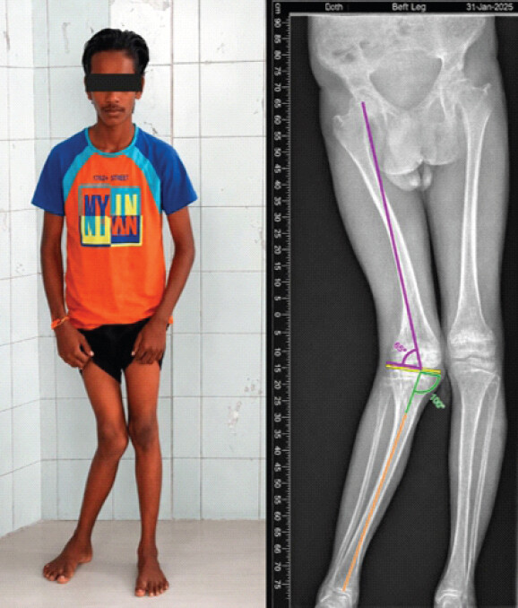
Pre-operative planning was performed to identify the site and magnitude of deformity and determine the correction required for restoration of normal lower-limb alignment. Clinical evaluation included assessment of knee range of motion, ligamentous stability, patellar tracking, and lower-limb rotational profile. The mechanical lateral distal femoral angle, medial proximal tibial angle, and joint line convergence angle were assessed. The reported normal reference values were 87° ± 3°, 87° ± 3°, and 0° ± 2°, respectively.
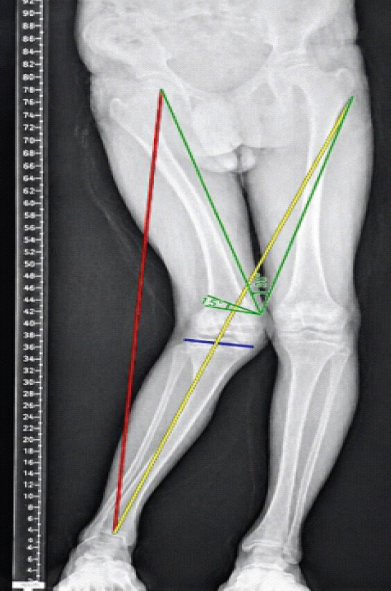
The reversed Miniaci method was used on long-leg standing radiographs to determine the correction angle and osteotomy parameters [5]. The desired post-operative weight-bearing line was planned to pass through the Fujisawa point, approximately 55% across the tibial plateau from the medial border. The correction angle, osteotomy level, hinge position, and required opening-wedge height were determined from this plan (Fig. 2).
Operative technique
The patient was positioned supine on a radiolucent operating table with the knee slightly flexed. A lateral approach to the distal femur was performed through an incision centered over the vastus lateralis. Following subperiosteal exposure of the supracondylar region, a 3.5-mm Steinmann pin was placed as an osteotomy cutting guide, obliquely from the lateral supracondylar area toward the adductor tubercle. A thinner 2.5-mm Steinmann pin was placed in the hinge area as an internal stop to limit saw or chisel penetration and protect the medial cortical hinge.
Following completion of the distal femoral osteotomy, gradual opening of the lateral wedge was performed using a bone spreader under fluoroscopic guidance until the planned correction was achieved. Mechanical alignment was assessed intraoperatively and confirmed radiographically. Stable fixation was obtained using a contoured lateral distal femoral buttress/locking plate with proximal and distal locking screws. The osteotomy gap was filled with autologous cancellous iliac crest bone graft. The hinge-protection Steinmann pin was then removed, and fluoroscopic assessment confirmed preservation of the medial cortical hinge, maintenance of correction, and overall construct stability (Fig. 3).
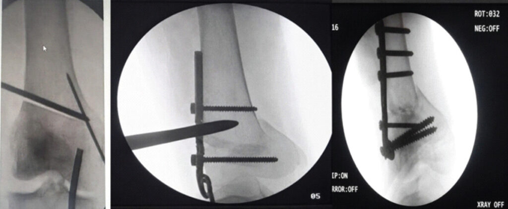
Soft-tissue balancing was assessed, including iliotibial-band tension and medial soft-tissue tension. Selective release of tight lateral soft-tissue structures was performed where required. After irrigation, a closed-suction drain was placed and the wound was closed in layers. Final fluoroscopic images demonstrated satisfactory implant position and restoration of mechanical alignment.
Post-operative protocol
Post-operative rehabilitation consisted of continuous passive motion initiated within 24–48 h and partial weight-bearing of 15–20 kg for the first 6–8 weeks, followed by gradual progression according to radiographic healing. Clinical and radiological follow-up was performed at 6 weeks, 3 months, 6 months, and 12 months. Union was defined as bridging callus across three cortices on plain radiographs. Return to recreational and occupational activity was planned according to healing and functional recovery.
Results
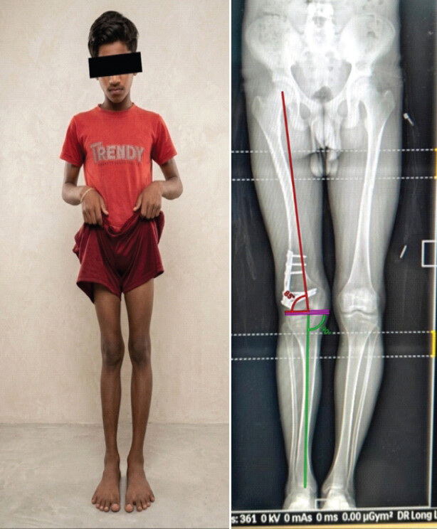
Post-operative radiographs demonstrated restoration of the mechanical axis restored to near-neutral alignment with maintenance of correction during follow-up. The osteotomy site showed satisfactory apposition without evidence of cortical hinge fracture or implant-related complication. Radiographic union was achieved within the expected follow-up period. The patient demonstrated improvement in gait pattern, resolution of symptoms, and improved cosmetic appearance. Functional evaluation at 6 months showed an American Knee Society Score >85, indicating excellent functional recovery. No infection, loss of correction, implant loosening, or neurovascular deficit was observed (Fig. 4, Table 1).
Radiographic parameters were as follows:
| Parameter | Pre-operative | Post-operative |
|---|---|---|
| mLDFA | 65° | 85° |
| MPTA | 100° | 90° |
| Mechanical axis deviation | 32 mm | 6 mm |
| Tibiofemoral angle | 38° | 10° |
mLDFA: Mechanical lateral distal femoral angle, MPTA: Medial proximal tibial angle
Discussion
Distal femoral osteotomy remains an established joint-preserving procedure for correction of symptomatic genu valgum and restoration of lower-limb alignment in young and active patients. Accurate deformity analysis and correction planning are essential for achieving favorable outcomes [3,4,5]. Both medial closing-wedge and lateral opening-wedge techniques can provide satisfactory clinical and radiographic correction. Lateral opening-wedge osteotomy offers advantages, including preservation of limb length and flexibility in achieving the desired correction.
The reversed Miniaci method is a graphical planning technique that allows calculation of the correction angle by transferring the desired post-operative mechanical axis onto a full-length weight-bearing radiograph [12]. The method uses the intended weight-bearing line and simulates correction around the planned osteotomy hinge point, facilitating estimation of the opening-wedge size and correction angle while reducing the risk of undercorrection or overcorrection [10,11].
Preservation of hinge integrity is critical because the intact cortex contributes to rotational stability and maintenance of the planned correction during early healing. Hinge disruption can lead to loss of correction, delayed union, non-union, and compromised fixation stability. The present case emphasizes the value of a deliberate hinge-protection strategy using a Steinmann pin during lateral opening-wedge distal femoral osteotomy.
The case also demonstrates the importance of comprehensive radiographic analysis when deformity involves more than one segment. The pre-operative Mechanical lateral distal femoral angle and medial proximal tibial angle indicated contributions from both the distal femur and proximal tibia. The decision to correct the distal femoral component with an opening-wedge osteotomy was based on the deformity analysis and the planned mechanical-axis correction.
The principal limitation of this report is that it describes a single patient and therefore cannot establish comparative efficacy of the technique. Longer follow-up and additional cases would be required to determine the reproducibility of the observed functional and radiographic outcomes.
Conclusion
Lateral opening-wedge distal femoral osteotomy planned with the reversed Miniaci method provided accurate correction of severe genu valgum in this patient. Deliberate medial cortical hinge protection, controlled wedge opening, stable lateral plate fixation, and appropriate rehabilitation were associated with maintenance of alignment, radiographic union, and satisfactory functional recovery. Further clinical experience is required to establish the reproducibility of this approach in similar deformities.
Clinical Message
In severe valgus deformity, accurate deformity localization and mechanical-axis planning are essential. A reversed Miniaci plan combined with controlled wedge opening, medial hinge protection, stable plate fixation, and appropriate rehabilitation can achieve reliable correction and osteotomy union in a symptomatic adolescent.
Conflict of Interest:
Nil
Source of Support:
Nil
Consent
The authors confirm that informed consent was obtained from the patient for publication of this article
How to Cite this Article
Zade SG, Khan HA, Mehraj P, Mohod S, Elahi S, Meghana B. A Rare Case of Developmental Lateral Femoral Condyle Hypoplasia Managed with Lateral Distal Femoral Opening-Wedge Osteotomy using the Reversed Miniaci Method: Journal of Orthopaedic Case Reports 2026 November;16(11):367-372. doi: 10.13107/jocr.2026.v16.i11.8500.
References
- Rerucha CM, Dickison C, Baird DC. Lower extremity abnormalities in children. Am Fam Physician 2017;96:226-33. [Google Scholar] | [PubMed]
- Buchan S, Bennet S, Barry M. Genu valgum in children. Orthop Trauma 2022;36:311-6. [Google Scholar] | [PubMed]
- Naik AB, Shah DS. Two-level osteotomy for genu valgum with tibia valga: Surgical planning and execution. J Orthop Case Rep 2021;11:33-7. [Google Scholar] | [PubMed]
- Omidi-K ashani F, Hasankhani IG, Mazlumi M, Ebrahimzadeh MH. Varus distal femoral osteotomy in young adults with valgus knee. J Orthop Surg Res 2009;4:15. [Google Scholar] | [PubMed]
- Ferreira A, Jacquet C, Guy S, Khakha R, Kley K, Argenson JN. Distal femoral osteotomy planning: Reversed Miniaci's method is more accurate than Dugdale and Paley methods. Orthop Traumatol Surg Res 2024;110:103697. [Google Scholar] | [PubMed]
- Tschinkel K, Gowland R. Knock-knees: Identifying genu valgum and understanding its relationship to vitamin D deficiency in 18th to 19th century Northern England. Int J Osteoarchaeol 2020;30:891-902. [Google Scholar] | [PubMed]
- Sutherland DH, Olshen R, Cooper L, Woo SL. The development of mature gait. J Bone Joint Surg Am 1980;62:336-53. [Google Scholar] | [PubMed]
- Ferguson DO, Fernandes JA. Children's orthopaedics: Lower limb alignment. Orthop Trauma 2016;30:539-46. [Google Scholar] | [PubMed]
- Patel M, Nelson R. Genu valgum. In: StatPearls. Treasure Island, FL. StatPearls Publishing 2023. [Google Scholar] | [PubMed]
- Sabharwal S, Zhao C. Pediatric lower limb deformities: Principles of evaluation and management. Indian J Orthop 2008;42:387-97. [Google Scholar] | [PubMed]
- Scheid DL, Sager DA. Evaluation and management of genu valgum in children. J Pediatr Orthop 2011;31:738-43. [Google Scholar] | [PubMed]
- Zajonz D, Schumann E, Wojan M, Kübler FB, Josten C, Bühligen U. Treatment of genu valgum in children by means of temporary hemiepiphysiodesis using eight-plates: Short-term findings. BMC Musculoskelet Disord 2017;18:456. [Google Scholar] | [PubMed]
© 2026 Journal of Orthopaedic Case Reports - Published by Indian Orthopaedic Research Group
About the Authors
-

Samir Gajanan Zade
-

Hisham Ali Khan
-

Pagidala Mehraj
-

Shubham Mohod
-

Shaharyar Elahi
-

B Meghana
Related Articles in Journal of Orthopaedic Case Reports
 June 1, 2026 Sequential Development and Staged Management of Distal Femoral Osteochondroma with Progressive Bilateral Genu Valgum: A Case Report
 August 1, 2025 Combined Sulcus Deepening Trochleoplasty, Varus Distal Femoral Osteotomy, and Medial Patellofemoral Ligament Reconstruction for Recurrent Patellar Instability with Trochlear Dysplasia, Genu Valgum, and Medial Patellofemoral Ligament Tear
 April 1, 2025 Genu Valgum and Lamellar Ichthyosis: Insights into a Rare Presentation
 February 1, 2025 Combined Arthroscopic Anterior Cruciate Ligament Reconstruction and Distal Femoral Osteotomy in a Patient of Bilateral Genu Valgum – A Rare Case Report