


Introduction
Femoral fractures in the pediatric age group account for about 1.6% of all bony injuries and are more common in males than females with an incidence of 2.6:1, respectively [1]. Pediatric tibial shaft fractures are the 2nd most common injuries after femoral fractures with their peak incidence between 10 and 14 years of age with a ratio of 2:1 for males to females, respectively [2]. For the past 2 decades, there has been an increased trend towards operative treatment in children using external fixation, locked intramedullary nailing, flexible intramedullary nails, and submuscular plate fixation for lower-limb fractures [3]. Treatment options for femoral shaft fractures for infants and up to 2 years of age are Pavlik harness, hip spica cast, and traction followed by spica cast. For children up to 5 years of age, treatment modalities are early spica cast, traction followed by spica cast, very rarely external fixation, and flexible intramedullary nails [4]. Most of the tibial diaphysis fractures were managed conservatively by applying a long leg cast. Even for completely displaced diaphyseal tibia fractures, conservative management is enough to achieve the union. Considering the physical and psychosocial burden on the child, various treatment modalities are now being utilized. These recent advances have reduced the family burden of care during the recovery period as well as early disabilities. Among these treatment modalities, flexible intramedullary nails have advantages over others because the chances of pin tract infection, refracture, physeal injuries, and excessive periosteal stripping are less common.
Aim
The objective of this study was to evaluate the functional outcome of titanium elastic nailing (TENS) system in pediatric lower-limb fractures.
Materials and Methods
This is a hospital-based prospective analytical study conducted in the tertiary care center after ethical committee approval (No: outward/SMVMCH-ECO/AL/348/2019). Fifteen children of age group between 5 and 16 years with closed lower-limb fractures presenting to the hospital from November 2019 to April 2021 were recruited for elastic nailing. Patients were aged up to 16 years with displaced diaphyseal fractures of the femur and tibia and type 1 and type 2 compound diaphyseal fractures of the femur and tibia. Failed conservative treatments were included in the present study. Patients weighing more than 50 kg, fractures linked to neurovascular injuries, and type 3 compound diaphyseal fractures of the tibia and femur were eliminated. The Flynn et al. formula (diameter of nail = width of the narrowest point of medullary canal on anteroposterior and lateral views ×0.4 mm) was used to estimate nail diameter before surgery [5]. Of the fifteen youngsters, seven had tibial diaphyseal fractures and eight had femur shaft fractures. Seven patients with tibial diaphyseal fractures had antegrade nailing, while all eight patients with femur fractures received retrograde nailing. High groin slabs were used for external immobilization if reduction proved insufficient. After that, the patient was confined in a cast and not allowed to bear any weight for a month.
Exercises for knee range of motion and quadriceps were initiated early for tibial fractures. An X-ray was obtained after a month, and partial weight bearing was initiated once the callus was confirmed on the radiograph. Patients were evaluated clinically and radiologically at intervals of 2 weeks, 6 weeks, 3 months, and 6 months. They were also monitored for complications, range of motion, and union. The Flynn et al. grading method was used to analyze the results.
All patients were made to weight bear after 2 months of surgery with four cortices united on X-ray. Nails were removed after bone remodeling occurred between 9 months and 12 months. The independent and dependent variables were compared using Chi-square test for categorical variables and either Mann–Whitney U test or Kruskal–Wallis test for numerical variables. The correlation between two numerical variables was compared using Pearson’s correlation coefficient. For all the statistical tests, a P < 0.05 was considered significant.
Results
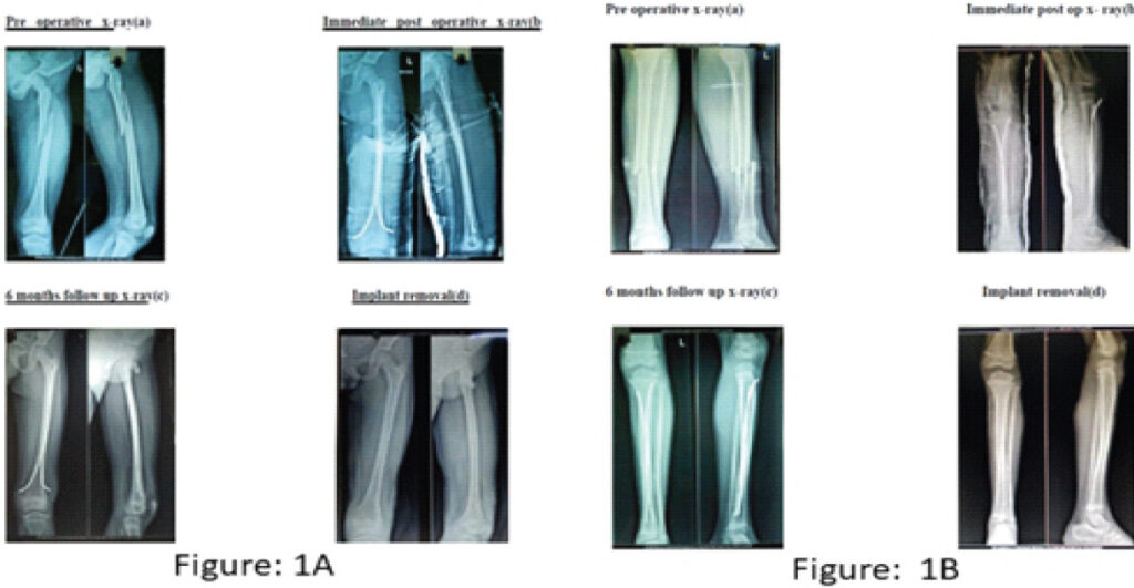
The results of the study are presented in the form of Tables 1, 2, 3, 4, 5 and Figs. 1, 2, 3, 4, and master chart. Among the fifteen children, 10 were males and 5 were females; the mean age of study participants was 8.5 years. The distribution of gender, side of fracture, type of fracture (open/closed), mode of injury, and pattern of fracture were not significant as tested by the test of proportions. Majority of the patients (53%) were operated on within the 2nd day of injury with an average duration of 3 days.
Flynn et al. scoring system
| Parameter | Excellent | Satisfactory | Poor |
|---|---|---|---|
| Limb length inequality | <1 cm | <2 cm | >2 cm |
| Malalignment | Up to 5° | 5–10° | >10° |
| Pain | None | None | Present |
| Complications | None | Minor | Major complications and or lasting morbidity |
Complications among the patients post-procedure (n=15)
| Complications | Number | Percentage |
|---|---|---|
| Nil | 12 | 80 |
| Knee stiffness and angulation | 1 | 6.7 |
| Nail irritation | 1 | 6.7 |
| Malunion | 1 | 6.7 |
Outcome among the patients following the procedure (n=15)
| Outcome post-procedure | Number | Percentage |
|---|---|---|
| Excellent | 12 | 80 |
| Satisfactory | 2 | 13.3 |
| Poor | 1 | 6.7 |
Comparison of the results
| Outcome | Flynn et al. | Ligier et al. | Goodwin et al. | Our study |
|---|---|---|---|---|
| No. of cases | 48 | 128 | 19 | 15 |
| Knee stiffness | – | 13 | – | 1 |
| Limb length discrepancy | – | 7 | – | 1 |
| Angulation | – | – | 2 (>10°) | – |
| Malunion | 2 | 14 | – | – |
Master chart of the study
| S.No. | Age | Sex | Mechanism of injury | Type of fracture | Associated injuries | Trauma to surgery (days) | Reduction | Side | Canal diameter (mm) | Nail size (mm) | LLD | Range of motion (knee/hip) | Angulation (AP view) | Bowing (lateral view) | Radiological union (weeks) | Full weight bearing (weeks) | Complications | Nail removed | Timing of nail removal (months) | Score | Group |
|---|---|---|---|---|---|---|---|---|---|---|---|---|---|---|---|---|---|---|---|---|---|
| 1 | 13 | M | Accidental fall | Short oblique | – | 3 | CR | RIGHT | 11 | 4 | – | FULL | – | – | 12 | 12 | – | YES | 12 | EXCELLENT | Femur |
| 2 | 7 | M | Accidental fall | Long oblique | – | 2 | CR | LEFT | 9 | 3.5 | – | FULL | – | – | 8 | 12 | – | YES | 12 | EXCELLENT | Femur |
| 3 | 11 | M | RTA | Transverse | – | 2 | CR | RIGHT | 10 | 3.5 | – | 0–20° | – | – | 12 | 12 | Knee stiffness, nail irritation | NO | – | SATISFACTORY | Femur |
| 4 | 10 | F | Accidental fall | Short oblique | – | 2 | CR | LEFT | 10 | 3.5 | – | FULL | – | – | 10 | 12 | – | NO | – | EXCELLENT | Femur |
| 5 | 13 | M | Accidental fall | Transverse | 0 | 2 | CR | RIGHT | 10 | 3.5 | – | FULL | – | – | 10 | 12 | – | NO | – | EXCELLENT | Femur |
| 6 | 9 | F | RTA | Transverse | – | 1 | CR | RIGHT | 10 | 3.5 | – | FULL | – | – | 10 | 12 | – | YES | 12 | EXCELLENT | Femur |
| 7 | 12 | F | RTA | Short oblique | – | 2 | CR | RIGHT | 10 | 3.5 | Shortening 1 cm | FULL | – | 20° | 10 | 12 | Angulation | YES | 12 | POOR | Femur |
| 8 | 5 | F | Accidental fall | Short oblique | – | 2 | CR | LEFT | 9 | 2.5 / 3.0 | – | FULL | – | – | 8 | 12 | – | YES | – | EXCELLENT | Femur |
| 9 | 5 | M | RTA | Transverse | Fracture BB leg | 1 | CR | RIGHT | 7 | 2.5 | – | FULL | – | – | 8 | 10 | – | YES | 8 | EXCELLENT | Tibia |
| 10 | 5 | F | RTA | Short oblique | Fracture BB leg | 2 | CR | RIGHT | 7 | 2.5 | – | FULL | – | – | 8 | 10 | – | YES | 18 | EXCELLENT | Tibia |
| 11 | 12 | M | Accidental fall | Short oblique | Fracture BB leg | 5 | CR | LEFT | 12 | 4 | – | FULL | – | – | 12 | 12 | – | YES | 10 | EXCELLENT | Tibia |
| 12 | 8 | M | RTA | Short oblique | Fracture BB leg | 1 | CR | RIGHT | 8 | 2.5 | – | FULL | 6° valgus | – | 10 | 10 | – | YES | 12 | SATISFACTORY | Tibia |
| 13 | 5 | M | RTA | Short oblique | Fracture BB leg | 2 | CR | LEFT | 7.5 | 3 | – | FULL | 3° valgus | – | 10 | 10 | – | YES | 10 | EXCELLENT | Tibia |
| 14 | 8 | M | RTA | Short oblique | Fracture BB leg | 3 | CR | RIGHT | 9 | 3.5 | – | FULL | – | – | 10 | 10 | – | YES | 12 | EXCELLENT | Tibia |
| 15 | 5 | M | Accidental fall | Short oblique | Fracture BB leg | 8 | CR | LEFT | 7 | 2.5 | – | FULL | – | – | 8 | 10 | – | YES | 10 | EXCELLENT | Tibia |
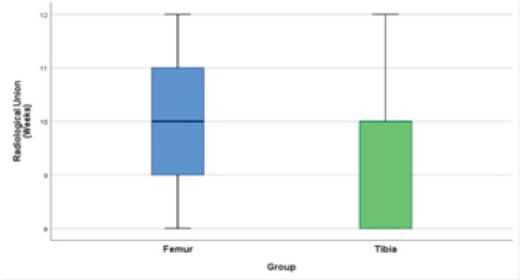
The average time for radiological union was 10.1 (±1.5) weeks for femur fractures and 9.4 (±1.5) weeks for tibial fractures. There was no significant difference (P > 0.05) in the time for radiological union among the fracture groups as calculated by Mann–Whitney U test.
Only one patient out of fifteen showed a limb length discrepancy (shortening of 1 cm). Due to nail irritation, one patient experienced knee stiffness with a range of motion of 0–20°. Nine of the fifteen patients began complete weight-bearing walking after 12 weeks. Two patients had valgus angulation, one patient had greater anterior femur angulation, and about 87% of patients achieved fracture union without angulation. Thirteen percent of the patients had adequate results, and over 80% had exceptional results. Just one patient (7%) experienced a bad outcome.
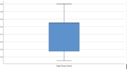
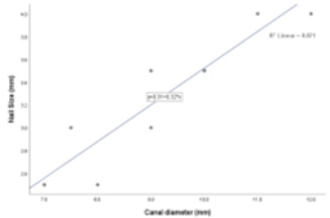
There was no significant association (P > 0.05) between age and the time for radiological union among the patients as tested by Chi-square test. There was no significant association (P > 0.05) between the age and the outcome among the patients. The average canal diameter and nail size were 9.1 (1.5) and 3.2 (0.5) mm. The mean and percentage of nail occupancy were 73.9 (8.5%).
The mean percentage of nail occupancy among patients with excellent, poor, and satisfactory outcomes was 75.4 (8.6), 70.0, and 66.3 (5.3%). There was no statistically significant difference (P > 0.05) in the distribution of nail as calculated by Kruskal–Wallis test [6]. All patients’ nail removal was done between 9 months and 1 year.
Discussion
Pediatric shaft fractures of the femur and tibia are treated by various methods such as traction, traction followed by spica cast, immediate spica cast, long leg cast, internal fixation with submuscular plate osteosynthesis, external fixator application, and flexible intramedullary nailing. For the past 20 years, there has been an increased trend toward operative treatment because of early mobilization and rehabilitation. Many studies and literature support the surgical treatment using titanium elastic nails as the best and safest method in treating pediatric lower-limb fractures.
Caglar et al. examined the average time to union for plating and flexible nail groups in femur shaft fractures in a randomized controlled trial [7]. Plate groups took 7.7 months to unite, while flexible nail groups took 4 months. The average union time for a femur fracture is 10 weeks, while the average union time for a tibia fracture is 8 weeks, according to the results of our study (P = 0.478). This demonstrated a flexible nailing technique for fracture treatment that minimized soft tissue dissection, prevented periosteal stripping, and conserved the fracture hematoma.
Two incidences of knee stiffness caused by nail irritation at the entrance site were described by Flynn et al. in their prospective study of 234 patients treated with titanium elastic nails for femur shaft fractures [5]. The most frequent side effect of titanium nails for femur fractures is nail irritation at the entry site, which accounts for 7–40% of cases [7]. Similarly, out of eight femur shaft fractures treated with titanium elastic nails, we found one incidence of knee stiffness as a result of nail irritation of the soft tissue. Occurrences of discomfort at the nail insertion site were reported in a retrospective study by Narayanan et al. on 79 patients with femur shaft fractures, with an overall acceptable result [8]. Six of the eight femur fractures in the present study had great outcomes, one was satisfactory, and one was unsatisfactory. Knee stiffness developed as a result of nail irritation of soft tissue, which might have been avoided with proper titanium nail clipping. An earlier study by Khazzam et al. documented 14 problems, including two delayed unions, three refractures, three occurrences of malalignment, five irritated nail tips, and one nail migration [9]. In a similar way, three patients in our study experienced angulation, nail tip discomfort, and malunion.
Flynn et al. reported no limb length discrepancy of not more than 1 cm in 49 patients with femur shaft fracture treated by titanium nails, but in our study, out of 8 patients with femur fracture, one patient had excessive femur anterior bowing and shortening of 1 cm [5]. Ideally, titanium nails should have three-point fixation in fracture reduction; this principle of fracture reduction failed in our patient which led to shortening and anterior bowing.
In 60% of instances, solitary tibial shaft fractures with an intact fibula result in varus angulation. Two of the 19 patients with tibial shaft fractures who had a mean follow-up of 13 months in the retrospective analysis by Goodwin et al. had angulation more than 10° [10]. The current study findings demonstrated that none of the seven tibial shaft fractures had more than 5° of valgus angulation since each of the seven tibial shaft fractures had corresponding fibula fractures.
In the earlier study by Sankar et al. reported of 19 patients with tibial shaft fractures, 11 found no limb length discrepancy and permitted full weight bearing after an average of 8.65 weeks; in our study, weight bearing was permitted after 3 months for femur shaft fractures and 10 weeks for tibial shaft fractures [11]. All of the patients in the present study were fixed with higher nail diameters that occupied more than 70% (interquartile range 70–77.8%) of the medullary canal, as described by Green et al. To preserve axial, rotational, flexural, and torsional stability, we thereby adhered to the symmetrical bracing principle [12].
Conclusion
A titanium elastic nail is the method of choice in pediatric lower-limb fractures due to its advantages over other methods. TEN system avoids excessive periosteal stripping with minimal soft tissue damage and preserves the fracture hematoma. Titanium nails provide micromotion at the fracture site, abundant callus formation, stability, and early mobilization. TENS is an easier and promising procedure with minimal complications, earlier post-operative rehabilitation, and better outcomes than any other procedure.
Clinical Message
Titanium elastic nails provide faster union with better psychosocial outcomes, which is a minimally invasive and functionally superior method for managing pediatric lower-limb fractures.
Conflict of Interest:
Nil
Source of Support:
Nil
Consent
The authors confirm that informed consent was obtained from the patient for publication of this article
How to Cite this Article
Velayudham R, Manjini M, Kumarasamy M, Mohanen P. Functional Outcome of Titanium Elastic Nailing in Pediatric Lower-Limb Fractures: A Prospective Study. J Orthop Case Rep 2026 November;16(11): 373-378. doi: 10.13107/jocr.2026.v16.i11.8502.
References
- Hedlund R, Lindgren U. The incidence of femoral shaft fractures in children and adolescents. J Pediatr Orthop 1986;6:47-50. [Google Scholar] | [PubMed]
- Galano GJ, Vitale MA, Kessler MW, Hyman JE, Vitale MG. The most frequent traumatic orthopaedic injuries from a national pediatric inpatient population. J Pediatr Orthop 2005;25:39-44. [Google Scholar] | [PubMed]
- Naranje SM, Stewart MG, Kelly DM, Jones TL, Spence DD, Warner WCJr. Changes in the treatment of pediatric femoral fractures: 15-year trends from United States kids' inpatient database (KID) 1997 to 2012. J Pediatr Orthop 2016;36:e81-5. [Google Scholar] | [PubMed]
- Kocher MS, Sink EL, Blasier RD, Luhmann SJ, Mehlman CT, Scher DM. Treatment of pediatric diaphyseal femur fractures. J Am Acad Orthop Surg 2009;17:718-25. [Google Scholar] | [PubMed]
- Flynn JM, Hresko T, Reynolds RA, Blasier RD, Davidson R, Kasser J. Titanium elastic nails for pediatric femur fractures: A multicenter study of early results with analysis of complications. J Pediatric Orthop 2001;21:4-8. [Google Scholar] | [PubMed]
- Kruskal WH, Wallis WA. Use of ranks in one-criterion variance analysis. J Am Stat Assoc 1952;47:583-621. [Google Scholar] | [PubMed]
- Caglar O, Aksoy MC, Yazc M, Surat A. Comparison of compression plate and flexible intramedullary nail fixation in pediatric femoral shaft fractures. J Pediatr Orthop B 2006;15:210-4. [Google Scholar] | [PubMed]
- Narayanan UG, Hyman JE, Wainwright AM, Rang M, Alman BA. Complications of elastic stable intramedullary nail fixation of pediatric femoral fractures, and how to avoid them. J Pediatr Orthop 2004;24:363-9. [Google Scholar] | [PubMed]
- Khazzam M, Tassone C, Liu XC, Lyon R, Freeto B, Schwab J. Use of flexible intramedullary nail fixation in treating femur fractures in children. Am J Orthop (Belle Mead NJ) 2009;38:E49-55. [Google Scholar] | [PubMed]
- Goodwin RC, Gaynor T, Mahar A, Oka R, Lalonde FD. Intramedullary flexible nail fixation of unstable pediatric tibial diaphyseal fractures. J Pediatr Orthop 2005;25:570-6. [Google Scholar] | [PubMed]
- Sankar WN, Jones KJ, David Horn B, Wells L. Titanium elastic nails for pediatric tibial shaft fractures. J Child Orthop 2007;1:281-6. [Google Scholar] | [PubMed]
- Green JK, Werner FW, Dhawan R, Evans PJ, Kelley S, Webster DA. A biomechanical study on flexible intramedullary nails used to treat pediatric femoral fractures. J Orthop Res 2005;23:1315-20. [Google Scholar] | [PubMed]
© 2026 Journal of Orthopaedic Case Reports - Published by Indian Orthopaedic Research Group
About the Authors
-

Raja Velayudham
-

Madhavan Manjini
-

Manikandan Kumarasamy
-

Pragash Mohanen
Related Articles in Journal of Orthopaedic Case Reports
 November 1, 2026 Long-Stem Bypass Fixation for Concomitant Tibial Stress Fractures and Advanced Knee Arthritis: A Prospective Clinical Study
 August 1, 2026 A Prospective Analysis of Physeal-Sparing Intramedullary Interlocking Nail for Adolescent Tibial Shaft Fractures
 August 1, 2026 Dual Fixation in Complex Long Bone Fractures: A Case Series of 30 Patients
 June 1, 2026 Comparison of Surgical Outcomes and Perioperative Laboratory Parameters Following Minimally Invasive Plate Osteosynthesis versus Conventional Plating in Long Bone Fractures