


Introduction
Acute compartment syndrome is a surgical emergency defined by elevated intracompartmental pressure compromising tissue perfusion, risking irreversible neuromuscular injury, limb loss, and death if fasciotomy is delayed [1]. Although trauma accounts for most cases, up to 30% occur without fracture or major injury and can arise from vascular injury, coagulopathy, thrombosis, infection, or iatrogenic causes [1]. The classic constellation of pain out of proportion to examination, tense swelling, paresthesia, and neurovascular compromise demands urgent recognition by the orthopedic surgeon, though atypical presentations are well documented [1,2].
Anticoagulation-associated spontaneous compartment syndrome, while uncommon, is increasingly recognized with both low molecular weight heparin (LMWH) and direct oral anticoagulants (DOACs) [3,4]. Hemorrhagic risk with these agents is not uniform and is shaped by a complex interaction of patient-specific factors including renal function, body composition, prior bleeding history, concomitant medications, and the degree of anticoagulation achieved [5]. Individual risk modifiers do not operate independently; their combined effect on hemostasis can be substantially greater than any single factor would predict, creating a clinical scenario in which seemingly manageable individual risks converge to become limb-threatening.
Current evidence primarily characterizes bleeding risk associated with single anticoagulant agents or specific drug combinations, and guidance for orthopedic surgeons managing patients with multiple simultaneous hemostatic modifiers remains limited. This case illustrates how the simultaneous presence of multiple hemostatic vulnerabilities, each individually recognized but not collectively assessed, produced a compartment syndrome requiring emergent fasciotomy and reoperation, and argues for systematic hemostatic risk assessment before and during anticoagulation in surgical patients.
Case Report
A 50-year-old male (weight 90.7 kg, body mass index 28.5 kg/m2) with coronary artery disease, hypertension, and a history of deep venous thrombosis (DVT) more than 10 years prior presented to the emergency department with acute left calf pain and swelling of approximately 24 h’ duration. He reported no recent trauma, prolonged immobility, or current tobacco use. His household had recently been ill with COVID-19, and he had returned from a Caribbean cruise within the preceding weeks. His prior DVT had occurred more than a decade earlier; available records did not confirm what anticoagulant, if any, he had received at that time, and he denied any prior DOAC use.
Medications on presentation included aspirin 81 mg orally once daily, carvedilol 25 mg orally twice daily, valsartan 320 mg orally once daily, buprenorphine-naloxone 8 mg/2 mg sublingually once daily, and eletriptan 40 mg orally as needed for migraine. He also reported chronic use of fish oil and high-dose cayenne pepper supplementation; doses were unspecified. He was receiving testosterone cypionate 100 mg (0.5 mL of a 200 mg/mL formulation) intramuscularly once weekly, obtained from a non-prescription source. He reported a history of prolonged bleeding after dental extraction but no prior surgical bleeding complications and no family history of bleeding disorders.
Collectively, these features established at least five hemostatic risk factors prior to any anticoagulation: antiplatelet therapy, platelet-modifying supplementation, exogenous testosterone with potential pharmacokinetic effects on DOACs, testosterone-associated erythrocytosis, and a personal bleeding history suggestive of a possible underlying diathesis.
Venous duplex ultrasonography revealed a non-occlusive thrombus in the mid-to-distal left popliteal vein. Laboratory studies obtained at this visit, including renal function, were not reviewed prior to discharge; creatinine was subsequently found to be mildly elevated at the time of the second presentation. The patient received a single dose of enoxaparin 90 mg subcutaneously (approximately 1 mg/kg) at 00:08 and was discharged with a prescription for rivaroxaban 15 mg orally twice daily, consistent with the standard DVT treatment induction regimen [6, 7]. Aspirin was not discontinued at discharge, and the patient was not counseled to discontinue fish oil, cayenne pepper, or testosterone prior to initiating anticoagulation.
No enoxaparin dose adjustment was made. Food and Drug Administration (FDA) labeling recommends dose reduction only for creatinine clearance below 30 mL/min [8]; however, pharmacokinetic data indicate that anti-factor Xa exposure is marginally elevated at creatinine clearance 50–80 mL/min, a range consistent with this patient’s subsequent calculated clearance of approximately 76 mL/min by Cockcroft–Gault equation. This mild impairment represented a sixth, incremental contributor to the overall hemostatic burden.
The patient took his first rivaroxaban 15 mg dose approximately 12.5 h after enoxaparin administration and presented to a different emergency department approximately 1 h later with worsening left calf pain radiating to the medial thigh. Repeat venous duplex ultrasonography demonstrated the known non-occlusive left popliteal DVT alongside a new 4.3 × 7.6 cm intramuscular hematoma. Non-contrast computed tomography (CT) of the lower extremity demonstrated rapid expansion to 9.6 × 4.1 × 17.4 cm, compressing the medial gastrocnemius muscle belly (Fig. 1 and 2). Physical examination at this time revealed tense swelling, pain out of proportion to examination, and neurovascular compromise consistent with acute compartment syndrome.
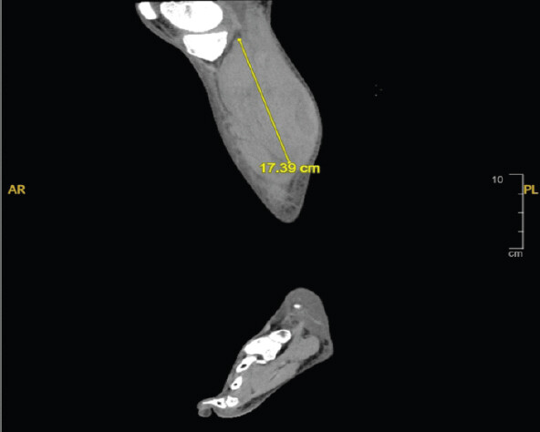
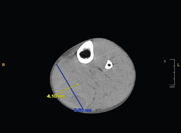
Laboratory studies showed hemoglobin 172 g/L, hematocrit 51.3%, platelet count 262 × 109/L, prothrombin time 15.5 s, international normalized ratio 1.3, and activated partial thromboplastin time 33.5 s. Creatinine was 133 µmol/L with an estimated glomerular filtration rate of 56 mL/min/1.73 m2. A heparin-calibrated anti-Xa assay drawn prior to fasciotomy returned >1.10 U/mL (therapeutic reference range 0.3–0.7 U/mL), reflecting combined activity of residual enoxaparin, rivaroxaban, and a heparin infusion started for ongoing DVT treatment; individual drug contributions could not be separated by this assay [9, 10]. During admission, testing for myeloproliferative disease and antiphospholipid antibody syndrome was negative, as was testing for von Willebrand disease and hemophilia A; platelet function studies, factor XI and XIII activity, and fibrinolytic assays were not obtained.
Surgical management
The patient underwent emergent four-compartment fasciotomy of the left lower leg. Intraoperative findings included approximately 200–300 mL of evacuated hematoma and an area of necrotic gastrocnemius and soleus muscle measuring approximately 2 × 3 × 3 cm. A wound vacuum-assisted closure (VAC) device was placed. Within 30 min postoperatively, significant hemorrhage through the wound VAC prompted emergent return to the operating room; re-exploration revealed no discrete vascular bleeding source, but diffuse soft-tissue oozing throughout the wound, consistent with pharmacologic coagulopathy, an intrinsic hemostatic defect, or both. Hemostatic agents were applied, and bleeding was controlled.
Andexanet alfa was considered for rivaroxaban reversal prior to reoperation. A rivaroxaban-calibrated anti-Xa assay returned <20 ng/mL, below the threshold at which reversal is generally recommended [6, 7]; andexanet alfa was therefore not administered. Rivaroxaban and aspirin were held throughout the remaining hospitalization. Given the simultaneous hemorrhagic and thrombotic complications (below), therapeutic anticoagulation was held altogether; a retrievable inferior vena cava (IVC) filter was placed, and prophylactic-dose unfractionated heparin was used instead.
Outcome and follow-up
Hemoglobin declined from 172 g/L at presentation to a nadir of 88 g/L on post-operative day 1; the patient was not transfused, and no further clinically significant oozing was documented after post-operative day 2. Repeat venous duplex imaging 4 days postoperatively revealed the known non-occlusive popliteal thrombus unchanged but with new acute thrombus extending into the femoral veins; CT pulmonary angiography was negative for pulmonary embolism. The patient required multiple wound washouts, tissue expander placement, and delayed primary closure with a skin substitute over a hospitalization of approximately 14 days. At 6-month follow-up, he was ambulating without neurologic deficit and had no wound complications; the IVC filter was subsequently retrieved. Outpatient hematology-oncology follow-up had been recommended at discharge to guide long-term anticoagulation management, but this follow-up did not take place, and the reason for this gap is not documented.
Discussion
For the orthopedic surgeon, this case underscores that acute compartment syndrome from spontaneous intramuscular hemorrhage rarely reflects a single causative factor. In this patient, at least six distinct patient-intrinsic hemostatic risk factors were simultaneously present at the time hemorrhage occurred: (1) concomitant antiplatelet therapy, (2) platelet-modifying supplementation, (3) a potential testosterone-mediated pharmacokinetic interaction with rivaroxaban, (4) testosterone-associated erythrocytosis, (5) mildly reduced renal clearance, and (6) a personal bleeding history consistent with an incompletely evaluated occult diathesis. Pharmacokinetic overlap between enoxaparin and rivaroxaban is considered separately below as an additional, contested variable, since the central argument of this report is that hemorrhage could plausibly have occurred even without any such overlap.
Bleeding within 1 h of a first rivaroxaban dose is plausible without anticoagulant overlap, particularly given the patient’s other risk factors. Rivaroxaban achieves near-maximal factor Xa inhibition within 2–4 h of ingestion [8], and the intraoperative finding of diffuse oozing without a discrete vessel raises the possibility that rivaroxaban alone, added to his other risk factors, was sufficient to precipitate hemorrhage independent of any residual enoxaparin activity. This distinction carries direct surgical implications: it suggests the anticoagulant transition itself was not necessarily the primary culprit and that a similar bleed could recur with rivaroxaban monotherapy in a patient with this risk profile.
Pharmacokinetic overlap between enoxaparin and rivaroxaban nonetheless remains a plausible contributing factor. Residual anti-factor Xa activity from enoxaparin would be expected at the 12.5-h interval observed here, given a half-life of 4.5–7 h and persistence of anti-Xa levels above 0.2 IU/mL for up to approximately 31.5 h after a treatment dose [3, 8]. FDA labeling for transitioning from a non-warfarin parenteral anticoagulant was designed for scheduled every-12-h LMWH dosing rather than a single dose, leaving its applicability here uncertain [8]. A rivaroxaban-calibrated anti-Xa assay was not available at symptom onset, precluding definitive characterization of the overlap contribution.
Concomitant aspirin increases bleeding risk with rivaroxaban independent of anticoagulation intensity; in the COMPASS trial, rivaroxaban plus aspirin was associated with significantly higher major bleeding than aspirin alone (hazard ratio 1.70; 95% confidence intervals 1.40–2.05) [11]. Fish oil-derived omega-3 fatty acids impair platelet aggregation via reduced thromboxane A2 synthesis, additive with aspirin [12]; high-dose capsaicin, the active component of cayenne pepper, has demonstrated similar inhibition of platelet aggregation and thromboxane formation [13]. Testosterone and its precursor androstenedione are endogenous substrates of P-glycoprotein, which also transports rivaroxaban [14]; a recent study of gender-affirming testosterone therapy found no significant effect on this pathway at physiologic replacement doses, though this patient’s supraphysiologic, non-prescription dosing was not represented in that data [15]. Separately, testosterone-associated erythrocytosis increases whole blood viscosity, promoting thrombosis through rheologic mechanisms while potentially impairing platelet margination at sites of microvascular injury.
The patient’s bleeding history and the intraoperative pattern of diffuse, non-focal oozing both raise concern for an underlying hemostatic defect that was not fully evaluated. Testing excluded von Willebrand disease and hemophilia A, but platelet function testing, factor XI and XIII activity, and fibrinolytic assays were not obtained; bleeding disorder of unknown cause is increasingly recognized as the most common outcome of hemostatic evaluation in patients with a clinically significant bleeding history and no identifiable molecular defect [16].
Notably, this patient’s known DVT extended into the femoral veins during the same admission in which he was hemorrhaging, illustrating that anticoagulation does not eliminate thrombotic risk even as severe bleeding develops and that surgeons managing these patients must weigh ongoing thrombotic and hemorrhagic risk simultaneously when deciding whether and when to resume anticoagulation postoperatively.
For orthopedic surgeons, the practical implication is that a full hemostatic risk inventory, antiplatelet use, platelet-modifying supplements, hormonal therapies, renal function, and personal bleeding history should inform both the decision to anticoagulate and the threshold for surgical exploration when a post-operative or post-fasciotomy patient continues to bleed without an identifiable vascular source.
Conclusion
This case illustrates that life-threatening hemorrhagic complications of anticoagulation are often the product of multiple converging hemostatic vulnerabilities rather than any single causative factor. The same outcome may plausibly have resulted from rivaroxaban alone, independent of any anticoagulant transition timing. Orthopedic surgeons should recognize this pattern both in surgical decision-making for compartment syndrome and in guiding perioperative anticoagulation planning; rigorous, individualized assessment of a patient’s cumulative hemostatic burden before initiating anticoagulation should be a standard component of orthopedic perioperative care.
Clinical Message
Orthopedic surgeons managing anticoagulated patients, particularly those consenting for or performing fasciotomy for spontaneous compartment syndrome, should systematically assess the full burden of concurrent hemostatic risk factors (antiplatelet therapy, platelet-modifying supplements, hormonal therapies, renal function, and personal bleeding history) rather than attributing hemorrhage to anticoagulant transition timing alone, since this affects both the surgical approach to intraoperative bleeding and post-operative anticoagulation planning.
Conflict of Interest:
Nil
Source of Support:
Nil
Consent:
The authors confirm that informed consent was obtained from the patient for publication of this article
How to Cite this Article
Griffin J, Komlos D. Acute Compartment Syndrome from Spontaneous Intramuscular Hemorrhage in the Setting of Converging Hemostatic Risk Factors: A Case Report. J Orthop Case Rep. 2026 November;16(11): 186-190. doi: 10.13107/jocr.2026.v16.i11.8434.
References
- Mabvuure NT, Malahias M, Hindocha S, Khan W, Juma A.. Acute compartment syndrome of the limbs: Current concepts and management. Open Orthop J 2012;6:535-43. [Google Scholar] | [PubMed]
- Ojike NI, Roberts CS, Giannoudis PV.. Compartment syndrome of the thigh: A systematic review. Injury 2010;41:133-6. [Google Scholar] | [PubMed]
- Kirchhof P, Radaideh G, Kim YH, Lanas F, Haas S, Amarenco P. Global prospective safety analysis of rivaroxaban. J Am Coll Cardiol 2018;72:141-53. [Google Scholar] | [PubMed]
- Rivera-Caravaca JM, Anguita Sanchez M, Sanmartín Fernández M, Rafols C, Barón-Esquivias G, Arribas Ynsaurriaga F. Adverse clinical outcomes and associated predictors in rivaroxaban-treated atrial fibrillation patients with renal impairment. Am J Cardiol 2023;203:122-7. [Google Scholar] | [PubMed]
- Goodman SG, Wojdyla DM, Piccini JP, White HD, Paolini JF, Nessel CC. Factors associated with major bleeding events: Insights from the ROCKET AF trial (rivaroxaban once-daily oral direct factor Xa inhibition compared with vitamin K antagonism for prevention of stroke and embolism trial in atrial fibrillation). J Am Coll Cardiol 2014;63:891-900. [Google Scholar] | [PubMed]
- Siegal DM, Curnutte JT, Connolly SJ, Lu G, Conley PB, Wiens BL. Andexanet alfa for the reversal of factor Xa inhibitor activity. N Engl J Med 2015;373:2413-24. [Google Scholar] | [PubMed]
- Connolly SJ, Crowther M, Eikelboom JW, Gibson CM, Curnutte JT, Lawrence JH. Full study report of andexanet alfa for bleeding associated with factor xa inhibitors. N Engl J Med 2019;380:1326-35. [Google Scholar] | [PubMed]
- Food and Drug Administration. Rivaroxaban (Xarelto) Prescribing Information 2025. Available from: https://dailymed.nlm.nih.gov/dailymed/drugInfo.cfm?setid=9178947d-9d58-4c1a-b158-6030e17556af. [Google Scholar] | [PubMed]
- Tomaselli GF, Mahaffey KW, Cuker A, Dobesh PP, Doherty JU, Eikelboom JW. 2017 ACC expert consensus decision pathway on management of bleeding in patients on oral anticoagulants: A report of the American college of cardiology task force on expert consensus decision pathways. J Am Coll Cardiol 2017;70:3042-67. [Google Scholar] | [PubMed]
- Tomaselli GF, Mahaffey KW, Cuker A, Dobesh PP, Doherty JU, Eikelboom JW. 2020 ACC expert consensus decision pathway on management of bleeding in patients on oral anticoagulants: A report of the American college of cardiology solution set oversight committee. J Am Coll Cardiol 2020;76:594-622. [Google Scholar] | [PubMed]
- Eikelboom JW, Connolly SJ, Bosch J, Dagenais GR, Hart RG, Shestakovska O. Rivaroxaban with or without aspirin in stable cardiovascular disease. N Engl J Med 2017;377:1319-30. [Google Scholar] | [PubMed]
- Mori TA, Beilin LJ, Burke V, Morris J, Ritchie J.. Interactions between dietary fat, fish, and fish oils and their effects on platelet function in men at risk of cardiovascular disease. Arterioscler Thromb Vasc Biol 1997;17:279-86. [Google Scholar] | [PubMed]
- Wang JP, Hsu MF, Teng CM.. Antiplatelet effect of capsaicin. Thromb Res 1984;36:497-507. [Google Scholar] | [PubMed]
- Yano K, Seto S, Kamioka H, Mizoi K, Ogihara T.. Testosterone and androstenedione are endogenous substrates of P-glycoprotein. Biochem Biophys Res Commun 2019;520:166-70. [Google Scholar] | [PubMed]
- Hunter M, Coig R, Risler L, Patton KK, Narla RR, Greene DN. Effect of testosterone therapy on cytochrome P450 3A and P-glycoprotein activities using midazolam and digoxin as probe substrates among transgender adults. Pharmacotherapy 2026;46:e70093. [Google Scholar] | [PubMed]
- Baker RI, O'Donnell JS.. How I treat bleeding disorder of unknown cause. Blood 2021;138:1795-804. [Google Scholar] | [PubMed]
© 2026 Journal of Orthopaedic Case Reports - Published by Indian Orthopaedic Research Group
About the Authors
-

Jade Griffin
-

Daniel Komlos
Related Articles in Journal of Orthopaedic Case Reports
 June 10, 2024 Two Very Rare Case of Simultaneous Bilateral Intertrochanteric Fracture Operated in Single Stage – A Case Report
 December 10, 2021 Management of Lateral Malleolus Giant Cell Tumor using A Reverse Proximal Fibular Graft-A Case Report
 June 1, 2026 Hang Fire – Acute Crush Injury of Fingers – Till Appropriate Intercede – A Case Report
 October 11, 2013 Asymmetrical Fracture Dislocation of Shoulder – A Case Report and Review of Literature