


Introduction
Nodular fasciitis is a benign, self-limited myofibroblastic proliferation initially described by Konwaler et al. in 1955 as subcutaneous pseudosarcomatous fibromatosis [1]. This lesion typically presents as a rapidly growing, solitary soft-tissue mass arising in the subcutaneous tissues or fascia of the upper extremities, trunk, or head and neck region of young adults [2,3]. Despite its benign nature, its clinical presentation and histologic features may be alarming, often mimicking sarcoma with rapid growth, high cellularity, and prominent mitotic activity [4]. Although nodular fasciitis most commonly arises in superficial soft tissues, intra-articular occurrence is exceedingly rare and is rarely reported in the literature. Among documented cases, the knee is the most frequently affected joint, representing approximately 27% of intra-articular nodular fasciitis cases [5,6,7]. The rarity of this presentation, combined with non-specific and occasionally aggressive imaging findings, creates significant diagnostic challenges and often raises concern for malignancy.
The understanding of nodular fasciitis has evolved from being considered a reactive process to recognition as a self-limited neoplasm. In 2011, Erickson-Johnson et al. identified the recurrent ubiquitin-specific protease 6 (USP6) gene rearrangements in nodular fasciitis, with MYH9-USP6 being the most common fusion product [8]. USP6 gene rearrangements are now detected in up to 92% of cases and serve as a valuable diagnostic tool, particularly in morphologically or clinically atypical cases [9,10].
We report a unique case of nodular fasciitis arising in the subpatellar region with erosion into the undersurface of the patella in a 67-year-old man. To our knowledge, this represents the first reported case of nodular fasciitis with intraosseous involvement of the patella. This case highlights the diagnostic complexity of intra-articular nodular fasciitis and the critical role of molecular confirmation.
Case Report
Patient consent was obtained to write this case report.
A 67-year-old male with a medical history significant for gout and stage IV follicular lymphoma presented to a sports medicine clinic with a 6-month history of progressively worsening right anterior knee pain. He had been previously treated at an outside hospital with corticosteroid injections and knee aspiration, which demonstrated no evidence of infection or gout. The patient described sharp, stabbing anterior knee pain that was exacerbated with knee flexion and walking.
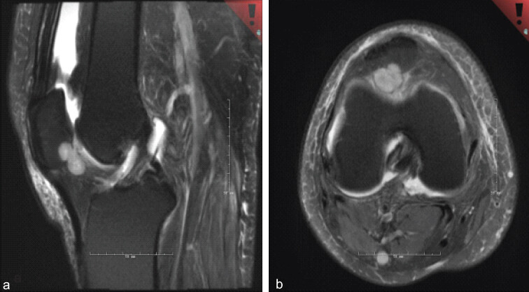
Magnetic resonance imaging with and without contrast was performed, revealing a 2.1 cm × 1.3 cm × 1.9 cm heterogeneous soft tissue mass in the subpatellar region of the right knee. The lesion was hyperintense on T2-weighted images with internal nodularity and peripheral enhancement. Notably, the mass demonstrated cortical erosion into the undersurface of the patella (Fig. 1). Given the patients’ oncologic history and the aggressive imaging features, the differential diagnosis included metastatic disease, primary soft-tissue neoplasms, such as tenosynovial giant cell tumor or synovial chondromatosis, gouty arthritis, and, less likely, degenerative arthropathy.
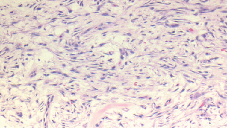
The magnetic resonance imaging (MRI) findings prompted a referral to orthopedic oncology, where, following a multidisciplinary tumor board meeting, the patient underwent an image-guided biopsy by interventional radiology. Histopathologic examination revealed a hypercellular lesion composed of spindled cells with mild atypia and minimal mitotic activity in a myxoid background that contained occasional erythrocytes and giant cells (Fig. 2). While the morphology was suggestive of nodular fasciitis, the intra-articular location and osseous involvement were highly atypical. Fluorescence in situ hybridization (FISH) for USP6 was therefore performed and demonstrated a rearrangement at 17p13, confirming the diagnosis of nodular fasciitis.
Given the patient’s refractory pain to conservative management and associated bony involvement, surgical excision was recommended. An incision was made over the anterior right knee, and a medial parapatellar arthrotomy was performed. Upon entering the joint, nodular synovitis and copious amounts of synovial fluid were found. The mass was identified arising from the subpatellar region and extending into the inferior portion of the patella. The lesion was excised using electrocautery and curettage of the patella. The joint was thoroughly irrigated and closed in layers.
Final pathology was consistent with the pre-operative diagnosis of nodular fasciitis involving both the subpatellar soft tissues and the intraosseous patellar component. At 2-week post-operative follow-up, the patient reported significant improvement in pain without complications.
Discussion
Nodular fasciitis is a benign, self-limited myofibroblastic proliferation that classically affects young adults between the second and fourth decades of life [2]. Presentations in patients over 60 years of age, as in our patient, are exceedingly uncommon. In the extremities, nodular fasciitis is typically found in the forearm (27%), thigh (17%), and upper arm (12%) [3]. Intra-articular occurrence is exceptionally rare, representing a diagnostic challenge for clinicians and pathologists.
A comprehensive review by Wang et al. identified only 21 reported cases of intra-articular nodular fasciitis in the literature [5]. Of these cases, the knee was the most commonly affected joint (67%), followed by the shoulder (14%), hand (10%), hip (5%), and ankle (5%). Of the lesions in the knee, the subpatellar fat pad appears to be a common site for nodular fasciitis to arise. The median reported age of 26 years (range 4–54 years) is substantially younger than in our case, underscoring the unusual demographic and clinical features of our patient [5,6,7,11].
The most striking and novel feature of our case is the presence of intraosseous erosion of the patella. While osseous involvement in nodular fasciitis has been rarely documented, it is typically seen in the cranial fasciitis variant, which affects the calvarium in young children [12]. A recent study by Zhang et al. reported rare cases of nodular fasciitis with bone involvement in locations other than the skull, including one case affecting the hallux [13,14]. However, to our knowledge, our patient represents the first reported case of nodular fasciitis with intraosseous involvement of the patella.
Radiographically, nodular fasciitis often demonstrates non-specific and at times aggressive features resembling those of malignancy. On MRI, lesions typically appear iso- to hypointense on T1-weighted images and hyperintense on T2-weighted images [15,16]. Heterogeneous enhancement is common, and some cases demonstrate aggressive features, including transcompartmental spread and osseous involvement [17]. In our patient, the presence of cortical erosion combined with his history of follicular lymphoma raised concern for malignancy, highlighting the importance of tissue diagnosis.
The histopathologic diagnosis of nodular fasciitis is characterized by a proliferation of plump, immature-appearing myofibroblasts arranged in short, irregular fascicles within a variably myxoid to collagenous stroma [18,19]. Scattered inflammatory cells, extravasated erythrocytes, and occasional multinucleated giant cells are common findings. Immunohistochemically, the cells typically stain positive for smooth muscle actin and vimentin but negative for desmin, distinguishing them from smooth muscle tumors [19].
The discovery of recurrent USP6 gene rearrangements has revolutionized our understanding of nodular fasciitis. In 2011, Erickson-Johnson et al. identified the MYH9-USP6 fusion gene as the primary genetic alteration in nodular fasciitis [8]. USP6 rearrangements are now detected in 86-92% of cases by FISH analysis, with a specificity approaching 100% [9,10]. The gene fusion results in overexpression of USP6, a deubiquitinase that promotes cell proliferation through activation of JAK-STAT and NF-kB signaling pathways [20]. In our patient, FISH confirmed USP6 (17p13) rearrangement, supporting the diagnosis of nodular fasciitis despite the unusual presentation with bone involvement.
Management of nodular fasciitis varies depending on the severity of symptoms and anatomic location. Although spontaneous regression has been reported, symptomatic lesions or those in critical locations often warrant surgical excision. Marginal resection is usually curative, and recurrence is exceedingly rare even with incomplete resection [2]. In our patient, the decision to proceed with surgical resection was based on the severity of symptoms refractory to conservative management and the presence of bony erosion into the patella.
This case highlights several important clinical principles. First, nodular fasciitis should be considered in the differential diagnosis of intra-articular knee masses, even in older patients and when aggressive features, such as bone erosion are present. Second, USP6 FISH analysis is an invaluable diagnostic tool when the morphologic diagnosis is uncertain or when the clinical presentation is atypical. Third, surgical treatment remains an appropriate option for symptomatic lesions despite the self-limited nature of nodular fasciitis, particularly when conservative management has failed. Ultimately, a close clinical correlation between imaging, histopathology, and molecular findings is crucial for accurate diagnosis and effective management.
Conclusion
We report the first documented case of nodular fasciitis with intraosseous involvement of the patella in a 67-year-old man. This unusual presentation highlights the importance of maintaining a broad differential diagnosis for intra-articular knee masses with bony erosion. Molecular confirmation through USP6 FISH analysis proved essential in establishing the correct diagnosis in this clinically challenging case. Surgical resection provided both diagnostic certainty and symptom relief for this patient with symptoms refractory to conservative management. This case expands our understanding of the anatomic diversity of nodular fasciitis and emphasizes the value of molecular diagnostics.
Clinical Message
Nodular fasciitis is a benign myofibroblastic proliferation that can closely mimic malignancy due to its rapid growth, high cellularity, and occasionally aggressive radiographic appearance. Although typically confined to superficial soft tissues, it can rarely present as an intra-articular knee mass with associated osseous erosion, creating significant diagnostic uncertainty, particularly in patients with a history of malignancy. This case emphasizes the importance of maintaining a broad differential diagnosis for intra-articular lesions with bone involvement and highlights the critical role of molecular confirmation using USP6 gene rearrangement analysis to establish an accurate diagnosis and avoid overtreatment. Recognition of this rare presentation allows for appropriate surgical management and symptom resolution while preventing misclassification as a malignant process.
Conflict of Interest:
Nil
Source of Support:
Nil
Consent:
The authors confirm that informed consent was obtained from the patient for publication of this article
How to Cite this Article
Riley DJ, Gonzalez FO, Blank AT. First Reported Case of Nodular Fasciitis with Intraosseous Patellar Involvement: A Case Report. J Orthop Case Rep. 2026 November;16(11):166-170. doi: 10.13107/jocr.2026.v16.i11.8426.
References
- Konwaler BE, Keasbey L, Kaplan L.. Subcutaneous pseudosarcomatous fibromatosis (fasciitis). Am J Clin Pathol 1955;25:241-52. [Google Scholar] | [PubMed]
- Bernstein KE, Lattes R.. Nodular (pseudosarcomatous) fasciitis, a nonrecurrent lesion: Clinicopathologic study of 134 cases. Cancer 1982;49:1668-78. [Google Scholar] | [PubMed]
- Shimizu S, Hashimoto H, Enjoji M.. Nodular fasciitis: An analysis of 250 patients. Pathology 1984;16:161-6. [Google Scholar] | [PubMed]
- Montgomery EA, Meis JM.. Nodular fasciitis. Its morphologic spectrum and immunohistochemical profile. Am J Surg Pathol 1991;15:942-8. [Google Scholar] | [PubMed]
- Wang W, Huang Y, Wang C, Hong J, Ma C, Lin N. Intra-articular nodular fasciitis: A rare lesion case report and an updated review of the literature. BMC Musculoskelet Disord 2019;20:5. [Google Scholar] | [PubMed]
- Buac NP, Lee L, Vijayakumar G, Gusho C, Blank AT.. Intra-articular nodular fasciitis of the knee, a rare clinical presentation and histological diagnosis: A case report. Orthop Rev (Pavia) 2024;16:94572. [Google Scholar] | [PubMed]
- Hagino T, Ochiai S, Sato E, Watanabe Y, Senga S, Kondo T. Intraarticular nodular fasciitis causing limitation of knee extension: A case report. Knee 2010;17:424-7. [Google Scholar] | [PubMed]
- Erickson-Johnson MR, Chou MM, Evers BR, Roth CW, Seys AR, Jin L. Nodular fasciitis: A novel model of transient neoplasia induced by MYH9-USP6 gene fusion. Lab Invest 2011;91:1427-33. [Google Scholar] | [PubMed]
- Wang JC, Li WS, Kao YC, Lee JC, Lee PH, Huang SC. Clinicopathological and molecular characterisation of USP6-rearranged soft tissue neoplasms: The evidence of genetic relatedness indicates an expanding family with variable bone-forming capacity. Histopathology 2021;78:676-89. [Google Scholar] | [PubMed]
- Amary MF, Ye H, Berisha F, Tirabosco R, Presneau N, Flanagan AM.. Detection of USP6 gene rearrangement in nodular fasciitis: An important diagnostic tool. Virchows Arch 2013;463:97-8. [Google Scholar] | [PubMed]
- Chan MF, Ong KO, Foo SS, Selvarajan S.. Intra-articular nodular fasciitis: An unexpected diagnosis for a joint lesion: A case report. Malays Orthop J 2014;8:63-5. [Google Scholar] | [PubMed]
- Lauer DH, Enzinger FM.. Cranial fasciitis of childhood. Cancer 1980;45:401-6. [Google Scholar] | [PubMed]
- Gan S, Xie D, Dai H, Zhang Z, Di X, Li R. Proliferative myositis and nodular fasciitis: A retrospective study with clinicopathologic and radiologic correlation. Int J Clin Exp Pathol 2019;12:4319-28. [Google Scholar] | [PubMed]
- Spagnolli G, Pasquali MP, Rosati M, Lisanti M.. Nodular fasciitis of the hallux: Difficulties in diagnosis. Acta Orthop Belg 1997;63:46-9. [Google Scholar] | [PubMed]
- Khuu A, Yablon CM, Jacobson JA, Inyang A, Lucas DR, Biermann JS.. Nodular fasciitis: Characteristic imaging features on sonography and magnetic resonance imaging. J Ultrasound Med 2014;33:565-73. [Google Scholar] | [PubMed]
- Dinauer PA, Brixey CJ, Moncur JT, Fanburg-Smith JC, Murphey MD.. Pathologic and MR imaging features of benign fibrous soft-tissue tumors in adults. Radiographics 2007;27:173-87. [Google Scholar] | [PubMed]
- Coyle J, White LM, Dickson B, Ferguson P, Wunder J, Naraghi A.. MRI characteristics of nodular fasciitis of the musculoskeletal system. Skeletal Radiol 2013;42:975-82. [Google Scholar] | [PubMed]
- Allen PW.. Nodular fasciitis. Pathology 1972;4:9-26. [Google Scholar] | [PubMed]
- Kayaselçuk F, Demirhan B, Kayaselçuk U, Ozerdem OR, Tuncer I.. Vimentin, smooth muscle actin, desmin, S-100 protein, p53, and estrogen receptor expression in elastofibroma and nodular fasciitis. Ann Diagn Pathol 2002;6:94-9. [Google Scholar] | [PubMed]
- Quick L, Young R, Henrich IC, Wang X, Asmann YW, Oliveira AM Jak1-STAT3 signals are essential 2016;76:5337-47. [Google Scholar] | [PubMed]
© 2026 Journal of Orthopaedic Case Reports - Published by Indian Orthopaedic Research Group
About the Authors
-

Dylan J Riley
-

Fernando O. Gonzalez
-

Alan T Blank
Related Articles in Journal of Orthopaedic Case Reports
 June 1, 2026 Arthroscopic-assisted Suture-based Fixation of Patellar Osteochondral Fractures using the DG-lock Suturing: A Novel Technique
 March 1, 2026 Single-Stage Treatment of a Patellar Osteochondral Defect in an Adolescent Using Autologous Minced Cartilage – Platelet-Rich Plasma Scaffold Technique: A Case Report and Technical Considerations
 January 1, 2026 Lipomas Eroding Adjacent Bone: Report of Three Cases
 January 1, 2026 Aneurysmal Bone Cyst-cloaked Codman’s Tumor in the Patella: Case Report of an Easily Misdiagnosed Entity