


Introduction
Total knee arthroplasty (TKA) has evolved considerably over the past few decades with respect to implant design and materials. Despite these advancements, fracture of the tibial baseplate remains a rare yet considerable challenge. While this has been reported in the literature in the past, these reports have primarily focused on cemented constructs in older implant designs that fractured many years after implantation due to fatigue failure resulting from osteolysis underneath the baseplate [1, 2, 3, 4, 5, 6, 7, 8, 9, 10, 11, 12]. In recent years, improvements in design and manufacturing capabilities have increased interest in cementless TKA with the goal of improving long-term fixation relative to cement [13]. While reports of cementless tibial baseplate fracture have been reported in certain designs, there are few reports to our knowledge of a tibial baseplate fracture in the most commonly used, modern cementless three-dimensional (3D)-printed porous titanium-coated design at short-term follow-up (Table 1) [1, 3, 4].
Reported tibial baseplate fracture cases following total knee arthroplasty
| Study (Tibial baseplate fracture) | Year published | Number of cases | Implant | Fixation type (cementless vs. cemented) | Time to fracture |
|---|---|---|---|---|---|
| Retrieval study of tibial baseplate fracture after total knee arthroplasty; Chatterji et al. [6] | 2005 | 25 | 10 kinematic, 6 Miller- Galante, 5 porous-coated anatomic, and 4 ortholoc II | Cemented (12 patients); Cementless (13 patients) | Mean, 10.8 months (range, 1–36 months) |
| Fracture of the metal tibial tray after kinematic total knee replacement; Abernethy et al. [20] | 1995 | 16 | Howmedica Kinematic prosthesis | Cemented | Mean 56.5 months (Range 23 months–105 months) |
| Implant fracture of the regenerex® modular metal tibial component: A report of three cases; Rytter et al. [4] | 2019 | 3 | Zimmer biomet; regenerex® modular TKR. | Cementless | 10, 12 and 23 months |
| Fracture of the tibial component in total knee arthroplasty: Report on two cases; Da Palma et al. [20] | 2015 | 2 | Howmedica PCA prosthesis | Cementless | 9 years, 2 years |
| Early tibial tray failure of a duracon knee with retrieval analysis; Ho et al. [7] | 2004 | 1 | Howmedica duracon prosthesis | Cemented | 3 years |
| Total knee replacement with an uncemented porous tantalum tibia component: A failure analysis; Fokter et al. [1] | 2022 | 1 | Zimmer NexGen© LPS-(Limb Preservation System) flex trabecular metal™ | Cementless | 3.6 years |
| Fracture of the tibial baseplate 16 years after Miller-Galante II total knee arthroplasty; Mineta et al . [11] | 2017 | 1 | Zimmer Miller-Galante II (MG II) | Cementless | 16 years |
| Tibial tray fracture in a modern prosthesis with retrieval analysis; Callaghan et al. [5] | 2018 | 1 | DePuy PFC (Press Fit Collar) sigma design | Cemented | 1.4 years (17 months) |
| Fracture of the tibial tray after total knee arthroplasty; Altintas et al. [8] | 1999 | 1 | Zimmer MillerGalante II (MGII) | Cemented | 6 years |
| Fracture of the tibial tray of the PCA knee: A case report of early failure caused by improper design; Flivik et al . [18] | 1990 | 1 | Howmedica PCA prosthesis | Cemented | 4 years |
| Fractured Whiteside Ortholoc II knee components; Swarts et al. [12] | 2001 | Whiteside Ortholoc II | Cementless | 9 years | |
| Early tibial component fractures in a cementless, 3D-printed, titanium implant; Lam and Duffy [31] | 2022 | 3 | Triathlon tritanium tibial baseplate; Stryker Orthopedics | Cementless | 3 years, 4 years, 1.4 years (17 months) |
| Catastrophic tibial baseplate failure of a modern cementless total knee arthroplasty implant; Scully et al. [3] | 2019 | 1 | Zimmer NexGen modular trabecular tibial tray | Cementless | 4 years |
PCA: Porous-coated anatomic, TKK: Total knee replacement, 3D: Three-dimensional
Case Report
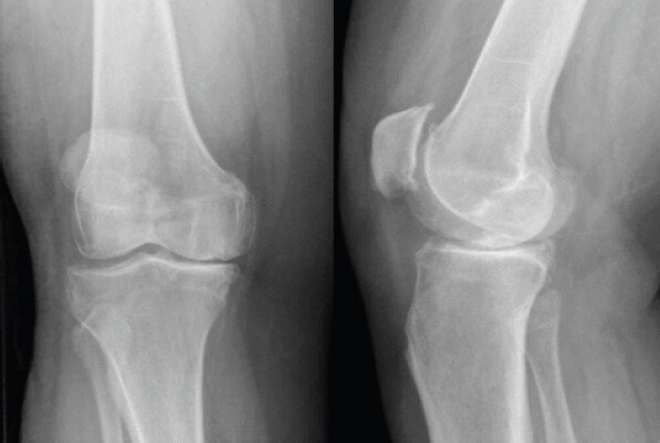
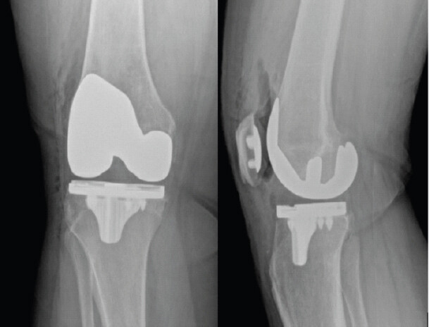
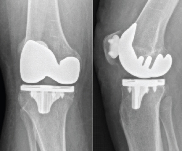
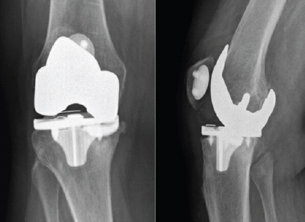
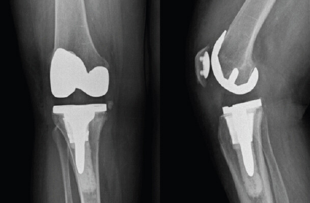
Verbal informed consent was obtained from the patient before submission of this report. A 70-year-old female underwent an uneventful right TKA utilizing conventional guides (Stryker Triathlon Cementless Knee System; Stryker Corporation, Kalamazoo, MI) at an outside hospital and was discharged home on post-operative day 1 (Fig. 1 and 2). We are not able to determine the degree of varus/valgus deformity pre-operatively, as full-length standing radiographs were not obtained at her original surgery. In the immediate post-operative period, her tibial component was in 3° of varus, femoral component was in 4° of varus, and tibial slope was 3° of posterior slope on short Anteroposterior and lateral radiographs (Fig. 2). The patient had a medical history notable for obesity (body mass index [BMI] 40.77 kg/m2), type 2 diabetes mellitus (hemoglobin A1C 6.1%), hypertension, hyperlipidemia, obstructive sleep apnea, chronic obstructive pulmonary disease, non-ST segment elevation myocardial infarction, and prior aortic valve replacement. One month post-operatively, she began to develop sharp right knee pain with ambulation and was referred to pain management. Her radiographs at that time did not show any clear signs of implant failure, and she was referred to pain management after unrevealing radiographic and infectious workups (Fig. 3). Over the course of the next year, she experienced progressively worsening medial knee pain preventing full weight-bearing. She presented for a second opinion to our institution 20 months post-operatively and was found to have catastrophic failure of the polyethylene insert and a fractured tibial baseplate (Fig. 4); she was indicated for revision surgery.
A standard midline medial parapatellar exposure was performed utilizing the patient’s prior incision. Upon entering the joint, both the patellar and femoral components were noted to be well-fixed and were left in situ to reduce her morbidity from the removal of well-fixed components. The polyethylene was visualized and noted to be fractured posteromedially. The posteromedial corner of the tibial baseplate demonstrated a fracture with loosening of this piece and impaction of the posteromedial tibia. The remaining tibial baseplate was grossly well-fixed. The remaining tibial component was removed using a combination of an oscillating saw and flexible osteotomes. Due to some bone loss following extraction of the primary baseplate, along with her body weight and relative osteopenia, a press-fit cone and short, fully cemented tibial stem construct were used for additional fixation (Stryker Triathlon Revision Knee System; Stryker Corporation, Kalamazoo, MI) (Fig. 5). Intraoperative cultures taken were ultimately negative for any signs of infection. She had no complications in the post-operative period. At her most recent follow-up, approximately 3.5 years after surgery, she was ambulating using a cane outdoors or without assistive devices around the house with resolution of her pre-operative pain.
Discussion
The use of cementless primary TKA has increased in recent years due to interest in the potential benefits of long-term biologic fixation, akin to the rise in cementless total hip arthroplasty [14]. While early studies of modern cementless TKA designs have shown successful outcomes and survivorship compared to cemented constructs, concerns remain about the possibility of early revision or changes in failure modes as the indications for cementless TKA expand [13, 15, 16]. In our case, the patient is a 70-year-old woman with early cementless Stryker Triathlon tibial baseplate failure consisting of posteromedial fracture of both the baseplate and polyethylene insert within 2 years after her index procedure, with symptoms that developed around 1 month after surgery. While her etiology of failure is not clear, two possibilities include: (1) Catastrophic failure of her highly cross-linked polyethylene insert leading to direct metal-on-metal contact resulting in baseplate fracture, or (2) fatigue/catastrophic failure of the tibial baseplate resulting in subsequent polyethylene fracture.
There have been a number of possible explanations to tibial baseplate fracture in previous studies (Table 1). In cemented designs, component fractures were often the result of either (1) progressive fatigue failure of the baseplate itself due to causes, such as osteolysis resulting in loss of tibial bone support or (2) polyethylene fracture leading to direct contact of the femoral component on the tibia component with subsequent fracture of the tibia secondary to metal-on-metal wear [1,4,5,6,7,8,11,12,17,18,19,20,21]. Contributing factors to baseplate fracture have also included design of the prosthesis, male gender, BMI, patient activity level, increased implantation time, and component under-sizing/malposition, such as residual varus alignment [6,12,21,22]. In our study, given her short time of implantation, polyethylene wear and chronic fatigue failure of the implant appear less likely as etiologies, suggesting that these modern designs of cementless TKA components may have unique failure modes relative to older cemented designs (Table 1).
One possible contribution to TKA failure includes technical considerations. Several radiostereometric analysis studies have demonstrated that there is a typical biphasic migration pattern with cementless components during the osseointegration period that is significantly longer than that of their cemented counterparts and can last up to 2 years [23,24]. It is possible that early micromotion is less well tolerated with titanium as the baseplate material, and this predisposed our patient to early fatigue failure. This could have been exacerbated by the patient’s pre-operative BMI of 42, which can predispose to medial tibial collapse in cemented TKA designs [25,26,27,28]. Several factors may contribute to this early failure of biologic fixation. While there were no full-length films pre-operatively, the implants appeared to be in some varus alignment, which may lead to overload of the medial side and predispose the implant to fatigue failure if it failed to osseointegrate, and this would more specifically affect the posteromedial portion during flexion [6,7,10,11,29]. Fatigue failure resulting in baseplate fracture can also occur in a well-aligned component that are not well-supported by host bone [6,21]. Sepehri and Masri report a case which they attribute to inadequate tibial baseplate size and positioning with insufficient cortical support [2]. In our case, the tibial baseplate sized at 3 appeared well-sized and appropriate, and the overall tibial bone stock appeared sufficient to support a cementless component, so this is less likely.
Biomaterial issues may also have possibly contributed to early tibial baseplate fracture in this design. Kurtz et al. found that in titanium alloy tibial baseplates, the presence of equiaxed or acicular microstructure formed during manufacturing influences fatigue fracture propagation [21]. The Stryker Triathlon is a 3D-printed highly porous titanium alloy with a sequentially annealed highly cross-linked polyethylene insert. The X3 polyethylene insert associated with the Stryker Triathlon system is a modern highly cross-linked polyethylene which may be predisposed to early catastrophic failure [30]. In addition, there have been case reports of early polyethylene fracture of sequentially annealed polyethylene in the setting of primary TKA [6,11,21]. Sonn and Meneghini describe a case requiring revision of a yellow-discolored polyethylene insert thought to be due to oxidation, which was found to have significant posteromedial polyethylene wear within just 5 years of implantation [30]. This significant wear precipitated the direct articulation of the femoral component with the tibial tray, leading to tibial component damage [30]. It is possible that this same mechanism of failure occurred in the cementless component used in this study, with polyethylene fracture followed by tibial component fracture due to direct articulation with the femoral component. There is a paucity of reports of cementless tibial baseplate fractures following primary TKA in the literature. Scully et al. described the first known case, although the time to fracture was 4 years from the index surgery [3]. Lam and Duffy reported the first case series of three patients with early tibial component fractures in a cementless, 3D-printed implant [31]. However, only one case they describe is within 2 years of the index procedure.
A notable limitation to our single case report is that the findings are based on one patient and cannot establish the incidence, risk factors, or generalizability of early tibial baseplate fracture. Further, while we propose the above failure mechanisms in this patient, the exact cause of failure remains unclear. In the present study, no detailed metallurgical, fractographic, or material analysis was performed to confirm the mechanism of component fracture. Due to limited assessment of implant positioning, the possible contribution of component malposition, inadequate cortical support, or impaired osseointegration cannot be definitively established. Our proposed explanation of early polyethylene fracture was not conclusively demonstrated, and the sequence of polyethylene and baseplate failure remains uncertain. As noted above, the unavailability of full-length standing radiographs limits accurate assessment of pre-operative deformity and post-operative mechanical alignment. Multiple risk factors, including obesity, possible varus alignment, osteopenia, and comorbidities, may confound the attribution of failure to implant design or material properties. The findings from this single reported cementless titanium baseplate design cannot be extrapolated to all cementless TKA systems or implant materials. Finally, our case report and literature review cannot establish comparative failure rates between cementless and cemented implants. Larger studies with systematic implant surveillance and retrieval analysis are required to identify risk factors and clarify failure mechanisms.
Conclusion
Cementless 3D-printed porous titanium tibial baseplate fracture is a rare and devastating complication of TKA. We report a case of early catastrophic failure occurring within 20 months of the index procedure. Based on previous literature, possible etiologies for such a component failure may include early fatigue failure of the titanium baseplate versus catastrophic failure of the polyethylene insert followed by baseplate fracture due to the articulating metal surfaces. Further studies will have to continue to follow the outcomes of cementless TKA with newer biomaterials and designs.
Clinical Message
TKA is evolving as a procedure with regard to the innovative types of materials and designs used for implants. While highly infrequent, orthopedic surgeons must be aware of the complications that can arise in modern cementless TKA implants to better plan revision surgeries and improve patient outcomes. Our case report adds to the scarce literature describing a tibial baseplate fracture in a modern cementless titanium 3D-printed porous implant in a short follow-up period from the index procedure.
Conflict of Interest:
Nil
Source of Support:
Nil
Consent:
The authors confirm that informed consent was obtained from the patient for publication of this article
How to Cite this Article
Jubril A, Denasty A, Krishnan R, Kaplan N, Ricciardi B. Early Post-operative Fracture of Titanium Cementless Tibial Baseplate: Case Report and Review of the Literature J Orthop Case Rep. 2026 November;16(11): 191-196. doi: 10.13107/jocr.2026.v16.i11.8436.
References
- Fokter SK, Gubeljak N, Punzón-Quijorna E, Pelicon P, Kelemen M, Vavpetič P. Total knee replacement with an uncemented porous tantalum tibia component: A failure analysis. Materials (Basel) 2022;15:2575. [Google Scholar] | [PubMed]
- Sepehri A, Masri BA.. Short-term tibial baseplate fracture after total knee arthroplasty in the absence of osteolysis and implant loosening: A case report. JBJS Case Connect 2020;10:e2000109. [Google Scholar] | [PubMed]
- Scully WF, Deren ME, Bloomfield MR.. Catastrophic tibial baseplate failure of a modern cementless total knee arthroplasty implant. Arthroplast Today 2019;5:446-52. [Google Scholar] | [PubMed]
- Rytter S, Madsen F, Jepsen CF, Stilling M.. Implant fracture of the regenerex® modular metal tibial component: A report of three cases. Knee 2019;26:1143-51. [Google Scholar] | [PubMed]
- Callaghan JJ, DeMik DE, Bedard NA, Odland AN, Kane WM, Kurtz SM.. Tibial tray fracture in a modern prosthesis with retrieval analysis. Arthroplast Today 2018;4:143-7. [Google Scholar] | [PubMed]
- Chatterji U, Ashworth MJ, Smith AL, Brewster N, Lewis PL.. Retrieval study of tibial baseplate fracture after total knee arthroplasty. J Arthroplasty 2005;20:101-7. [Google Scholar] | [PubMed]
- Ho TF, Tsai RY, Lee PY, Ku MC.. Early tibial tray failure of a Duracon knee with retrieval analysis. J Arthroplasty 2004;19:797-802. [Google Scholar] | [PubMed]
- Altintas F, Sener N, Ugutmen E.. Fracture of the tibial tray after total knee arthroplasty. J Arthroplasty 1999;14:112-4. [Google Scholar] | [PubMed]
- Worgul CA, Lentine B, Dicken QG, Freccero DM.. Catastrophic failure of the tibial component after total knee arthroplasty: Fracture and dissociation between the baseplate and stem. Arthroplast Today 2022;14:194-8. [Google Scholar] | [PubMed]
- Cankaya D, Yilmaz S, Deveci A, Ozkurt B.. Tibial baseplate fracture associated with polyethylene wear and osteolysis after total knee arthroplasty. Int J Surg Case Rep 2014;5:49-51. [Google Scholar] | [PubMed]
- Mineta K, Okada M, Matsumoto S, Hamada D, Goto T, Sairyo K.. Fracture of the tibial baseplate 16 years after Miller-Galante II total knee arthroplasty. Case Rep Orthop 2017;2017:4080816. [Google Scholar] | [PubMed]
- Swarts E, Miller SJ, Keogh CV, Lim G, Beaver RJ.. Fractured Whiteside Ortholoc II knee components. J Arthroplasty 2001;16:927-34. [Google Scholar] | [PubMed]
- Nam D, Lawrie CM, Salih R, Nahhas CR, Barrack RL, Nunley RM.. Cemented versus cementless total knee arthroplasty of the same modern design: A prospective, randomized trial. J Bone Joint Surg Am 2019;101:1185-92. [Google Scholar] | [PubMed]
- Haeberle HS, Salem HS, Ehiorobo JO, Sodhi N, Mont MA.. Newer generation of cementless total knee arthroplasty: A systematic review. Surg Technol Int 2020;36:351-9. [Google Scholar] | [PubMed]
- Pacoret V, Kalk E, Labattut L, Girardot G, Baulot E, Martz P.. Survival rate of cemented versus cementless tibial component in primary total knee arthroplasty over 5 years of follow-up: Comparative study of 109 prostheses. SICOT J 2020;6:36. [Google Scholar] | [PubMed]
- Quispel CR, Duivenvoorden T, Beekhuizen SR, Verburg H, Spekenbrink-Spooren A, Van Steenbergen LN. Comparable mid-term revision rates of primary cemented and cementless total knee arthroplasties in 201,211 cases in the dutch arthroplasty register (2007-2017). Knee Surg Sports Traumatol Arthrosc 2021;29:3400-8. [Google Scholar] | [PubMed]
- Rousseau MA, Lazennec JY, Catonné Y.. Early mechanical failure in total knee arthroplasty. Int Orthop 2008;32:53-6. [Google Scholar] | [PubMed]
- Flivik G, Ljung P, Rydholm U.. Fracture of the tibial tray of the PCA knee. A case report of early failure caused by improper design. Acta Orthop 1990;61:26-8. [Google Scholar] | [PubMed]
- Abernethy PJ, Robinson CM, Fowler RM.. Fracture of the metal tibial tray after Kinematic total knee replacement. A common cause of early aseptic failure. J Bone Joint Surg Br 1996;78:220-5. [Google Scholar] | [PubMed]
- Da Palma IM, Albuquerque RP, Barretto JM.. Fracture of the tibial component in total knee arthroplasty: Report on two cases. Rev Bras Ortop 2011;46:325-8. [Google Scholar] | [PubMed]
- Kurtz MA, Gilbert JL, Spece H, Klein GR, Cates HE, Kurtz SM.. Tibial baseplate microstructure governs high cycle fatigue fracture in vivo. J Biomed Mater Res B Appl Biomater 2024;112:e35507. [Google Scholar] | [PubMed]
- Constantinescu DS, Ross JA, Patel NK, Strong BM, Giambra LA, Golladay GJ.. Radiographic false evidence of a tibial baseplate fracture after total knee arthroplasty. Arthroplast Today 2021;8:114-7. [Google Scholar] | [PubMed]
- Nilsson KG, Henricson A, Norgren B, Dalén T.. Uncemented HA-coated implant is the optimum fixation for TKA in the young patient. Clin Orthop Relat Res 2006;448:129-39. [Google Scholar] | [PubMed]
- Van Hamersveld KT, Marang-Van De Mheen PJ, Nelissen RG, Toksvig-Larsen S.. Peri-apatite coating decreases uncemented tibial component migration: Long-term RSA results of a randomized controlled trial and limitations of short-term results. Acta Orthop 2018;89:425-30. [Google Scholar] | [PubMed]
- Sinicrope BJ, Feher AW, Bhimani SJ, Smith LS, Harwin SF, Yakkanti MR. Increased survivorship of cementless versus cemented TKA in the morbidly obese. A minimum 5-year follow-up. J Arthroplasty 2019;34:309-14. [Google Scholar] | [PubMed]
- bdel MP, Bonadurer GF 3rd, Jennings MT, Hanssen AD.. Increased aseptic tibial failures in patients with a BMI ≥35 and well-aligned total knee arthroplasties. J Arthroplasty 2015;30:2181-4. [Google Scholar] | [PubMed]
- Vazquez-Vela Johnson G, Worland RL, Keenan J, Norambuena N.. Patient demographics as a predictor of the ten-year survival rate in primary total knee replacement. J Bone Joint Surg Series B 2003;85:52-6. [Google Scholar] | [PubMed]
- Schreurs BW, Hannink G.. Total joint arthroplasty in younger patients: Heading for trouble?. Lancet 2017;389:1374-5. [Google Scholar] | [PubMed]
- Han CD, Han CW, Yang IH.. Femoral component fracture due to osteolysis after cemented mobile-bearing total knee arthroplasty. J Arthroplasty 2009;24:323.e7-12. [Google Scholar] | [PubMed]
- Sonn KA, Meneghini RM.. Early failure of sequentially annealed polyethylene in total knee arthroplasty. Arthroplast Today 2020;6:18-22. [Google Scholar] | [PubMed]
- Lam AD, Duffy GP.. Early tibial component fractures in a cementless, 3D-printed, titanium implant. Arthroplast Today 2022;18:31-8. [Google Scholar] | [PubMed]
© 2026 Journal of Orthopaedic Case Reports - Published by Indian Orthopaedic Research Group
About the Authors
-

Ayodeji Jubril
-

Adwin Denasty
-

Ravi Krishnan
-

Nathan Kaplan
-

Benjamin Ricciardi
Related Articles in Journal of Orthopaedic Case Reports
 October 1, 2026 Revision Total Knee Arthroplasty in a Case of Operated Right Total Knee Arthroplasty with Implant Loosening and Pigmented Villonodular Synovitis
 August 1, 2025 Polyethylene Insert Wear Leading to Early Aseptic Bilateral Total Knee Arthroplasty Failure: Case Report
 September 1, 2024 A Case of Varus Collapse and Bone Defects in Revision Total Knee Arthroplasty, after 12 Years of Primary Total Knee Replacement: Surgical Challenge and Bailout
 August 6, 2024 Mechanical Failure of SIGMA Total Condylar-III Revision Total Knee System Modular Femoral Component Due to Bolt Screw Breakage – Our Experience in Overcoming Broken Component Incarceration