


Introduction
Perilunate fracture-dislocations (PLFDs) represent severe disruptions of carpal biomechanics, accounting for 5–7% of wrist injuries [1]. The trans-styloid and trans-scaphoid variants involve radial styloid and scaphoid fractures with ligamentous injury, often resulting in carpal instability and median nerve compression [2,3,4]. Despite advancements in surgical techniques, these injuries remain challenging due to risks of non-union, avascular necrosis, and post-traumatic arthritis [5].
Recent literature highlights the superiority of open reduction and internal fixation (ORIF) with ligament repair [6], while arthroscopic-assisted techniques offer improved visualization [7], though their role in complex fracture-dislocations remains limited [8].
This case report describes the successful management of this rare and unstable injury pattern.
Case Report
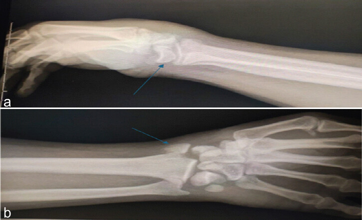
A 27-year-old right-hand–dominant male presented following a high-energy motor vehicle accident with severe wrist pain, swelling, and obvious deformity. Initial radiographs demonstrated a displaced scaphoid waist fracture, a radial styloid fracture, and perilunate dislocation. (Fig.1)
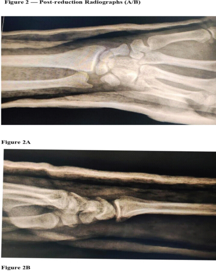
Closed reduction was attempted under conscious sedation using traction–counter-traction and volar-to-dorsal pressure on the lunate. A thumb spica splint was applied. Post-reduction assessment showed good distal vascularity, normal capillary refill, and full finger motion without numbness. Post-reduction radiographs confirmed satisfactory alignment of the radial styloid and lunate. (Fig. 2)
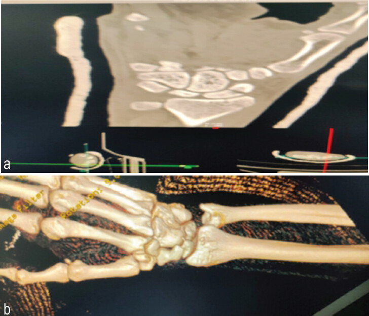
A computed tomography scan was subsequently performed to confirm reduction and assist in operative planning. (Fig. 3)
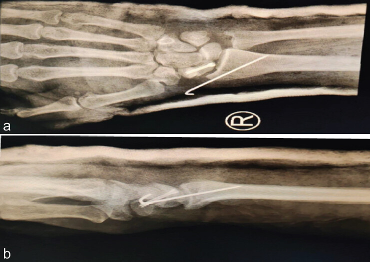
Definitive ORIF was performed through a dorsal approach. The scaphoid fracture was anatomically reduced and fixed with a Herbert screw, while the radial styloid fragment was stabilized with a Kirschner wire. Carpal stability was assessed intraoperatively, followed by repair of the injured dorsal ligaments, capsular closure, retinacular repair, and layered wound closure. A thumb spica splint was reapplied postoperatively. (Fig. 4)
The wrist remained immobilized until the 6-week follow-up, at which time the K-wire was removed, and physiotherapy was initiated – initially focusing on edema control, followed by progressive range-of-motion exercises. (Fig. 5)
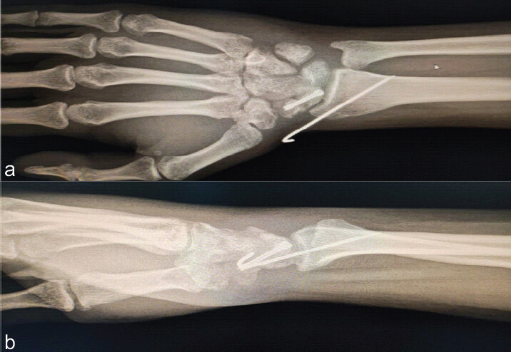
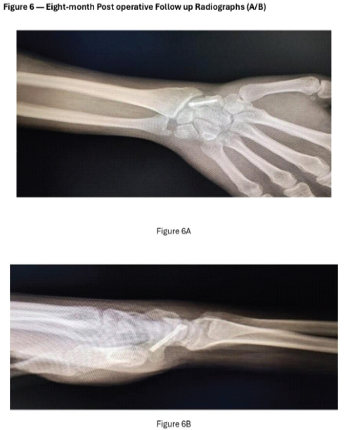
By 12 weeks from the time of injury, the patient had regained full, pain-free wrist motion with no radiographic evidence of instability. By 8 months after injury, complete healing on styloid radius and scaphoid. (Fig. 6)
Discussion
PLFDs are high-energy injuries produced by wrist hyperextension. Early recognition is essential for optimal outcomes [9]. Mayfield and Johnson described progressive perilunate instability through four stages, beginning radially and propagating ulnarly [10].
The trans-styloid trans-scaphoid variant is rare, previously reported only in isolated case reports and small series [2,4], making the 2026 study by Mazhar et al. the largest series to date [11].
Closed reduction alone has been associated with worse outcomes [12,13], while conservative treatment of trans-scaphoid PLFDs produced poor results [14]. The dorsal approach provides excellent visualization for scaphoid and lunate reduction [12]. Fixation with a Herbert screw remains the gold standard for scaphoid fractures, while K-wire fixation of the radial styloid restores radial column stability.
Arthroscopic techniques offer improved visualization and minimally invasive advantages [5,11], but open approaches remain preferred for complex fracture-dislocations.
Conclusion
Trans-styloid trans-scaphoid PLFDs are highly unstable injuries requiring urgent surgical intervention. Early anatomical reduction and rigid internal fixation restore carpal alignment and stability, providing excellent functional recovery and minimizing long-term complications.
Clinical Message
Trans-styloid trans-scaphoid perilunate fracture-dislocations (PLFDs) represent complex, high-energy wrist injuries that are easily missed on initial radiographic evaluation. This case highlights the pivotal role of advanced imaging, such as CT scans, in delayed or ambiguous presentations to ensure accurate diagnosis. Achieving optimal clinical outcomes and preserving wrist function depend on prompt open reduction, precise internal fixation of the radial column using Herbert screws and Kirschner wires, which subsequently facilitates spontaneous lunate reduction and mitigates the risk of avascular necrosis (AVN).”
Conflict of Interest:
Nil
Source of Support:
Nil
Consent
The authors confirm that informed consent was obtained from the patient for publication of this article
How to Cite this Article
Elmeligy RA, Sayed M. Combined Radial Styloid and Scaphoid Fractures with perilunate Dislocation. J Orthop Case Rep. 2026 November;16(11): 88-91. doi: 10.13107/jocr.2026.v16.i11.8398.
References
- Herzberg G, Comtet JJ, Linscheid RL, Amadio PC, Cooney WP, Stalder J. Perilunate dislocations and fracture-dislocations: A multicenter study. J Hand Surg Am 1993;18:768-79. [Google Scholar] | [PubMed]
- Frane N, Regala P, Klein B, Mitgang J, Katsigiorgis G. Case study: Trans-styloid, trans-scaphoid, trans-triquetral, and perilunate dislocation. J Orthop Case Rep 2021;11:63-7. [Google Scholar] | [PubMed]
- Liechti R, Merky DN, Grobbelaar AO, Van De Wall BJ, Vögelin E, Hirsiger S. Outcomes of acute perilunate injuries-a systematic review. Eur J Trauma Emerg Surg 2023;49:2071-84. [Google Scholar] | [PubMed]
- Alzahrani AA, Alzahrani MH, Alzahrani TA, Alzahrani MA, Alsubaei DM. A variant of perilunate dislocation: A case report and literature review. Radiol Case Rep 2024;19:3870-3. [Google Scholar] | [PubMed]
- Park MJ, Ahn JH. Arthroscopically assisted reduction and percutaneous fixation of dorsal perilunate dislocations and fracture-dislocations. Arthroscopy 2005;21:1153.e1-9. [Google Scholar] | [PubMed]
- Van Der Oest MJ, Duraku LS, Artan M, Hundepool CA, Power DM, Rajaratnam V. Perilunate injury timing and treatment options: A systematic review. J Wrist Surg 2022;11:164-76. [Google Scholar] | [PubMed]
- Alyamani AM, Alfawzan MF, Alhassan TS, Almeshal OM. Trans-styloid, trans-hamate dorsal lunate dislocation: A case report. Int J Surg Case Rep 2019;61:96-8. [Google Scholar] | [PubMed]
- Agha RA, Mathew G, Rashid R, Kerwan A, Al-Jabir A, Sohrabi C. Revised preferred reporting of case series in surgery (PROCESS) guideline: An update for the age of artificial intelligence. Prem J Sci 2025;10:100080. [Google Scholar] | [PubMed]
- Johnson RP. The acutely injured wrist and its residuals. Clin Orthop Relat Res 1980;149:33-44. [Google Scholar] | [PubMed]
- Mayfield JK, Johnson RP, Kilcoyne RK. Carpal dislocations: Pathomechanics and progressive perilunar instability. J Hand Surg Am 1980;5:226-41. [Google Scholar] | [PubMed]
- Mazhar FN, Zareie B, Nasab OM, Shariatzade H, Pourmahmoudian M, Kafiabadi MJ. Concomitant trans-styloid and trans-scaphoid perilunate fracture-dislocations: Evaluation of surgical outcomes in the largest reported series. Int J Surg Case Rep 2026;138:316-21. [Google Scholar] | [PubMed]
- Hildebrand KA, Ross DC, Patterson SD, Roth JH, MacDermid JC, King GJ. Dorsal perilunate dislocations and fracture-dislocations: Questionnaire, clinical, and radiographic evaluation. J Hand Surg Am 2000;25:1069-79. [Google Scholar] | [PubMed]
- Melone CP Jr , Murphy MS, Raskin KB. Perilunate injuries. Repair by dual dorsal and volar approaches. Hand Clin 2000;16:439-48. [Google Scholar] | [PubMed]
- Morawa LG, Ross PM, Schock CC. Fractures and dislocations involving the navicular. Clin Orthop Relat Res 1976;118:58-65. [Google Scholar] | [PubMed]
© 2026 Journal of Orthopaedic Case Reports - Published by Indian Orthopaedic Research Group
About the Authors
-

Rashwan Adel Elmeligy
-

Mustafa Sayed
Related Articles in Journal of Orthopaedic Case Reports
 September 1, 2026 Concomitant Distal Radius Fracture with Scaphoid Fracture-Dislocation and Carpal Instability: A Case Report
 April 10, 2024 Volar Lunate Dislocation: A Case Report
 September 10, 2022 Trans-Scaphoid Perilunate Fracture Dislocation Managed With Open Reduction: A Case Report
 September 1, 2026 Proximal Row Carpectomy for Established Post-Traumatic Radiocarpal Arthritis Following Distal Radius Volar Rim and Scaphoid Fractures: A Case Report