


Introduction
Fracture-dislocations of the proximal interphalangeal (PIP) joint [1] represent complex injuries of the finger in which restoration of articular congruity, stability, and functional motion is essential. When treatment is delayed, the injury may become increasingly difficult to manage due to fibrosis, callus formation, articular incongruity, and persistent limitation of motion.
The volar lip of the middle phalanx [2] plays an important role in maintaining PIP joint stability. Substantial loss of this articular surface can compromise joint congruity and make conventional reduction or soft-tissue procedures insufficient for reconstruction.
Hemi-hamate arthroplasty uses an osteochondral graft [3,4] harvested from the hamate to reconstruct the deficient volar articular surface of the middle phalanx. By restoring the articular contour, the procedure aims to provide a stable and congruent PIP joint while preserving useful digital motion.
We report a case of a chronic fracture-dislocation of the PIP joint of the right ring finger treated with hemi-hamate arthroplasty, with post-operative assessment demonstrating substantial improvement in PIP joint motion and favorable functional scores.
Case Report
A 29-year-old male presented with a 1-month history of an injury to the right ring finger sustained while playing cricket. The injury occurred when the patient was struck by a cricket ball. Following the injury, he consulted several doctors and was treated with a finger cast. He subsequently underwent aggressive physiotherapy.
Despite these measures, he continued to experience restricted movement of the right ring finger and difficulty with activities of daily living. The hospital record documented a 1-month-old fracture-dislocation of the PIP joint of the right ring finger (Fig. 2).
On pre-operative assessment, PIP joint flexion and extension were recorded as 20°–75°. The measurements recorded above and below the PIP joint were 7 cm and 8 cm, respectively. The patient had persistent limitation of PIP joint motion. Pre-operative radiographs demonstrated a chronic PIP fracture-dislocation with substantial volar articular surface deficiency and dorsal subluxation of the middle phalanx (Fig. 1).
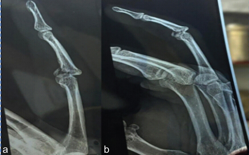
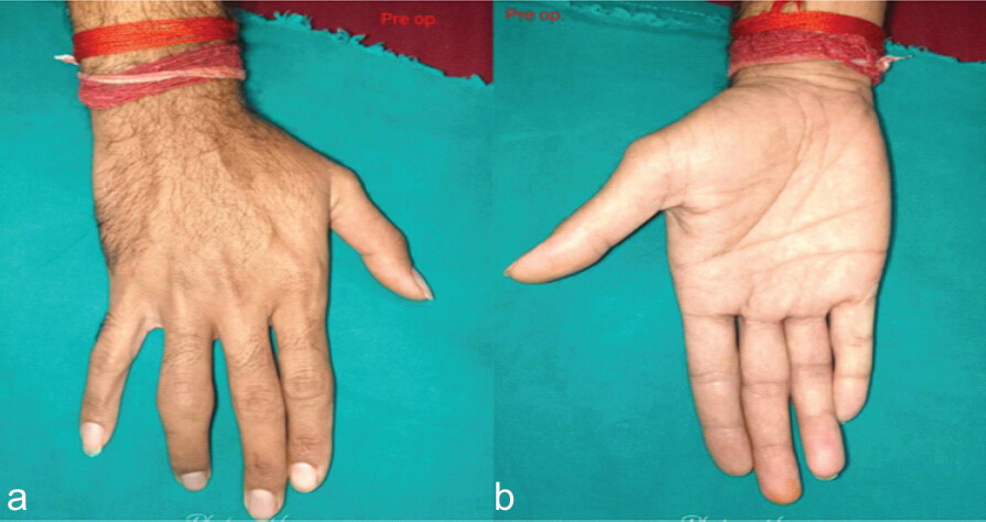
In view of the chronic fracture-dislocation and persistent functional limitation, operative reconstruction was planned.
Surgical technique
The patient underwent hemi-hamate arthroplasty of the right ring-finger PIP joint under regional anesthesia. The patient was positioned supine with the right upper limb placed on a hand table. The limb was prepared and draped in the standard sterile manner and a tourniquet was inflated.
A volar Bruner incision was made over the PIP joint. The digital neurovascular bundles were identified and protected. The flexor tendon sheath was opened, and the flexor tendons were retracted. The volar plate was then incised to expose the PIP joint.
A chronic fracture-dislocation with a volar lip defect was identified. Fibrous tissue and callus were thoroughly debrided. The base of the middle phalanx was prepared to create a recipient bed for the graft.
A separate dorsal incision was made over the hamate. An osteochondral graft was harvested from the dorsal distal hamate. The graft was contoured to match the defect at the base of the middle phalanx and was subsequently inset and fixed under image intensifier guidance (Fig. 3). Joint stability was confirmed intraoperatively before wound closure. An extension-block slab was applied at the completion of the procedure.
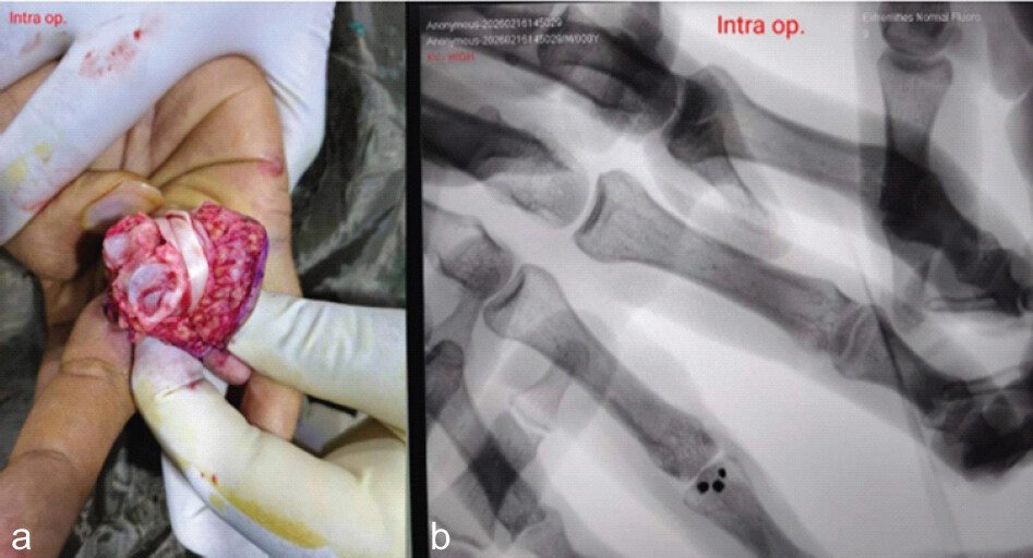
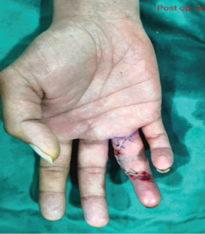
Post-operative management
Postoperatively, the finger was maintained in an extension-block slab. The documented post-operative plan included immobilization for 6–8 weeks, followed by finger mobilization and active PIP joint movement. After removal of the slab, active and active-assisted physiotherapy was advised. A Capener splint or finger cot was recommended following slab removal. The patient was advised to avoid using the right ring finger for work and to avoid driving for 2–3 months.
Clinical outcome
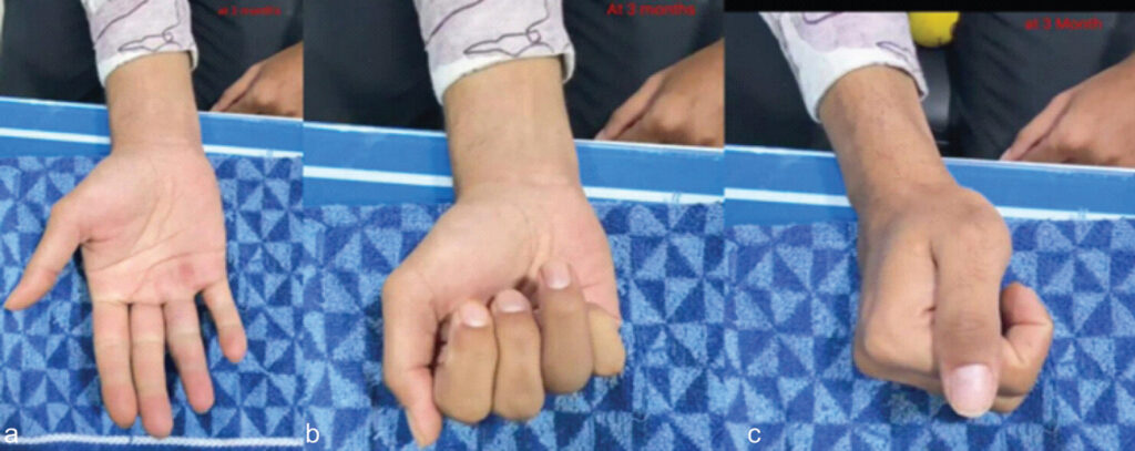
Post-operative assessment demonstrated improvement in PIP joint motion. The PIP joint achieved a range of 0°–110°, compared with the pre-operative range of 20°–75°.
The pre-operative flexion arc was therefore 55°, while the post-operative flexion arc was 110°, representing a 55° increase in the measured arc of motion.
Post-operative grip strength was 31.3 kg.
Functional assessment using the Disabilities of the Arm, Shoulder and Hand (DASH) [5] questionnaire demonstrated a total score of 4.55. The work module score was 6.25, and the sports module score was 12.5.
Discussion
Chronic PIP joint fracture-dislocations [6] can be difficult to treat because delayed presentation may result in persistent articular incongruity, fibrosis, callus formation, and restricted joint motion. In the present case, the patient presented approximately 1 month after a cricket-ball injury despite previous immobilization and physiotherapy. He continued to have restricted PIP joint motion and difficulty with activities of daily living.
Intraoperative assessment demonstrated a chronic fracture-dislocation associated with a defect of the volar lip. Fibrous tissue and callus were debrided, and the base of the middle phalanx was prepared for reconstruction.
The treatment of chronic PIP fracture-dislocations must address both joint stability and the articular surface. When there is substantial loss of the volar articular surface, reconstruction with an osteochondral graft can restore the contour of the joint and provide a stable articular platform [7].
In this case, a hemi-hamate osteochondral graft [8] was harvested from the dorsal distal hamate [9] and contoured to reproduce the deficient portion of the middle phalanx. The graft was then fixed under image intensifier guidance, and joint stability was confirmed intraoperatively.
The post-operative outcome was favorable. PIP joint motion [10] improved from 20° to 75° preoperatively to 0°–110° postoperatively. The flexion arc consequently increased from 55° to 110°. In addition, the patient demonstrated a grip strength of 31.3 kg and a DASH score of 4.55.
This case demonstrates that hemi-hamate arthroplasty can be considered for reconstruction of selected chronic PIP fracture-dislocations associated with a volar articular defect. Restoration of the articular surface in this patient was associated with substantial improvement in PIP joint motion and a favorable functional outcome.
Conclusion
Hemi-hamate arthroplasty provided a useful reconstructive option in this case of a chronic fracture-dislocation of the right ring-finger PIP joint with a volar lip defect. Following reconstruction, PIP joint motion improved from 20°–75° to 0°–110°, with a post-operative grip strength of 31.3 kg and a DASH score of 4.55.
The procedure may be considered in selected chronic PIP fracture-dislocations in which restoration of the deficient volar articular surface is required.
Clinical Message
Chronic PIP joint fracture-dislocations with substantial volar articular surface deficiency can result in persistent stiffness and functional limitation. Hemi-hamate arthroplasty allows reconstruction of the deficient articular surface using an osteochondral hamate graft and may restore useful PIP joint motion and hand function.
Conflict of Interest:
Nil
Source of Support:
Nil
Consent
The authors confirm that informed consent was obtained from the patient for publication of this article
How to Cite this Article
Maheshwari K, Patel H, Sathwara H, Parmar P, Bhimani N, Musar K. Hemi-Hamate Arthroplasty for Chronic Proximal Interphalangeal Joint Fracture-Dislocation with Volar Lip Deficiency Following a Cricket Injury: J Orthop Case Rep. 2026 November;16(11): 83-87. doi: 10.13107/jocr.2026.v16.i11.8396.
References
- Leibovic SJ, Bowers WH. Anatomy of the proximal interphalangeal joint. Hand Clin 1994;10:169-78. [Google Scholar] | [PubMed]
- Punekar AD, Ghoti SD, Das J, Rathod AS, Jain H, Shetty RS. Surgical management of volar lip fractures of the middle phalanx: A case series on diverse techniques and outcomes, do acute and chronic presentations differ in outcome? J Orthop Case Rep. 2026;16:282-9. [Google Scholar] | [PubMed]
- Calfee RP, Kiefhaber TR, Sommerkamp TG, Stern PJ. Hemi-hamate arthroplasty provides functional reconstruction of acute and chronic proximal interphalangeal fracture-dislocations. J Hand Surg Am 2009;34:1232-41. [Google Scholar] | [PubMed]
- Branam GM, Saber AY. Osteochondral Autograft Transplantation. In: StatPearls. Treasure Island, FL:StatPearls Publishing;2026. Available from: http://www.ncbi.nlm.nih.gov/books/nbk560655 [Last accessed on 2026 Aug 14]. [Google Scholar] | [PubMed]
- Gummesson C, Atroshi I, Ekdahl C. The disabilities of the arm, shoulder and hand (DASH) outcome questionnaire: Longitudinal construct validity and measuring self-rated health change after surgery. BMC Musculoskelet Disord 2003;4:11. [Google Scholar] | [PubMed]
- Elfar J, Mann T. Fracture-dislocations of the proximal interphalangeal joint. J Am Acad Orthop Surg 2013;21:88-98. [Google Scholar] | [PubMed]
- Xie Y, Seth I, Munasinghe N. Osteochondral autograft from the second toe for complex proximal interphalangeal joint fracture-dislocations: A case report and literature review. Ann Jt 2025;10:20. [Google Scholar] | [PubMed]
- Marcoccio I, Civitenga C, Maffeis J, Minini A, Riccio M, Gravina P. A hemi-hemi-hamate osteochondral graft: A modified hemi-hamate technique for a unicondylar defect. J Hand Surg Glob Online 2023;6:236-40. [Google Scholar] | [PubMed]
- Hacking C. Hamate Radiology. Radiopaedia. Available from: https://www.radiopaedia.org/articles/hamate [Last accessed on 2026 Aug 14]. [Google Scholar] | [PubMed]
- Park J, Lee CH, Choi Y, Joo IH, Lee KH, Kim SJ. Extension mechanism of the proximal interphalangeal joint of the human phalanx: A cadaveric biomechanical study. Biomed Res Int 2020;2020:7585976. [Google Scholar] | [PubMed]
© 2026 Journal of Orthopaedic Case Reports - Published by Indian Orthopaedic Research Group
About the Authors
-

Karn Maheshwari
-

Hiren Patel
-

Happy Sathwara
-

Poojan Parmar
-

Nikita Bhimani
-

Kana Musar
Related Articles in Journal of Orthopaedic Case Reports
 February 1, 2026 Surgical Management of Volar Lip Fractures of the Middle Phalanx: A Case Series on Diverse Techniques and Outcomes, Do Acute and Chronic Presentations Differ in Outcome?
 October 1, 2026 A Functional Outcome of Closed Intra-articular Fracture of Proximal Interphalangeal Joint of Hand Managed By Suzuki Frame – A Longitudinal Study
 July 1, 2026 When Fat Takes Over: A Progressively Enlarging Giant Hand Lipoma
 January 1, 2026 Ten Foundational Principles in Hand Surgery: A Primer for Orthopedic Practice