


Introduction
Periprosthetic joint infection (PJI) remains one of the most serious complications following total knee arthroplasty, often resulting in implant failure, prolonged morbidity, and impaired function. Accurate and timely identification of rare causative pathogens is challenging but critical. Matrix-assisted laser desorption ionization–time of flight mass spectrometry (MALDI-TOF MS) has emerged as a state-of-the-art tool in advanced microbiology laboratories, allowing rapid, species-level identification even in culture-negative or chronic infections [1,2].
Management of extensive PJI in frail or elderly patients with significant soft-tissue damage, patellar dislocation, and rupture of the arthrotomy poses additional surgical challenges. In cases where conventional revision strategies are inadequate due to severe soft-tissue or bone compromise, novel surgical techniques such as K-nail arthrodesis provide a reliable limb-salvage option. This technique offers stable fixation, facilitates local antifungal delivery, and provides functional support while reducing the risk of reinfection [3,4,5,6].
While bacterial pathogens account for the majority of PJIs, fungal infections are uncommon, representing <1% of cases, and are associated with delayed diagnosis, higher recurrence rates, and poorer outcomes [7,8].
Among fungal pathogens, Candida albicans is most frequently reported, followed by other non-albicans Candida (NAC) species [9,10]. Candida viswanathii is an exceptionally rare organism, previously described only in a few clinical scenarios, including non-orthopedic cases such as fatal meningitis [11]. To the best of our knowledge, no cases of periprosthetic knee joint infection caused by C. viswanathii have been reported to date.
This case report illustrates the diagnostic complexity and surgical decision-making involved in managing a periprosthetic knee infection caused by C. viswanathii, highlighting the role of advanced microbiological identification and innovative arthrodesis techniques.
Case Report
An 80-year-old, well-nourished, non-immunocompromised male underwent bilateral total knee arthroplasty for advanced osteoarthritis. The initial post-operative course was uneventful. However, 2 months following surgery, the patient developed persistent knee swelling and recurrent joint effusion. He underwent multiple joint aspirations and debridement procedures at an outside institution. Initial microbiological cultures were negative, and empirical antibiotic therapy was administered.
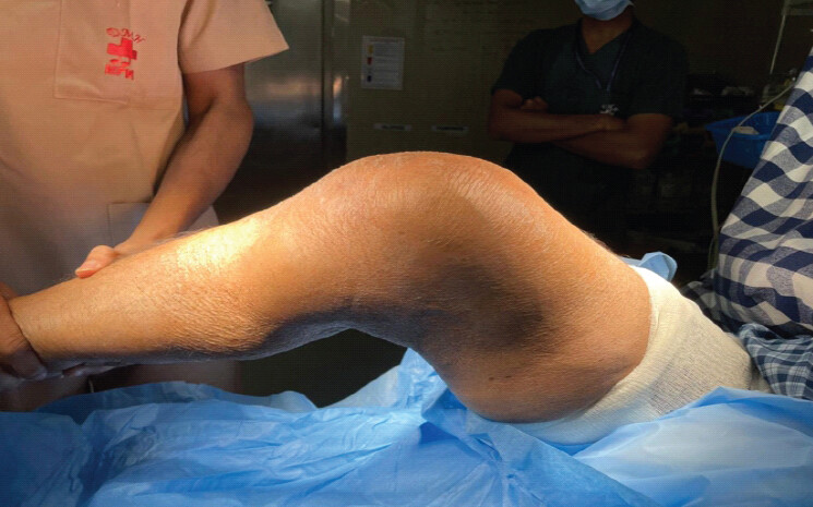
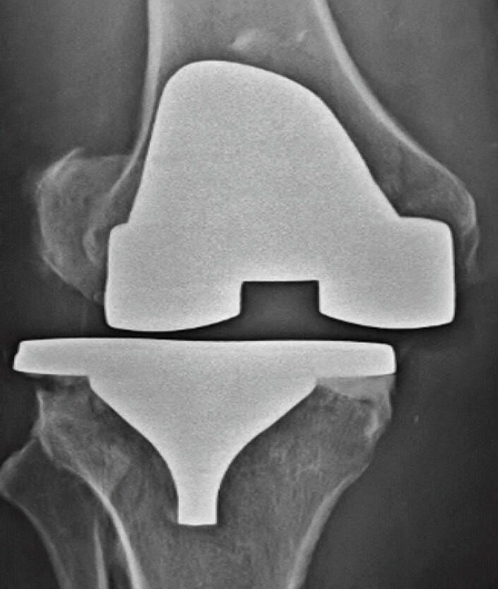
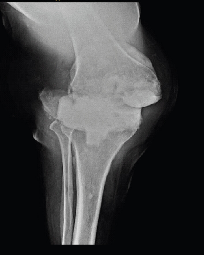
Five months postoperatively, the patient presented to our institution with worsening pain, swelling, patellar dislocation, arthrotomy rupture, and significant functional limitation. Clinical examination revealed poor quadriceps function and reduced range of motion, with marked soft-tissue compromise, including arthrotomy rupture and patellar dislocation (Fig. 1). Radiological evaluation demonstrated prosthetic instability with extensive periarticular soft-tissue damage (Fig. 2).
Antibiotics were withheld for 2 weeks, and repeat joint aspiration was performed. Fungal cultures revealed growth of C. viswanathii, which was confirmed using MALDI-TOF MS. This established the diagnosis of fungal PJI and represented the first reported case of C. viswanathii infection following total knee arthroplasty.
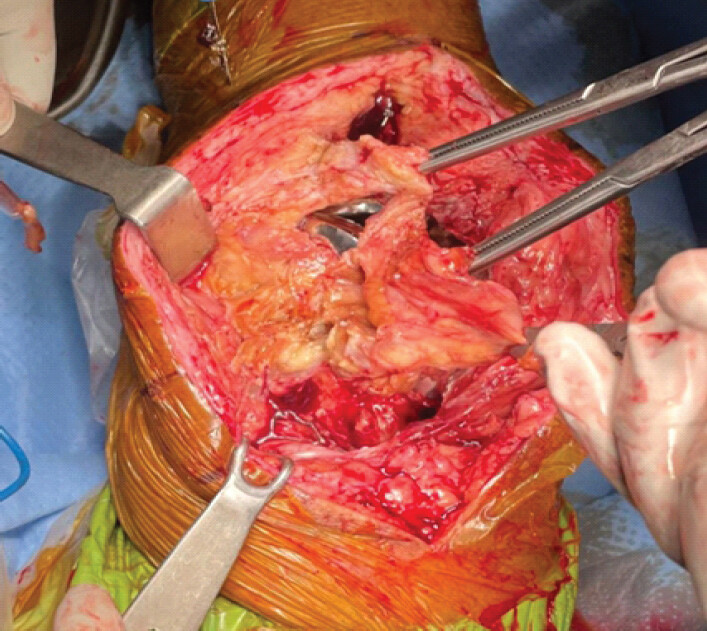
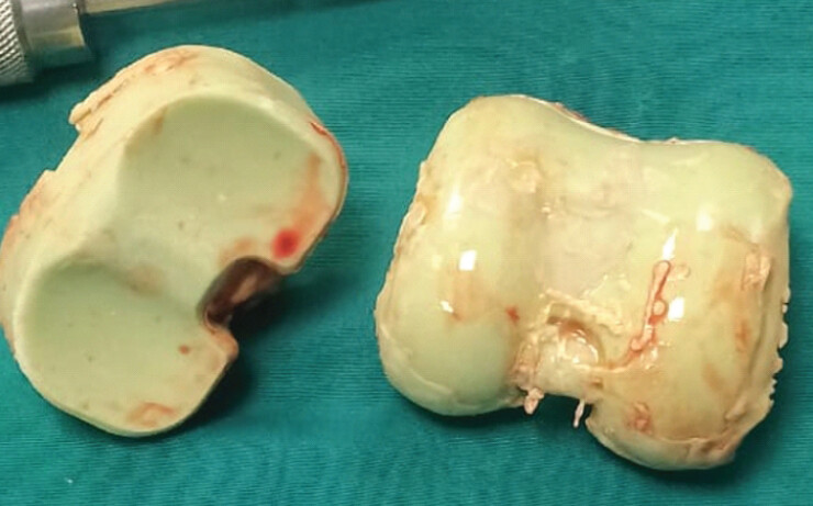
The patient underwent first-stage revision arthroplasty with removal of the prosthesis (Fig. 3) and placement of a dynamic spacer impregnated with amphotericin B (Fig. 4) to achieve high local antifungal concentrations. Under infectious disease specialist guidance, systemic antifungal therapy was initiated with oral posaconazole and later changed to voriconazole and 5-fluorocytosine. On review 3 months postoperatively, the patient was noted to have recurrent patellar dislocation associated with arthrotomy dehiscence (Fig. 5). Erythrocyte sedimentation rate (ESR) was 116 mm/h, and C-reactive protein (CRP) level was 11.1 mg/L.
Despite aggressive management, progressive soft-tissue destruction was noted. Given the persistence of infection, severity of soft-tissue loss and instability, and the overall frail clinical condition of the patient, revision to a static spacer was planned. A novel cement rod (K-nail) technique was used, with the construct fixed into the distal femur and proximal tibia using amphotericin B–impregnated cement.
Novel Surgical Management Technique – Knee Arthrodesis with Intramedullary Polymethylmethacrylate (PMMA) Cemented K-Nail Construct – Described by Author Dr Suresh Patil
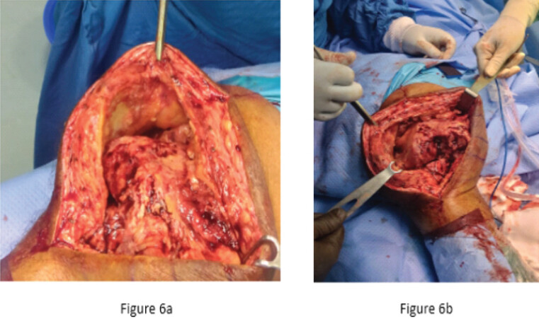
Antimicrobial therapy was withheld prior to surgery to aid intraoperative microbiological diagnosis, and antifungal treatment was initiated only after obtaining adequate deep tissue and fluid samples. Following surgical exposure, the femoral, tibial, and patellar components were removed, and all residual cement was meticulously cleared. Extensive intra-operative soft-tissue destruction and bone loss were noted (Fig. 6a and b). An extensive synovectomy was performed, along with thorough debridement of necrotic bone and soft tissue until healthy bleeding tissue was encountered after tourniquet release. The femoral and tibial intramedullary canals were opened, and multiple intraoperative samples were obtained for fungal culture and histopathological examination. The canals were subsequently brush-irrigated to remove infected and devitalized material.
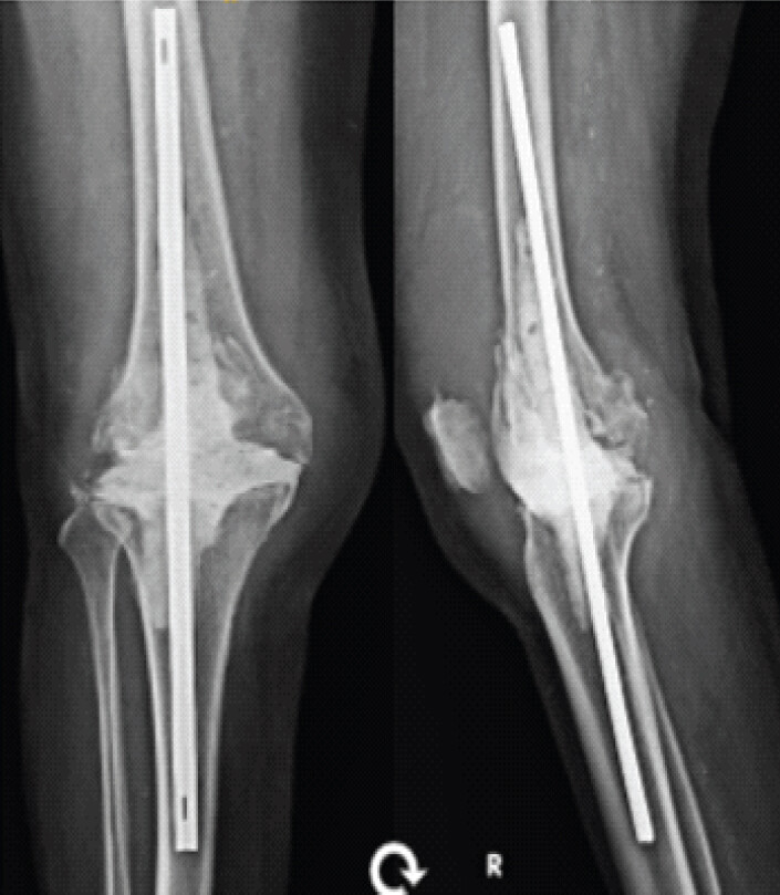
A cement-coated rod (K-nail) construct was then fashioned using a metallic rod of appropriate length to achieve stable intramedullary fixation within the femur and tibia while maintaining slight valgus and flexion alignment. Amphotericin B–impregnated PMMA cement was hand-mixed and applied once a suitable working consistency was achieved. The rod was inserted retrograde into the femur and antegrade into the tibia, ensuring central positioning across the knee joint.
Additional antifungal-loaded cement was applied around the rod within the metaphyseal regions of the femur and tibia to enhance construct stability and provide high local antifungal delivery. A flat cement spacer was molded within the joint space to minimize soft-tissue adherence and facilitate potential future exposure. Limb alignment and distraction were maintained until complete cement polymerization, with continuous saline irrigation used to limit thermal injury and protect posterior neurovascular structures.
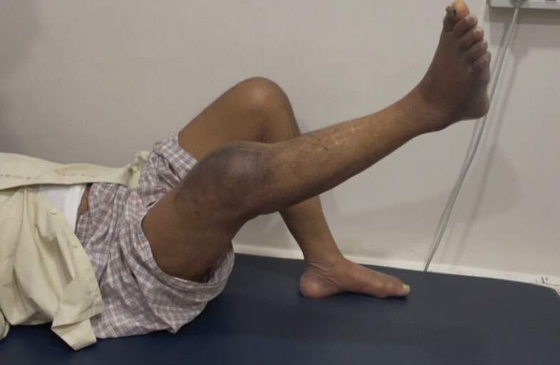
After cement hardening, the construct was found to be stable. Wound closure was performed over deep suction drains in layers, with satisfactory soft-tissue coverage achieved without undue tension. Post-operative radiographic imaging was performed, and a stable construct was noted (Fig. 7). Postoperatively, the patient was mobilized with assistive devices based on clinical stability and soft-tissue condition. Systemic antifungal therapy was continued under infectious disease guidance, with inflammatory markers monitored during follow-up. Intravenous micafungin 150 mg once a day for 6 months was advised. The biomarkers of the patient immediately postoperatively were as follows: CRP (4.2 mg/L), ESR (96 mm/h). On regular 3-monthly follow-up examinations, the patient demonstrated clinical improvement with reduction in pain and swelling and normalization of inflammatory markers, including CRP. At 3 months following surgery, the biomarkers for this patient were as follows: CRP (4.7 mg/L), ESR (34 mm/h). At 7 months following surgery, the biomarkers for this patient were as follows: CRP (4.2 mg/L), ESR (28 mm/h). At 1-year follow-up, the patient was able to perform a successful straight leg raise (Fig. 8).
Discussion
Fungal PJI and C. viswanathii
Fungal PJI is a rare but devastating complication of total knee arthroplasty and is associated with significant diagnostic delays and poor outcomes [7,8]. Approximately 90% of invasive candidiasis is caused by five common species (Candida albicans, Candida tropicalis, Candida glabrata, Candida krusei, and Candida parapsilosis). The incidence of PJI due to NAC species is rising and is often associated with aggressive local tissue destruction and marked antifungal resistance.
C. viswanathii is a rare pathogen, with limited literature describing its virulence, clinical manifestations, mycological properties, and management. First described in India in 1959 [11], the incidence of C. viswanathii remains sporadic. Most documented cases originate from South and Southeast Asia. The largest published case series of C. viswanathii infections, spanning 4 years in India, documented 23 cases [12]. A major challenge associated with C. viswanathii is accurate and timely diagnosis. Non-specific clinical presentation and low sensitivity of routine laboratory markers pose challenges in early recognition and treatment. A high index of suspicion is required even in immunocompetent individuals. Uncommon Candida species may be absent from routinely used laboratory databases, leading to delayed identification or inadequate therapy. Culture of multiple joint aspirates and intraoperative tissue samples remains the gold standard, while advanced identification techniques such as MALDI-TOF MS are crucial for detecting rare fungal organisms.
Data on the antifungal susceptibility of C. viswanathii (2015) are shown in Table 1 [12].
Demonstrating antifungal susceptibility of Candida viswanathii through minimum inhibitory concentration (MIC) range for various antifungal agents
| Organism/Year (n) | Antifungal agent | MIC range | MIC50 | MIC90 |
|---|---|---|---|---|
| Candida viswanathii 2015 (n=16) | Amphotericin B | 0.06–0.25 | 0.125 | 0.25 |
| Itraconazole | 0.03–1 | 0.25 | 0.25 | |
| Fluconazole | 0.5–64 | 64 | 64 | |
| Voriconazole | 0.03–8 | 1 | 2 | |
| Micafungin | 0.125 | 0.125 | 0.125 | |
| Caspofungin | 0.125–1 | 0.5 | 0.5 | |
| Anidulafungin | 0.125–1 | 0.5 | 1 | |
| Posaconazole | 0.0321–0.5 | 0.25 | 0.25 |
MALDI-TOF MS in identification of C. viswanathii
Uncommon organisms may be misidentified or remain unidentified unless expanded or custom reference libraries are used, including curated online resources such as MicrobeNet [1,2]. Although published data specifically addressing MALDI-TOF MS identification of C. viswanathii are limited, available reports suggest that this species can be distinguished from closely related taxa, such as C. tropicalis, when relevant spectra are present in the database [1,2]. MALDI-TOF MS has become an important tool in clinical mycology for rapid species-level identification of yeast isolates, including Candida species [13,14]. The technique identifies organisms by analyzing characteristic protein spectra from cultured isolates and matching them with reference databases, allowing timely species-level identification in routine practice. While this tool demonstrates high accuracy for common Candida species when appropriate reference spectra are available, identification of rare or emerging species may be limited by incomplete database representation (Fig. 9) [15].
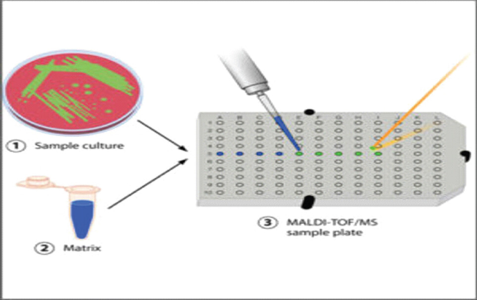
This highlights the value of MALDI-TOF MS in recognizing rare fungal pathogens in prosthetic joint infections, provided adequate reference data are available, and underscores the importance of correlating microbiological findings with clinical and operative features.
Susceptibility of C. viswanathii to antimicrobial therapy
C. viswanathii has been associated with elevated minimum inhibitory concentrations to fluconazole, increasing the risk of treatment failure with routine antifungal drugs [12]. Resistance or reduced susceptibility to commonly used antifungals may contribute to persistent infection, invasive disease, and potential nosocomial transmission, particularly in vulnerable populations. These factors highlight the importance of maintaining a high index of suspicion for persistent invasive infections not responding to antibiotic therapy, prompting early and precise mycological diagnosis combined with antifungal susceptibility testing. Prompt recognition of resistance patterns allows timely modification of antifungal regimens, which is crucial for optimizing outcomes and preventing complications [7,8].
Management of chronic fungal PJI remains challenging due to delayed diagnosis, aggressive local tissue destruction, and high rates of recurrence. Persistent intramedullary infection, soft-tissue destruction, and failure of staged debridement and spacer placement precluded reimplantation with a revision prosthesis. Knee arthrodesis thus remained a pragmatic and effective limb salvage strategy. Intramedullary fixation using cemented (PMMA) “K-nail” constructs provides stability, allows early ambulation, and facilitates canal debridement and high local delivery of antimicrobial agents, which is crucial for clearing persistent intramedullary fungal pathogens. Amphotericin B remains the most commonly recommended agent for local delivery due to its broad antifungal spectrum and heat-stable characteristics. Fusion rates are comparable – reported between 70% and 90% in chronically infected total knee arthroplasty. The use of a cement rod arthrodesis construct in our case allowed effective infection control while maintaining limb length and alignment, despite extensive soft-tissue compromise and failure of initial spacer-based management. Our case further supports the role of this technique as a definitive second-stage procedure following unsuccessful initial revision in rare and aggressive fungal infections.
Conclusion
Fungal PJI caused by C. viswanathii is extremely rare and poses significant diagnostic and therapeutic challenges. This case emphasizes the importance of maintaining a high index of suspicion in persistent or culture-negative PJIs. Advanced microbiological identification using MALDI-TOF MS assay, aggressive surgical management, antifungal-loaded cement, and prolonged systemic antifungal therapy are essential for successful outcomes.
Clinical Message
Persistent or culture-negative periprosthetic knee joint infections should prompt consideration of rare fungal pathogens, including C. viswanathii, especially in frail elderly patients. Early use of advanced diagnostic tools such as MALDI-TOF MS requires state-of-the-art laboratories, microbiological expertise, and reference databases for accurate identification and targeted antifungal therapy. In cases with severe soft-tissue compromise or failed revision surgery, antifungal-loaded cement knee arthrodesis using a novel fusion technique can serve as an effective limb-salvage strategy.
Conflict of Interest:
Nil
Source of Support:
Nil
Consent
The authors confirm that informed consent was obtained from the patient for publication of this article
How to Cite this Article
Joshi S, Patil S, Kulkarni M, Shaligram M, Muthiah S, Joshi R. First Reported Case of Periprosthetic Fungal Knee Joint Infection Caused by Candida viswanathii: A Case Report. J Orthop Case Rep. 2026 November;16(11): 92-98. doi: 10.13107/jocr.2026.v16.i11.8400.
References
- Centers for Disease Control and Prevention (CDC). MicrobeNet:Bacterial and Fungal Identification Database. Available from: https://www.cdc.gov/microbenet/. [Last accessed on 2026 Oct 12]. [Google Scholar] | [PubMed]
- Bader O. MALDI-TOF-MS-based species identification and typing approaches in medical mycology. Proteomics 2013;13:788-99. [Google Scholar] | [PubMed]
- Anagnostakos K, Kelm J, Schmitt E, Jung J. Fungal periprosthetic hip and knee joint infections: Clinical experience with a 2-stage treatment protocol. J Arthroplasty 2012;27:293-8. [Google Scholar] | [PubMed]
- Marra F, Robbins GM, Masri BA, Duncan C, Wasan KM, Kwong EH. Amphotericin B-loaded bone cement to treat osteomyelitis caused by Candida albicans. Can J Surg 2001;44:383-6. [Google Scholar] | [PubMed]
- Hwang BH, Yoon JY, Nam CH, Jung KA, Lee SC, Han CD. Fungal peri-prosthetic joint infection after primary total knee replacement. J Bone Joint Surg Br 2012;94:656-9. [Google Scholar] | [PubMed]
- Conway JD, Mont MA, Bezwada HP. Arthrodesis of the knee. J Bone Joint Surg Am 2004;86:835-48. [Google Scholar] | [PubMed]
- Kuiper JW, Van Den Bekerom MP, Van Der Stappen J, Nolte PA, Colen S. 2-stage revision recommended for treatment of fungal hip and knee prosthetic joint infections. Acta Orthop 2013;84:517-23. [Google Scholar] | [PubMed]
- Azzam K, Parvizi J, Jungkind D, Hanssen A, Fehring T, Springer B. Microbiological, clinical, and surgical features of fungal prosthetic joint infections: A multi-institutional experience. J Bone Joint Surg Am 2009;91(Suppl 6)142-9. [Google Scholar] | [PubMed]
- Ramamohan N, Zeineh N, Grigoris P, Butcher I. Candida glabrata infection after total hip arthroplasty. J Infect 2001;42:74-6. [Google Scholar] | [PubMed]
- Phelan DM, Osmon DR, Keating MR, Hanssen AD. Delayed reimplantation arthroplasty for candidal prosthetic joint infection: A report of 4 cases and review of the literature. Clin Infect Dis 2002;34:930-8. [Google Scholar] | [PubMed]
- Randhawa HS, Mishra SK, Damodaran VN, Prakash A, Chowdhary A, Khan ZU. Pathogenicity of Candida viswanathii for normal and cortisone-treated mice. J Mycol Med 2015;25:287-92. [Google Scholar] | [PubMed]
- Shankarnarayan SA, Rudramurthy SM, Chakrabarti A, Shaw D, Paul S, Sethuraman N. Molecular typing and antifungal susceptibility of Candida viswanathii, India. Emerg Infect Dis 2018;24:1956-8. [Google Scholar] | [PubMed]
- Marklein G, Josten M, Klanke U, Müller E, Horré R, Maier T. Matrix-assisted laser desorption ionization-time of flight mass spectrometry for fast and reliable identification of clinical yeast isolates. J Clin Microbiol 2009;47:2912-7. [Google Scholar] | [PubMed]
- Dhiman N, Hall L, Wohlfiel SL, Buckwalter SP, Wengenack NL. Performance and cost analysis of matrix-assisted laser desorption ionization-time of flight mass spectrometry for routine identification of yeast. J Clin Microbiol 2011;49:1614-6. [Google Scholar] | [PubMed]
- Sendid B, Ducoroy P, François N, Lucchi G, Spinali S, Vagner O. Evaluation of MALDI-TOF mass spectrometry for the identification of medically important yeasts in the clinical laboratories of Dijon and Lille hospitals. Med Mycol 2013;51:25-32. [Google Scholar] | [PubMed]
© 2026 Journal of Orthopaedic Case Reports - Published by Indian Orthopaedic Research Group
About the Authors
-

Satyajeet Joshi
-

Suresh Patil
-

Mahesh Kulkarni
-

Megh Shaligram
-

Shivaram Muthiah
-

Rasika Joshi
Related Articles in Journal of Orthopaedic Case Reports
 November 1, 2026 Chronic Bilateral Knee Pain Following Total Knee Arthroplasty Due to Titanium Allergy: A Case Report
 November 1, 2026 Management of Periprosthetic Total Knee Infection with Partial Hardware Removal: A Case Report
 November 1, 2026 Intraoperative Proximal Fibula Fracture during Total Knee Arthroplasty for Moderate Valgus Knee Deformity: A Case Report
 October 1, 2026 Intraoperative Discovery of Incorrect Femoral Component Packaging during Bilateral Total Knee Arthroplasty: A Near-Miss Case Report