


Introduction
Total knee arthroplasty (TKA) remains a standard surgical treatment for advanced knee osteoarthritis, providing substantial improvements in pain and function. It provides substantial pain relief, improved function, and durable implant survival [1]. The medial parapatellar method remains the cornerstone surgical approach, offering consistent joint exposure and allowing effective mobilization of the patella. However, this technique requires incision through the extensor mechanism and may potentially compromise the patellar blood supply [2]. In contrast, the subvastus method involves dissection beneath the vastus medialis yet maintains the quadriceps tendon and extensor mechanism. This preservation has the potential to facilitate early quadriceps recovery and rehabilitation [3]. Previous comparative studies have reported advantages of the subvastus method in select early post-operative findings, such as quadriceps recovery, range of motion (ROM), and early functional recovery. However, the subvastus method can be technically more challenging and may provide more limited exposure and difficulty with patellar eversion, particularly in patients with large or stiff knees.
Despite the hypothetical advantages of maintaining the extensor mechanism, the clinical superiority of the subvastus method over the conventional medial parapatellar method remains uncertain. In a study of 90 patients, Bourke et al. [4] showed no significant difference in functional scores between the two methods. However, patients assigned to the subvastus method achieved straight-leg raising earlier, although the method was associated with greater technical difficulty for surgeons. Similarly, systematic reviews and meta-analyses have demonstrated some early post-operative advantages with the subvastus method but have not consistently demonstrated clinically important differences in overall functional outcomes [5]. Furthermore, evidence directly evaluating both functional and radiological outcomes remains limited. This uncertainty provides a rationale for further prospective comparative evaluation of these two commonly used methods. This study compared functional, pain, and radiological outcomes among the subvastus and medial parapatellar methods.
Materials and Methods
Study design and patient population
A randomized controlled trial included patients diagnosed with primary or secondary osteoarthritis who underwent TKA at a major medical center in India from July 2023 to September 2025. Patients were randomly allocated to either the subvastus (Group 1) or medial parapatellar (Group 2) method. Patients with a history of prior knee surgeries or extensor mechanism disruption were excluded from the trial. The study received approval from the Institutional Ethics Committee (HHL/LH/F149). The study protocol was approved by the Institutional Ethics Committee, and written informed consent was obtained from all patients before inclusion in the study.
Sample size
The required sample size was determined using G*Power 3.1. A medium effect size (Cohen’s d = 0.5), a two-sided α of 0.05, and an independent-samples t-test were assumed. The calculation yielded 100 patients, with 50 patients required per group, to achieve 70% power. This sample size was considered feasible and comparable to that used in previous studies of TKA.
Study procedure and study endpoints
All surgical and radiographic assessments were performed by the same team to minimize interobserver variability. Intraoperative parameters such as blood loss and complications were recorded immediately. Pain levels were evaluated using the Visual Analog Scale (VAS). Functional recovery was evaluated using the Knee Society Score (KSS) and ROM measurements. Radiological assessment included standardized anteroposterior and lateral radiographs for assessing coronal and sagittal component alignment using the α (femoral coronal), β (tibial coronal), γ (femoral sagittal), and δ (tibial sagittal) angles. The hip-knee-ankle (HKA) angle was measured on standing long-leg radiographs to assess mechanical axis restoration.
Data collection and follow-up
Data were prospectively collected from all patients enrolled in the trial. Following enrollment, each patient was randomly allocated to either (i) group 1 or (ii) group 2 using a unique hospital identification (UHID) number – odd numbers for the subvastus group and even numbers for the medial parapatellar group. Baseline demographic details and pre-operative deformities were collected and documented. Pre-operative assessments of pain using the VAS, ROM, and the objective and functional components of the KSS were performed. Post-operative complications such as wound dehiscence, wound infection, deep vein thrombosis (DVT), and blisters were documented throughout the follow-up duration. Post-operative assessments were conducted at 2 weeks, 6 weeks, 3 months, and 6 months.
Statistical analysis
Statistical analyses were performed using the Statistical Package for the Social Sciences version 20.0 (IBM Corp.). Continuous variables are expressed as mean ± standard deviation, whereas categorical variables are reported as counts and percentages. Differences in continuous variables between groups were assessed using the independent-samples Student’s t-test, whereas categorical variables were analyzed using the Chi-square test. All tests were two-sided, and P < 0.05 was considered indicative of statistical significance.
Results
Demographic details of study patients
The baseline demographics were comparable between the two groups. Mean age was 62.50 ± 5.06 years in Group 1 and 61.54 ± 5.20 years in Group 2 (P = 0.352). Similarly, the mean body mass index was comparable between the groups, i.e., 24.42 ± 3.92 in Group 1 and 23.75 ± 4.42 kg/m2 in Group 2 (P = 0.422). Females constituted the majority of participants in both groups (P = 0.349). The demographics of the trial patients are elaborated in Table 1.
Demographic characteristics of study patients
| Variable | Subvastus (n =50) | Medial parapatellar (n =50) | P-value |
|---|---|---|---|
| Age, years | 62.50±5.06 | 61.54±5.20 | 0.352 |
| Body mass index, kg/m2 | 24.42±3.92 | 23.75±4.42 | 0.422 |
| Males, n (%) | 14 (28.0) | 10 (20.0) | 0.349 |
| Females, n (%) | 36 (72.0) | 40 (80.0) |
P<0.05 was considered statistically significant
Pre-operative clinical and radiological parameters of study patients
Pre-operative clinical and radiological parameters were comparable among the two groups. There were no significant differences in VAS scores (P = 0.172), ROM (P = 0.393), KSS (objective) (P = 0.630), KSS (functional) (P = 0.402), or HKA alignment (P = 0.867). The pre-operative clinical and radiological parameters of the study patients are detailed in Table 2.
Pre-operative functional and radiological variables of study patients
| Variable | Subvastus (n =50) | Medial parapatellar (n =50) | P-value |
|---|---|---|---|
| Visual Analog Scale | 6.74±0.94 | 7.00±0.95 | 0.172 |
| Range of motion | 84.82±14.84 | 87.00±10.15 | 0.393 |
| Knee society score (objective) | 40.30±6.40 | 39.70±6.00 | 0.63 |
| Knee society score (functional) | 19.70±5.48 | 18.80±5.21 | 0.402 |
| Hip-knee-ankle | 167.93±2.80 | 168.03±2.69 | 0.867 |
P<0.05 was considered statistically significant
Post-operative complications of the study patients
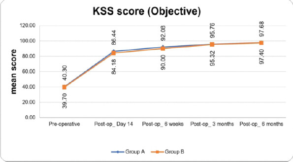
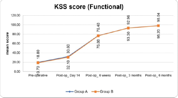
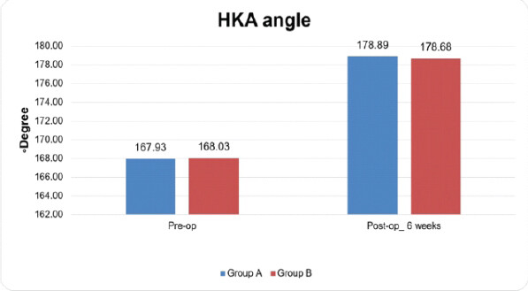
Post-operative complications were infrequent and comparable between the two groups. Wound dehiscence occurred in 1 (2.0%) patient in each group. No cases of DVT were reported in either group. Blister formation occurred in 1 (2.0%) patient in each group, indicating a similar overall complication profile between the two methods. The post-operative complications of the study patients are demonstrated in Table 3 and Figs. 1–3.
Post-operative complications of the study patients
| Complications | Subvastus (n=50) (%) | Medial parapatellar (n=50) (%) |
|---|---|---|
| Deep wound infection | 2 (4.0) | 3 (6.0) |
| Wound dehiscence | 1 (2.0) | 1 (2.0) |
| Deep vein thrombosis | 0 (0) | 0 (0) |
| Blister | 1 (2.0) | 1 (2.0) |
Discussion
Post-operative pain plays a critical role in early recovery and rehabilitation after TKA. The VAS score was 6.74 ± 0.94 and 7.00 ± 0.95 (P = 0.172) in the subvastus and medial parapatellar groups. Patients in the subvastus group had considerably reduced VAS scores during the initial 3 post-operative days compared with those in the medial parapatellar group (P <0.05). Similarly, another study found a trend of lower post-operative VAS scores in subvastus patients compared to medial parapatellar patients up to 6 months [6,7]. The study by Shah et al. [8] reported significantly lower VAS scores among patients undergoing the subvastus method. Moreover, patients treated with this technique required approximately 40% less post-operative analgesic compared to those who underwent the conventional medial parapatellar method. The meta-analysis of 10 studies revealed that patients undergoing the subvastus method required less analgesic medication within 24 h [9,10] and experienced less pain during the first 1–3 post-operative days [11,12], although two studies did not observe a contrast in post-operative pain scores between the methods [12,13]. The decreased pain observed with the subvastus method may be ascribed to the maintenance of the extensor mechanism and the avoidance of quadriceps tendon transection, thereby reducing soft-tissue trauma [14].
Post-operative ROM is an influential indicator of functional recovery following TKA. In our trial, at the 2-week follow-up, the subvastus patients demonstrated greater mean ROM of 82.56° ± 9.11° compared with 78.02° ± 7.33° in the medial parapatellar patients (P = 0.007). This advantage remained at 6 weeks (subvastus: 98.14° ± 10.38° and medial parapatellar: 92.86° ± 5.20°; P = 0.002). However, at 3 months, ROM was comparable between the two groups (P = 0.333), suggesting that the subvastus method may primarily offer an advantage in early post-operative functional recovery. These outcomes are in line with those of Bridgman et al. [13], who stated better ROM for subvastus patients 1 week postoperatively. Similarly, Bourke et al. [4] reported better mean flexion of subvastus patients compared to medial parapatellar patients up to discharge, while Lingayat et al. [15] observed better ROM at 3 months. The preservation of the quadriceps integrity in the subvastus method likely contributes to reduced post-operative stiffness and earlier initiation of active knee motion.
The objective and functional KSS values improved progressively after surgery in both patient groups. At the 6-month follow-up, the groups showed comparable outcomes, with no statistically significant difference (P = 0.280). Our study findings are supported by those of Mathew et al. [16]. Bourke et al. [4] conducted a study with longer-term follow-up and reported that the American KSS functional scores at 12 (P = 0.032) and 18 months (P = 0.028) favored the medial parapatellar method. Similarly, Teng et al., [17] in their meta-analysis, reported significantly better KSS functional scores at 4–6 weeks and higher knee scores at 12 months among patients treated with the subvastus approach. These findings suggest that the early advantage of the subvastus method arises from enhanced quadriceps activation and reduced trauma to the extensor mechanism, promoting faster restoration of independent ambulation.
Radiological findings revealed comparable results between the subvastus and medial parapatellar groups regarding coronal or sagittal alignment. Similarly, post-operative HKA angles were comparable, indicating accurate mechanical axis restoration in both groups. Thus, surgical proficiency and attention to technique are critical determinants of component placement accuracy, irrespective of the method.
Post-operative complications were minimal and comparable between the two methods. Deep wound infections occurred in 2 (4.0%) subvastus patients and 3 (6.0%) medial parapatellar patients. Wound dehiscence occurred in 1 (2.0%) patient in both the subvastus and medial parapatellar groups. No cases of DVT were reported. These outcomes agree with observations of Wu et al., [18] who also observed comparable rates of infection and DVT between the methods. The findings of Hu et al. [14] emphasized the role of surgeon experience in preventing complications, particularly in obese patients or patients with limited pre-operative knee flexion.
This study has several limitations. First, the relatively small sample size (100 patients) may have limited statistical power to detect differences in clinical outcomes and complications. Second, the single-center design may limit generalizability to other institutions and patient populations. Third, the absence of reported blinding may have introduced assessment or reporting bias. Fourth, allocation based on odd and even UHID numbers was predictable and may have introduced selection bias. Fifth, the 6-month follow-up was insufficient to evaluate long-term functional outcomes, implant survival, and revision rates. Sixth, radiological measurements were assessed by a single observer, preventing evaluation of interobserver reliability and reproducibility. Seventh, the reported statistical power of 70% may have been inadequate to detect smaller but clinically meaningful differences. Eighth, the effects of surgeon experience, the learning curve, and technical difficulty were not formally assessed. Ninth, the small sample size and low complication rates limited reliable comparisons of uncommon adverse events between the two approaches. Larger multicenter studies with longer follow-up, concealed random allocation, blinded outcome assessment where feasible, and standardized radiological evaluation are warranted.
Conclusion
The subvastus method was associated with better early post-operative pain relief and ROM, while functional and radiological outcomes were comparable at later follow-up.
Clinical Message
Both the subvastus and medial parapatellar methods are viable methods for total knee arthroplasty. Selection of methods should be tailored to the individual patient, taking into account patient-specific factors, the operator’s experience, and the extent of exposure required for optimal implant positioning.
Conflict of Interest:
Nil
Source of Support:
Nil
Consent
The authors confirm that informed consent was obtained from the patient for publication of this article
How to Cite this Article
Solanki J, Yadav M, Jha AK, Patel S, Singh SK. Functional and Radiological Results of the Subvastus Versus Medial Parapatellar Method in Total Knee Arthroplasty: A Randomized Controlled Cohort. J Orthop Case Rep. 2026 November;16(11):457-462. doi: 10.13107/jocr.2026.v16.i11.8524.
References
- Tampere T, Arnout N, Victor J. Total knee arthroplasty: The need for better patient selection. Knee Surg Sports Traumatol Arthrosc 2025;33:784-8. [Google Scholar] | [PubMed]
- Geng L, Fu J, Xu C, Ren P, Wang YM, Ji QB. The comparison between mini-subvastus approach and medial parapatellar approach in TKA : A prospective double-blinded randomized controlled trial. Orthop Surg 2022;14:2878-87. [Google Scholar] | [PubMed]
- Gupta G, Gupta AR, Aggarwal S, Raghuraj A. A comparative study between sub-vastus and medial parapatellar approaches in total knee arthroplasty: A prospective study. J Clin Orthop Trauma 2025;70:103209. [Google Scholar] | [PubMed]
- Bourke MG, Jull GA, Buttrum PJ, FitzPatrick PL, Dalton PA, Russell TG. Comparing outcomes of medial parapatellar and subvastus approaches in total knee arthroplasty: A randomized controlled trial. J Arthroplasty 2012;27:347-53.e1. [Google Scholar] | [PubMed]
- World Medical Association World medical association declaration of Helsinki: Ethical principles for medical research involving human subjects. JAMA 2013;310:2191-4. [Google Scholar] | [PubMed]
- Dixon JRJr. The international conference on harmonization good clinical practice guideline. Quality Assurance 1999;6:65-74. [Google Scholar] | [PubMed]
- Narayanan AS, Karuppanan S, Sivanandan MH, Mathew N, Maligi VB, Baskar N. A comparative study of functional outcome of subvastus Vs medial parapatellar approaches in total knee arthroplasty procedure. Int J Drug Deliv Technol 2026;16:139-46. [Google Scholar] | [PubMed]
- Shah P, Singh R, Shanwal PK, Yadav DK, Jain R. Comparing the subvastus and medial parapatellar approaches in total knee arthroplasty. J Orthop Case Rep 2026;16:346-50. [Google Scholar] | [PubMed]
- Roysam GS, Oakley MJ. Subvastus approach for total knee arthroplasty: A prospective, randomized, and observer-blinded trial. J Arthroplasty 2001;16:454-7. [Google Scholar] | [PubMed]
- Sastre S, Sanchez MD, Lozano L, Orient F, Fontg F, Nuñez M. Total knee arthroplasty: Better short-term results after subvastus approach: A randomized, controlled study. Knee Surg Sports Traumatol Arthrosc 2009;17:1184-8. [Google Scholar] | [PubMed]
- Pan WM, Li XG, Tang TS, Qian ZL, Zhang Q, Zhang CM. Mini-subvastus versus a standard approach in total knee arthroplasty: A prospective, randomized, controlled study. J Int Med Res 2010;38:890-900. [Google Scholar] | [PubMed]
- Van Hemert WL, Senden R, Grimm B, Van Der Linde MJ, Lataster A, Heyligers IC. Early functional outcome after subvastus or parapatellar approach in knee arthroplasty is comparable. Knee Surg Sports Traumatol Arthrosc 2011;19:943-51. [Google Scholar] | [PubMed]
- Bridgman SA, Walley G, MacKenzie G, Clement D, Griffiths D, Maffulli N. Sub-vastus approach is more effective than a medial parapatellar approach in primary total knee arthroplasty: A randomized controlled trial. Knee 2009;16:216-22. [Google Scholar] | [PubMed]
- Hu W, Nie Y, Li Y, Yang B, Su Z, Zhu Z. A modified minimally invasive subvastus approach shows superior early postoperative clinical outcomes compared to the medial parapatellar approach in total knee arthroplasty for osteoarthritis patients: A comparative study. BMC Musculoskelet Disord 2025;26:1117. [Google Scholar] | [PubMed]
- Lingayat MB, Vimal PV, Nawkhare R, Sharma R, Lakde S, Rashinkar OK. Comparing the subvastus and medial parapatellar approaches in total knee replacement. Int J Res Orthop 2024;10:1296-302. [Google Scholar] | [PubMed]
- Mathew AJ, Hugh C, Philip SG, Mathew T, Theodore L. Comparative study with 2 years follow up on the functional outcome of subvastus versus medial parapatellar approach in total knee arthroplasty in a tertiary care center in South India. Int J Res Orthop 2025;12:152-6. [Google Scholar] | [PubMed]
- Teng Y, Du W, Jiang J, Gao X, Pan S, Wang J. Subvastus versus medial parapatellar approach in total knee arthroplasty: Meta-analysis. Orthopedics 2012;35:e1722-31. [Google Scholar] | [PubMed]
- Wu Y, Zeng Y, Bao X, Xiong H, Hu Q, Li M. Comparison of mini-subvastus approach versus medial parapatellar approach in primary total knee arthroplasty. Int J Surg 2018;57:15-21. [Google Scholar] | [PubMed]
© 2026 Journal of Orthopaedic Case Reports - Published by Indian Orthopaedic Research Group
About the Authors
-

Jayvirsinh Solanki
-

Mukesh Yadav
-

Amit Kumar Jha
-

Swaroop Patel
-

S K Singh
Related Articles in Journal of Orthopaedic Case Reports
 September 1, 2026 Comparative Study of Post-operative Pain and Functional Outcomes in Robotic-assisted Total Knee Replacement versus Cruciate-retaining Manual Total Knee Replacement
 October 1, 2025 Radiological and Functional Assessment of Tibial Plateau Fractures Managed with Locking Compression Plates: A Prospective Study
 October 1, 2025 Outcomes of Proximal Humeral Fracture Fixation Using Minimally Invasive Plate Osteosynthesis: A Prospective Clinical and Radiographic Study
 November 1, 2026 Predictors of Functional Recovery Following Primary Ilizarov External Fixation in Compound Distal Third Tibial Fractures