

Physeal-sparing intramedullary interlocking nailing can provide stable fixation, reliable fracture union, and excellent functional recovery in adolescents with tibial shaft fractures while minimizing the risk of growth plate injury and growth-related complications.
Dr. Anil Kumar Karpetee, Department of Orthopaedics, Netaji Subhash Chandra Bose Medical College and Hospital, Jabalpur, Madhya Pradesh, India. E-mail: anilkarpeti47@gmail.com
Abstract
Introduction: Management of tibial shaft fractures in adolescents with open physes remains challenging because stable fixation must be achieved without compromising physeal integrity. This study evaluated the clinical, radiological, and functional outcomes of physeal-sparing intramedullary interlocking nailing in adolescent tibial shaft fractures.
Material and Methods: A prospective descriptive interventional cohort study was conducted in the Department of Orthopaedics at a tertiary care teaching hospital between May 2024 and April 2026. Twenty-six adolescents aged 13–17 years with tibial shaft fractures and open physes underwent physeal-sparing intramedullary interlocking nailing. Patients were assessed using Visual Analog Scale (VAS) for pain, tibial fracture healing score (TFHS) for functional outcome, and radiographic union scale for tibial fractures (RUST) for radiological healing. Follow-up evaluations were performed at 2, 6, 12, and 24 weeks postoperatively.
Results: The mean age was 15.23 ± 1.18 years, and 76.9% of patients were male. Road traffic accidents accounted for 69.2% of injuries. Closed fractures constituted 76.9% of cases, while 53.8% were AO/OTA type 42-A fractures. Median VAS pain scores improved from 5.0 at 2 weeks to 0.0 at 12 and 24 weeks. Median TFHS total score improved from 2.50 at 6 weeks to 1.00 at 24 weeks. Mean RUST scores increased significantly from 5.08 ± 0.74 at 6 weeks to 8.42 ± 0.99 at 12 weeks and 11.54 ± 0.58 at 24 weeks (P < 0.001). Radiographic union was achieved in 26.9% of patients at 12 weeks and in 100% by 24 weeks, with a mean union time of 14.23 ± 2.23 weeks. Complications included superficial infection (3.8%), anterior knee pain (7.7%), and delayed union (7.7%), with no cases of deep infection, nonunion, malunion, implant failure, or growth disturbance.
Conclusion: Physeal-sparing intramedullary interlocking nailing provides reliable fracture union, excellent functional recovery, and a low complication rate in adolescents with tibial shaft fractures while preserving physeal integrity.
Keywords: Adolescent tibial fracture, physeal-sparing nailing, intramedullary interlocking nail, fracture union, radiographic union scale for tibia score, functional outcome.

Tibial shaft fractures are among the most frequently encountered long-bone injuries in the pediatric and adolescent population and account for a substantial proportion of trauma-related orthopedic admissions. Although many fractures can be managed successfully with non-operative methods, unstable fracture patterns, open injuries, polytrauma, and failure of conservative treatment often necessitate surgical stabilization [1].
Several operative techniques have been described for adolescent tibial shaft fractures, including elastic stable intramedullary nailing (ESIN), external fixation, plate osteosynthesis, and intramedullary interlocking nailing. ESIN remains a commonly utilized option in skeletally immature patients because it minimizes physeal injury and provides satisfactory fracture healing. However, complications such as implant irritation, loss of reduction, delayed mobilization, and the need for secondary implant removal continue to be reported [2,3].
Rigid intramedullary interlocking nailing is considered the standard treatment for adult tibial shaft fractures because it provides stable fixation, permits early mobilization, and demonstrates high union rates. Nevertheless, its use in adolescents with open physes has traditionally been limited because of concerns regarding potential injury to the proximal tibial growth plate and the consequent risk of growth disturbance or angular deformity [4].
To address these concerns, physeal-sparing intramedullary nailing techniques have been developed in which the nail entry point is positioned distal to the proximal tibial physis while maintaining alignment with the medullary canal. Early clinical studies have suggested that this technique can provide stable fixation without evidence of physeal arrest, while also allowing earlier weight bearing and reduced immobilization compared with traditional methods [4,5].
Recent literature has highlighted the increasing interest in optimizing fixation strategies for tibial shaft fractures and improving functional outcomes while minimizing complications and reoperations [6]. However, evidence regarding the safety and effectiveness of physeal-sparing intramedullary interlocking nailing in adolescents remains limited, particularly in developing-country settings. Therefore, the present study was undertaken to evaluate the clinical, radiological, and functional outcomes of physeal-sparing intramedullary interlocking nailing in adolescent tibial shaft fractures.
Ethical considerations
The study was conducted in accordance with ethical principles governing human research. Ethical approval was obtained from the Institutional Ethics Committee (No. IEC/2024/4539-120, dated May 16, 2024). Written informed consent was obtained from all patients and their guardians before enrolment. Participation was voluntary, and confidentiality of patient information was maintained throughout the study period.
Study design and setting
This prospective study was conducted to evaluate the clinical, radiological, and functional outcomes of physeal-sparing intramedullary interlocking nailing in adolescent tibial shaft fractures. The study was carried out in the Department of Orthopaedics, Netaji Subhash Chandra Bose Medical College and Hospital, Jabalpur, Madhya Pradesh, a tertiary care teaching institution serving a large population of trauma patients from both urban and rural areas.
Study population
Adolescent patients presenting with tibial shaft fractures during the study were screened for eligibility. A total of 26 patients fulfilling the selection criteria were enrolled after obtaining written informed consent from the patients and their guardians.
Inclusion criteria
Patients meeting the following criteria were included:
- Age between 13 and 17 years
- Tibial shaft fracture with radiologically open proximal tibial physis
- Closed fractures and Gustilo–Anderson Grade I or Grade II open fractures
- Willingness to undergo surgical treatment and participate in follow-up assessments.
Exclusion criteria
Patients were excluded if they had:
- Gustilo–Anderson Grade III open fractures
- Associated intra-articular fractures involving the proximal or distal tibia
- Pathological fractures
- Medical contraindications to surgery
- Refusal to provide consent for operative intervention.
Sample size
The study included 26 patients. Sample size estimation was performed using the formula: n = Z²pq/d²; where Z = 1.96 at a 95% confidence interval, P = 0.0027, q = 1 − P = 0.9973, and d = 0.02. Based on these assumptions, the calculated sample size was 26 patients.
Preoperative assessment
All patients underwent detailed clinical and radiological evaluation before surgery. Demographic information, mechanism of injury, and relevant medical history were recorded. A comprehensive musculoskeletal examination was performed to identify associated injuries and assess neurovascular status.
Radiographic evaluation included standard anteroposterior and lateral radiographs of the affected tibia, incorporating one joint above and one joint below the fracture site. Limb elevation and temporary stabilization were provided until definitive fixation was undertaken.
Surgical technique
All patients underwent fixation using a physeal-sparing intramedullary interlocking nail. Surgery was performed with the patient in the supine position on a radiolucent operating table. Adequate knee flexion of at least 90° was obtained to facilitate nail insertion. Fracture reduction was achieved under fluoroscopic guidance and maintained by closed or mini-open techniques as required.
A longitudinal skin incision was made along the medial border of the tibial tubercle. The entry point was established approximately 1–2 cm distal to the proximal tibial physis to avoid physeal injury. Using a bone awl, an entry portal was created, and a 3.2-mm guidewire was advanced into the medullary canal in line with the tibial shaft axis. The guidewire was directed at a relatively steep angle to protect the posterior tibial cortex and maintain a physeal-sparing trajectory.
Sequential reaming was subsequently performed, generally 1.5 mm larger than the selected nail diameter. An appropriately sized tibial interlocking nail, most commonly 8 mm in diameter, was inserted over the guidewire. In patients with a narrow medullary canal or shorter tibial length, a suitably sized humeral interlocking nail was used when conventional tibial nails were unavailable. Proximal and distal locking screws were inserted under fluoroscopic guidance to achieve stable fixation.
Implant system
The instrumentation used during surgery included a tibial interlocking nail, locking bolts, 3.2-mm guidewire, 3.2-mm and 4.0-mm drill bits, depth gauge, reamers, insertion handle, battery-operated power drill, protection sleeve, bone awl, screwdriver, connecting bolt, holding sleeve, drill sleeve, and trocar.
Postoperative rehabilitation
Following surgery, the operated limb was maintained in an elevated position. Rehabilitation protocols were individualized according to fracture configuration, fixation stability, bone quality, and patient compliance.
Isometric quadriceps strengthening exercises and ankle pump exercises were initiated on the 2nd postoperative day. Progressive knee mobilization was encouraged as tolerated. Both active and passive range-of-motion exercises, including straight-leg raising, were performed. During the initial 48 h, emphasis was placed on ankle pumping exercises and static quadriceps strengthening. Routine wound inspection was performed during dressing changes. Surgical drains were not routinely used because most procedures were completed through a closed technique.
Outcome measures
Patients were assessed using clinical and radiological parameters throughout the follow-up period. The primary outcome measures included fracture union and functional recovery.
The following variables were evaluated:
- Demographic characteristics (age and sex)
- Mechanism and mode of injury
- Fracture characteristics, including location, morphology, and open/closed status
- Radiological healing assessed using the radiographic union scale for tibial fractures (RUST)
- Functional outcome assessed using the tibial fracture healing score (TFHS)
- Pain severity assessed using the Visual Analog Scale (VAS)
- Postoperative complications include superficial or deep infection, delayed union, nonunion, malunion, implant failure, limb length discrepancy, and growth-related disturbances.
Follow-up protocol
Patients were reviewed at 2, 6, 12, and 24 weeks postoperatively. At each visit, clinical examination and radiographic evaluation were performed (Figures 1-5). Pain scores, functional recovery, progression of fracture healing, and any complications were systematically documented.
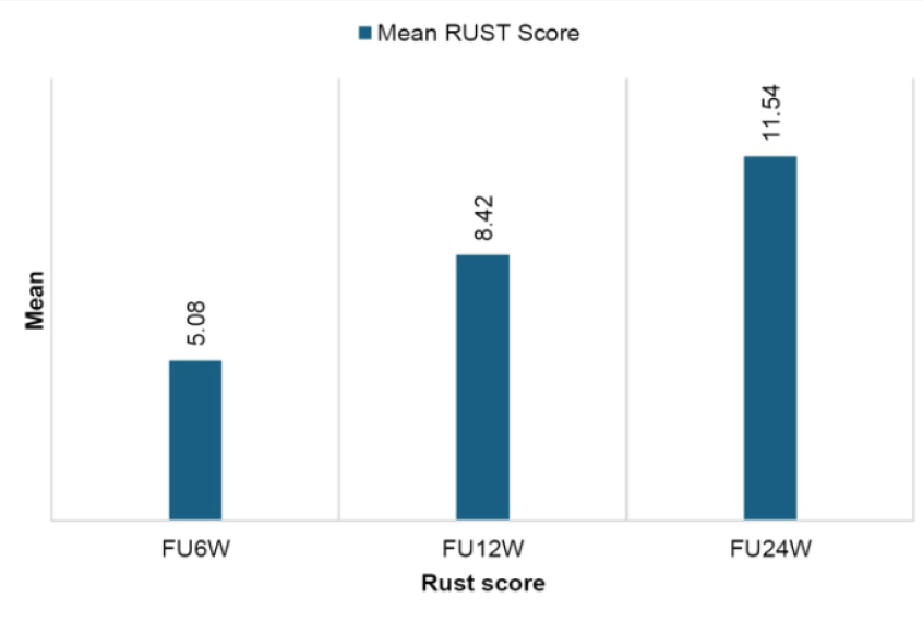
Figure 1: Mean radiographic union scale for tibial fractures score progression.
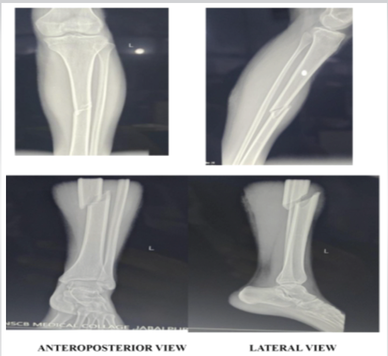
Figure 2: Pre-operative X-rays.
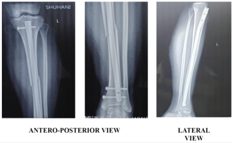
Figure 3: Immediate post-operative X-rays.
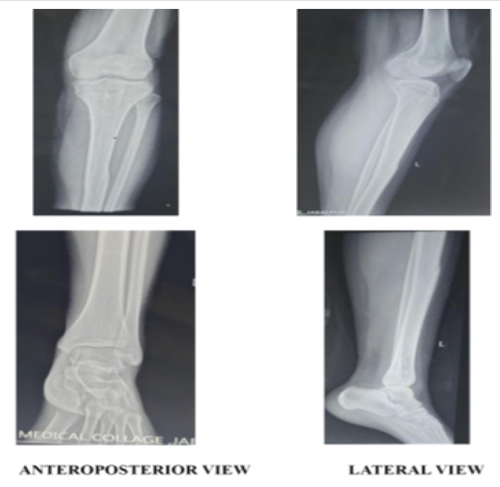
Figure 4: Implant removal done at 12 months.
Figure 5: Clinical images at 12 months follow up.
Statistical analysis
Data were compiled and analyzed using the Statistical Package for the Social Sciences (SPSS) software for Windows. Continuous variables were expressed as mean ± standard deviation or median with interquartile range (IQR), depending on data distribution. Categorical variables were presented as frequencies and percentages. Appropriate inferential statistical tests were applied where necessary, and a P < 0.05 was considered statistically significant.
For the 26 enrolled patients, the mean age was 15.23 ± 1.18 years (range: 13–17 years). Most of the participants belonged to the 15–16-year age group (57.7%), followed by 13–14 years (26.9%) and 17 years (15.4%). Males constituted 76.9% of the study population, while females accounted for 23.1%. The mean height and weight were 166.35 ± 5.17 cm and 55.46 ± 6.88 kg, respectively. Based on body mass index (BMI), 76.9% of patients had normal BMI, 19.2% were underweight, and 3.8% were overweight (Table 1).
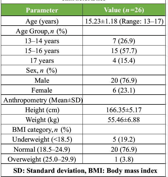
Table 1: Patient demographics and baseline characteristics
Road traffic accidents were the predominant mechanism of injury, accounting for 69.2% of cases, whereas sports-related injuries and falls each contributed 15.4%. Closed fractures were observed in 76.9% of patients, while 23.1% sustained Gustilo type I or II open fractures. The middle third of the tibial shaft was the most frequently affected site (57.7%), followed by the lower third (23.1%) and upper third (19.2%). According to the AO/OTA classification, simple (42-A) fractures represented 53.8% of cases, wedge (42-B) fractures 30.8%, and complex (42-C) fractures 15.4%. Comminuted fractures were the most common fracture pattern (30.8%), followed by spiral (26.9%), short oblique (23.1%), and transverse fractures (19.2%) (Table 2).
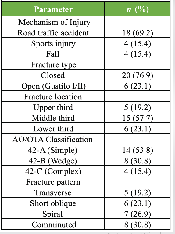
Table 2: Injury and fracture morphology
Closed reduction was achieved in 84.6% of patients, while a mini-open reduction technique was required in 15.4%. Reaming was performed in 88.5% of procedures. The mean nail diameter and length were 8.13 mm and 308.46 mm, respectively. A single proximal locking screw was used in 65.4% of patients, whereas two proximal screws were used in 34.6%. Distal fixation with two locking screws was performed in 92.3% of cases. The median operative duration was 91 minutes, the median estimated blood loss was 121 mL, and the median fluoroscopy time was 96 s. The mean distance between the guidewire entry point and the proximal tibial physis was 2.92 ± 0.28 cm (range: 2.6–3.5 cm) (Table 3).
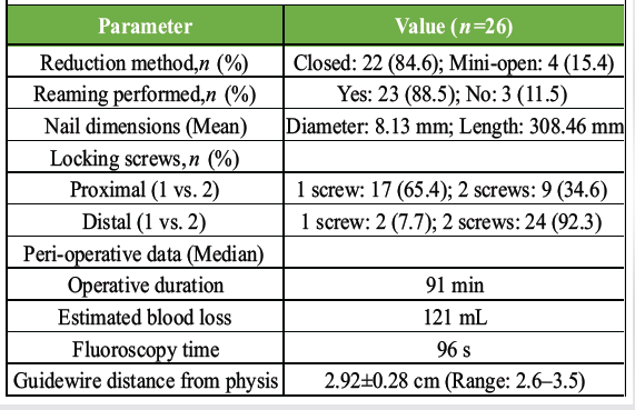
Table 3: Operative details and measurements
Clinical outcomes demonstrated progressive improvement throughout the follow-up period. The median VAS pain score decreased from 5.0 (IQR: 2) at 2 weeks to 2.0 (IQR: 2) at 6 weeks and reached 0.0 at both 12 and 24 weeks. Similarly, components of the tibia-femur-hindfoot score (TFHS) showed continuous improvement over time. The median total TFHS improved from 2.50 (IQR: 0.5) at 6 weeks to 1.63 (IQR: 0.75) at 12 weeks and further to 1.00 (IQR: 0.5) at 24 weeks, indicating progressive functional recovery (Table 4).
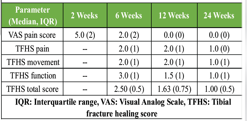
Table 4: Clinical outcomes over time (VAS and TFHS)Table 4: Clinical outcomes over time (VAS and TFHS)
Radiographic assessment revealed no cases of fracture union at 6 weeks. By 12 weeks, radiographic union was achieved in 7 patients (26.9%), whereas complete union was observed in all patients by 24 weeks. The mean RUST score increased significantly from 5.08 ± 0.74 at 6 weeks to 8.42 ± 0.99 at 12 weeks and further to 11.54 ± 0.58 at 24 weeks (P < 0.001 for both intervals). The mean time to radiographic union was 14.23 ± 2.23 weeks (Table 5). The progressive increase in mean RUST scores over the follow-up period is illustrated in Fig. 1.
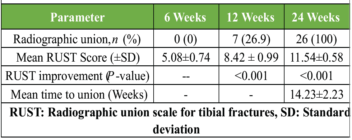
Table 5: Radiographic union and RUST score progression
Complications were infrequent. Superficial infection occurred in one patient (3.8%) and resolved with appropriate antibiotic therapy. Anterior knee pain was reported in two patients (7.7%). Delayed union was observed in two patients (7.7%), both of whom subsequently achieved fracture union without additional surgical intervention. No cases of deep infection, nonunion, malunion, implant failure, growth disturbance, or limb length discrepancy were encountered during the study period (Table 6).
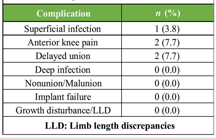
Table 6: Complications and adverse outcomes
The median interval from injury to surgery was 18 h (IQR 9). Following surgery, the median duration of postoperative antibiotic administration was 4 days. All patients were initiated on ankle pump and static quadriceps exercises within 48 h. Progressive weight-bearing was guided by radiographic healing, with 26.9% of patients achieving clinical and radiological union by 12 weeks and the entire cohort reaching full union by 24 weeks.
Pre-operative neurovascular status was normal in all 26 patients, and no cases of suspected compartment syndrome were documented. Crucially, for this adolescent cohort, no growth disturbances or limb length discrepancies were observed during the follow-up period, supporting the safety of the physeal-sparing approach.
The management of tibial shaft fractures in adolescents remains challenging because treatment must provide adequate mechanical stability while preserving the integrity of the proximal tibial physis. The present study evaluated the outcomes of physeal-sparing intramedullary interlocking nailing in 26 adolescents and demonstrated favorable clinical, radiological, and functional results, with complete fracture union achieved in all patients and no evidence of growth-related complications.
The study population consisted predominantly of male adolescents, with a mean age of 15.23 years, and road traffic accidents represented the most common mechanism of injury. Similar demographic patterns have been reported in recent pediatric trauma literature, where adolescent males constitute the majority of patients sustaining high-energy tibial shaft fractures owing to greater participation in outdoor activities and vehicular travel [7].
A major finding of the present study was the achievement of radiographic union in all patients, with a mean union time of 14.23 ± 2.23 weeks. Progressive improvement in RUST scores was observed throughout follow-up, increasing from 5.08 at 6 weeks to 11.54 at 24 weeks. These findings are consistent with contemporary reports demonstrating reliable fracture healing following intramedullary fixation in pediatric and adolescent tibial fractures [8]. The use of the RUST scoring system provided a standardized and reproducible assessment of fracture healing, which has been shown to possess good reliability and validity in the evaluation of tibial union [9].
Pain and functional recovery improved substantially during the follow-up period. Median VAS scores declined from 5.0 at 2 weeks to 0 by 12 weeks, while TFHS values demonstrated progressive functional improvement. Early restoration of function following stable intramedullary fixation has been reported by previous investigators, who noted that intramedullary devices permit controlled mobilization while maintaining fracture alignment and promoting biological healing [10].
The physeal-sparing technique used in the present study was specifically designed to avoid violation of the proximal tibial growth plate. No patient developed growth disturbance, angular deformity, or limb-length discrepancy during follow-up. Similar observations were reported by Williams et al., who described a physeal-sparing rigid intramedullary nailing technique in adolescents and found no evidence of physeal arrest or fixation failure while achieving satisfactory fracture healing [11].
Complication rates in the current study were low. Superficial infection occurred in one patient, anterior knee pain in two patients, and delayed union in two patients. Importantly, no cases of deep infection, nonunion, malunion, or implant failure were encountered. Comparable low complication rates have been reported in recent studies evaluating operative management of pediatric tibial fractures, supporting the safety of intramedullary fixation when appropriate surgical principles are followed [8,12].
The operative technique also demonstrated practical advantages. Closed reduction was possible in most patients, minimizing soft-tissue disruption and preserving the fracture biology. Stable fixation achieved through interlocking nail constructs likely contributed to the excellent union rates and favorable functional outcomes observed in this cohort. Recent comparative studies have suggested that intramedullary fixation techniques may facilitate earlier mobilization and satisfactory healing compared with prolonged immobilization or external fixation in selected pediatric patients [13].
The present study has certain limitations. The sample size was relatively small, the study was conducted at a single center, and there was no comparison group treated with alternative fixation methods, such as elastic stable intramedullary nailing or plating. In addition, the duration of follow-up may not be sufficient to detect very late growth-related complications. Nevertheless, the prospective design and standardized clinical and radiological assessment strengthen the validity of the findings.
Physeal-sparing intramedullary interlocking nailing appears to be a safe and effective treatment option for tibial shaft fractures in adolescents with open physes. The technique provided stable fixation, facilitated progressive pain relief and functional recovery, and achieved reliable fracture union in all patients without evidence of growth disturbance, limb length discrepancy, malunion, or implant failure. The low complication rate and favorable radiological and clinical outcomes observed in this study suggest that physeal-sparing interlocking nailing is a viable alternative to traditional treatment methods for appropriately selected adolescent tibial shaft fractures.
Physeal-sparing intramedullary interlocking nailing is a valuable treatment option for adolescent tibial shaft fractures with open physes, offering stable fixation without violating the proximal tibial growth plate. The technique facilitates predictable fracture union, progressive pain reduction, and early functional recovery while maintaining alignment. In this study, no growth disturbances, limb length discrepancies, malunions, or implant failures were observed. Careful attention to the entry point and surgical technique can help achieve excellent clinical and radiological outcomes with a low complication rate.
References
- 1. Hogue GD, Wilkins KE, Kim IS. Management of pediatric tibial shaft fractures. J Am Acad Orthop Surg 2019;27:769-78. [Google Scholar] [PubMed]
- 2. Landau AJ, Oladeji AK, Cummings JL, Goldstein R, Lin A, Hosseinzadeh P. Outcomes of elastic stable intramedullary nailing for surgical treatment of pediatric tibial shaft fractures. J Am Acad Orthop Surg Glob Res Rev 2023;7:e23.00031. [Google Scholar] [PubMed]
- 3. Hong P, Rai S, Tang X, Liu R, Li J. External fixation versus elastic stable intramedullary nailing in the treatment of open tibial shaft fractures in children. J Orthop Surg Res 2021;16:528. [Google Scholar] [PubMed]
- 4. Williams KA, Thier ZT, Mathews CG, Locke MD. Physeal-sparing rigid intramedullary nailing in adolescent tibial shaft fractures: A pilot study. Cureus 2021;13:e13893. [Google Scholar] [PubMed]
- 5. Burkus M, Tömböl F, Wiegand N, Kretzer A. Physeal-sparing unreamed locked intramedullary nailing for adolescent tibial fractures. Injury 2021;52 Suppl 1:S67-73. [Google Scholar] [PubMed]
- 6. Tamburini L, Zeng F, Neumann D, Jackson C, Mancini M, Block A, et al. A review of tibial shaft fracture fixation methods. Trauma Care 2023;3:202-11. [Google Scholar] [PubMed]
- 7. Marengo L, Enrietti E, Piccinno M, Ceroni L, Marre’ Brunenghi G, Boero S, et al. Casting, elastic intramedullary nailing or external fixation in pediatric tibial shaft fractures: Which is the most appropriate treatment? A multicenter study. J Pediatr Orthop B 2025;34:27-32. [Google Scholar] [PubMed]
- 8. Kubiak EN, Egol KA, Scher D, Wasserman B, Feldman D, Koval KJ. Operative treatment of tibial fractures in children: Are elastic stable intramedullary nails an improvement over external fixation? J Bone Joint Surg Am 2005;87:1761-8. [Google Scholar] [PubMed]
- 9. Leow JM, Clement ND, Tawonsawatruk T, Simpson CJ, Simpson AH. The radiographic union scale in tibial (RUST) fractures: Reliability of the outcome measure at an independent centre. Bone Joint Res 2016;5:116-21. [Google Scholar] [PubMed]
- 10. Chen X, Lu M, Xu W, Wang X, Xue M, Dai J, et al. Treatment of pediatric femoral shaft fractures with elastic stable intramedullary nails versus external fixation: A meta-analysis. Orthop Traumatol Surg Res 2020;106:1305-11. [Google Scholar] [PubMed]
- 11. Pennock AT, Bastrom TP, Upasani VV. Elastic intramedullary nailing versus open reduction internal fixation of pediatric tibial shaft fractures. J Pediatr Orthop 2017;37:e403-8. [Google Scholar] [PubMed]
- 12. Duan X, Al-Qwbani M, Zeng Y, Zhang W, Xiang Z. Intramedullary nailing for tibial shaft fractures in adults. Cochrane Database Syst Rev 2012;1:CD008241. [Google Scholar] [PubMed]
- 13. Brandt MD, Barnum MS, Herrera-Soto JA. Suprapatellar tibial nailing in skeletally immature patients: A case series. J Orthop Case Rep 2026;16:440-5. [Google Scholar] [PubMed]
Related Articles in Journal of Orthopaedic Case Reports
 October 1, 2026 Clinical and Functional Outcomes of Closed Intramedullary Interlocking Nailing in Extra-articular Distal Tibial Fractures: A Prospective Observational Study
 October 1, 2026 Association between Coronal Mechanical Axis Correction and Early Functional Outcomes after Primary Total Knee Arthroplasty: A Retrospective Observational Study
 October 1, 2026 Minimally Invasive J-Nail Fixation for Neer Type 2, 3, and 4 Proximal Humerus Fractures in Elderly Patients: A Prospective Clinical and Radiological Outcome Study
 October 1, 2026 A Study of Spinopelvic Relationship and its Impact on Functional Outcome of Total Hip Arthroplasty