


Introduction
Over the last 20 years, shoulder arthroplasty has increased at an exponential rate, and the reverse total shoulder arthroplasty (rTSA) has become the preferred arthroplasty for a wide range of conditions, including glenohumeral osteoarthritis with rotator cuff deficiency, proximal humeral fractures, tumour reconstruction and failed prior arthroplasty. In population-level shoulder registries, the number of rTSA procedures is estimated to have increased by 400% (2002–2017) due to both increasing numbers of people receiving an initial implant and inherent failure modes found in shoulder prostheses. The most impactful failure mode of the primary rTSA is the loosening of the glenoid component, which has been reported to be between 5.7% and 17% of all primary rTSAs at medium-term follow-up, with re-revision rates as high as 21% after conversion surgery. With this unprecedented burden, revision surgery is at a turning point in the discipline where the technical requirement to provide a more precise surgical outcome is outstripping the accuracy of traditional surgical instruments [1, 2].
The glenoid baseplate is the basic part of rTSA, which is extremely sensitive to positional error. Planned version and/or inclination deviations over 5° have been correlated with increased scapular notching, glenosphere impingement, aseptic loosening and early mechanical failure [3]. In revision surgery, these tolerances are even stricter. A significant portion of glenoid bone loss, such as is described in Walch B2/B3 and Favard E2/E3 classes in primary disease and Sears and Antuña schemes in post-arthroplasty defects, changes the landmarks that surgeons use to guide instruments for an accurate arthroplasty [4, 5, 6]. The coracoid may be grossly distorted, the geometry of the glenoid may be abnormal and tactile and visual cues may be unreliable due to the scapular plane that may be abnormal. This leads to an established difference between the digital plan and the implementation during surgery, which is repeatedly greater than acceptable limits of tolerance when using traditional freehand methods [7].
Several technological solutions have been developed to help bridge this planning-to-execution gap. Patient-specific instrumentation (PSI) is based on three-dimensional (3D) computed tomography (CT) data to create customized guide blocks to fit specific anatomical contact points on the glenoid face. In revision cases, those contact points are often missing or distorted, due to previous reaming, bone cement and peri-implant osteolysis [8]. Optical computer navigation provides real-time angular feedback but requires a fixed scapular reference tracker, line-of-sight limitations, a secondary monitor that distracts the surgeon’s eyesight, and an increase in operative time [9]. The use of robots in elective primary arthroplasty has been shown to provide the highest level of accuracy. Still, it has not been specifically validated for revision glenoid reconstruction in cases of severe bone loss and requires significant capital investment [10].
In contrast, augmented reality (AR) and mixed reality (MR) technologies are a completely different approach. AR overlays digital content onto the real world (transparency) and MR anchors digital holograms to physical space (dynamic changes according to the observer’s movement and position). The dominant hardware modality for surgical AR/MR is head-mounted displays (HMDs), which display a registered patient-specific hologram directly in the surgeon’s line of sight, bypassing the need to redirect the gaze to a remote screen [11]. Recent cadaveric validation studies have reported angular deviations as small as 0.9° (4 times more accurate) for the inclination and 1.8° (18 times more accurate) for the version between the HMD-guided and freehand techniques. To the best of our knowledge, however, the use of AR/MR HMDs for revision shoulder arthroplasty, which is further complicated by distorted anatomy, irregular bone stock, and the added complexity of the hardware removal procedure, has not been systematically reviewed [12].
The purpose of the present narrative review is to summarise existing literature on the MR and AR HMD technology used in glenoid baseplate positioning during revision shoulder surgery for patients with complex glenoid bone loss, critically analyse the accuracy measures, clinical outcomes, human factors considerations and the regulatory situation, and identify knowledge gaps that need to be filled before a broad implementation of AR and MR HMD technology.
Materials and Methods
A literature search was performed using a narrative approach in four major biomedical databases, which are PubMed/MEDLINE, Embase, Cochrane Library and Scopus. The search was performed in September 2026 and encompassed publications from January 2010 to September 2026. The lower date limit was determined to identify the first feasibility studies on AR in shoulder arthroplasty; a supplemental search was conducted for landmark classification papers on glenoid bone loss, which are still standard references in the field.
The main search terms adopted in PubMed were (“augmented reality” OR “mixed reality” OR “head-mounted display” OR “holographic navigation”) AND (“shoulder arthroplasty” OR “reverse shoulder arthroplasty” OR “glenoid” OR “shoulder replacement”) AND (“navigation” OR “positioning” OR “accuracy” OR “bone loss”). Secondary search words were specific pieces of hardware: (“HoloLens” OR “Magic Leap” OR “Pixee Medical” OR “Novarad VisAR” OR “Stryker Blue” OR “NextAR” OR “Xvision”) AND (“orthopaedic” OR “shoulder” OR “glenoid”) as comparator technologies, tertiary searches considered PSI, optical navigation and robotic arthroplasty. The results were compared with a manual search of the Journal of Shoulder and Elbow Surgery, Journal of Bone and Joint Surgery, EFORT Open Reviews, Bone and Joint Journal and the American Shoulder and Elbow Surgeons and European Society for Surgery of the Shoulder and the Elbow proceedings.
Inclusion criteria were: (1) Peer-reviewed original studies, systematic reviews, or consensus statements; (2) studies focused on AR/MR or HMD technology for glenoid component positioning; (3) quantitative accuracy metrics reported (version, inclination, and/or deviation from entry-point); and (4) studies on glenoid bone loss in primary or revision shoulder arthroplasty. Exclusion criteria were: (1) Studies where only VR was reported without AR/MR overlay on real anatomy; (2) non-peer-reviewed conference abstracts, opinion pieces, preprints that were not afterwards indexed in PubMed, Embase or Scopus; (3) animal and/or in-silico studies used as primary clinical evidence; (4) sources published in predatory journals (defined by Beall’s List framework).
The review adopted the Scale for the Assessment of Narrative Review Articles (SANRA) [13]. The criteria of SANRA that were approached are: Justification of the review (clinical importance), specification of objectives, description of literature search, reference to scientific rigour of included studies, appropriate referencing, and scientific reasoning.
Results
Glenoid bone-loss classifications, incidence of loosening and consequences of malposition
Proper glenoid bone loss classification is the foundation for proper surgical planning in revision shoulder surgery. Native glenoid morphology, originally described for primary glenohumeral osteoarthritis, consists of Walch Type A (concentric wear), Type B (posterior erosion and subluxation), Type C (dysplastic retroversion) and Type D (anterior glenoid erosion) [3]. Primary rTSA’s most technically difficult deformities are Walch B2 (biconcave erosion with posterior subluxation) and B3 (monoconcave erosion with posterior subluxation >15° of retroversion or >70% humeral head subluxation), and require corrective reaming or an angled augmented baseplate to restore the scapular neutral axis [14]. Glenoid erosion was classified on a 5-point scale (E0–E4) according to the Favard classification and the appearance on the magnetic resonance imaging, with grades E2/E3 indicating the highest risk of baseplate malpositioning [6]. The Sears classification of post-arthroplasty glenoid defects in revision surgery includes central, peripheral and combined defects, categorized as mild, moderate and severe [4]. The Antuña classification is a topographic and volumetric classification of glenoid defects, useful for planning glenoid reconstruction (Table 1) [5].
Glenoid bone-loss classification systems relevant to revision shoulder arthroplasty
| Classification | Author/Year | Setting | Major categories | Surgical relevance | References |
|---|---|---|---|---|---|
| Walch | Walch et al., 1999 | Primary glenohumeral OA | A1/A2 (central), B1/B2/B3 (posterior erosion/subluxation), C (dysplastic), D (anterior) | B2/B3: High risk for version error; requires corrective reaming or augmented baseplate | [3] |
| Favard | Favard et al., 2011 | rTSA for cuff-tear arthropathy | E0-E4 (superior glenoid erosion severity) | E2/E3: Compromised superior baseplate fixation; risk of baseplate tilt and early loosening | [6] |
| Sears | Sears et al ., 2012 | Post-arthroplasty glenoid defect | Central, peripheral, combined; mild/moderate/severe | Guides bone graft versus augmented implant versus custom implant decision in revision | [4] |
| Antuna | Antuna et al ., 2001 | Revision shoulder arthroplasty | Location (central/peripheral/combined) + severity | Foundation for the surgical reconstruction algorithm in revision glenoid surgery | [5] |
OA: Osteoarthritis, rTSA: Reverse total shoulder arthroplasty
Population-based data show that glenoid-related complications are the primary failure mode for shoulder arthroplasty. In medium-term analyses, the incidence of glenoid component loosening varies from 5.7% to 17% of rTSA failures [15, 16]. A recent study of 127 consecutive revision rTSAs for anatomic glenoid component loosening found that 13% of patients underwent re-revision for baseplate loosening at a mean follow-up of 35 months, and the overall radiographic baseplate loosening rate was 17% [17]. At 5 years post-revision, a 21% baseplate loosening rate was noted, and implant survivorship decreased from 97% at 2 years to 75% at 5 years in a matched cohort study with rTSA for glenoid loosening [18]. These malpositions combine to worsen the problems: version error >10° leads to scapular notching by accentuating inferior glenosphere impingement during adduction; inclination error leads to superior tilting of the metaglene with an increase in tensile forces acting on the locking mechanism, and predisposes to early loosening; entry-point offset >3 mm shifts the force vector away from the central post, compromising screw trajectory and bone-ingrowth surface contact [19]. Summary of pivotal studies on AR/MR HMD guidance for glenoid baseplate positioning in shoulder arthroplasty is given in Table 2.
Summary of pivotal studies on AR/MR HMD guidance for glenoid baseplate positioning in shoulder arthroplasty
| Author (Year) | Design | n | Intervention | Comparator | Primary outcome | Follow-up | Key finding |
|---|---|---|---|---|---|---|---|
| Kriechling et al. [31] (2021) | Cadaveric feasibility | 10 scapulae | AR-HMD (HoloLens-based) guidewire | None | Trajectory and entry-point deviation | Single session | Trajectory 2.7°±1.3°; entry 2.3 mm±1.1 mm |
| Rojas et al. [21] (2022) | Retrospective clinical | 17 rTSA | AR-HMD (NextAR, Medacta) | Intraop measurements | Version, inclination, entry point | – Postop CT | Inclination 1.0°±0.7°; retroversion 1.8°±1.3°; entry 1.1 mm±0.4 mm |
| Can Kolac et al. [11] (2024) | Narrative review | N/A | 3D planning, NAV, PSI, MR | — | Accuracy, outcomes, future directions | N/A | MR is identified as an emerging technology with a strong accuracy profile |
| Rojas et al. [22] (2025) | Proof-of-concept clinical | 17 rTSA | Navigated AR-HMD (NextAR) | Preop plan | Plan-to-postop deviation | Postop CT | Zero outliers intraop-to-postop; 3 outliers plan-to-postop |
| Dordain et al. [12] (2025) | Cadaveric comparative | 10 shoulders | AR-HMD (Pixee Medical) | Freehand conventional | Inclination and version absolute error | Single session | AR-HMD inclination 0.9°±1.6° versus freehand 5.1°±3.7° (P =0.007) |
| Lee et al. [20] (2026) | Systematic review and meta-analysis | 14 studies | PSI versus NAV versus MR | Conventional | Version, inclination, entry offset | Variable | PSI/NAV Conventional; MR comparable; PSI versus CON inclination P =0.030 |
| Imbert et al. [32] (2026) | Cadaveric comparative | 60 3D-printed scapulae | Stryker Blue MR Guidance | Freehand senior versus junior | Entry-point, pin orientation | Single session | MR equalized accuracy; freehand version 17°±12° reduced comparably |
| Segers et al. [34] (2026) | Prospective clinical series | 15 shoulders (BIO-RSA) | Navigated AR | Preop plan | Version, inclination, offset | 6 months | Version 4.1°±3.4°; inclination 1.9°±2.4°; 100% graft incorporation |
AR: Augmented reality, MR: Mixed reality, HMD: Head-mounted display, rTSA: Reverse total shoulder arthroplasty, PSI: Patient-specific instrumentation, NAV: Navigation, BIO-RSA: Bony-increased offset reverse shoulder arthroplasty, Postop: Postoperative, Preop: Preoperative, CT: Computed tomography
Traditional strategies: PSI, optical navigation, and robotic assistance
Three main technological aids have been created to enhance the accuracy of glenoid baseplate placement: PSI, optical electromagnetic navigation (NAV), and robotic-assisted systems.
PSI uses the CT-segmentation of a patient’s bone to produce patient-specific guide blocks that mark the precise contact footprints on the glenoid face and surrounding anatomy of the scapula. Meta-analytic data support that PSI is effective at minimising version deviation from conventional instrumentation (weighted mean difference of about 2.5°) and is especially successful at deformity correction [20, 21]. In the revision setting, PSI has important limitations, however. Residual cement, previous glenoid reaming, osteolytic cavitation, and hardware retention destroy the contact point architecture upon which PSI blocks rely for registration. Thus, the accuracy benefits of PSI are significantly reduced when used for malformed glenoid anatomy, and PSI cannot give real-time intraoperative feedback to detect intraoperative drift. Furthermore, it takes 5–10 working days before the guide is ready to be fabricated, which is not practical if a guide needs to be revised urgently [22].
Optical computer navigation is based on a rigid reference array mounted on the scapular spine, and an infrared camera measures the position of the instrument against a preregistered computer-generated anatomical model. Randomized cadaveric trials showed navigation to be superior to conventional instrumentation in terms of inclination accuracy, and clinical series showed reduced outlier rates with navigation [23, 24]. The disadvantages are that a reference tracker needs to be fixed and stationary; it must be free of any obstacles, such as other instruments and assistants to the operator, and the operator must pause the operative procedure to check the remote display. Fractures of the coracoid are reported in 0.05% of cases, which is due to the fixation of the tracker and is therefore a limitation. During longer revision procedures, the extra registration time of 8–30 min is a significant issue, and periscapular scarring can compromise tracker stability during revision surgery [25].
Currently, the technology of robotic-assisted arthroplasty is the frontier of glenoid precision in primary TSA. Still, glenoid clearances for shoulders are not yet available in most jurisdictions, and there are no dedicated validation series for revision glenoid reconstruction with severe bone defects [26]. Although the terms AR and MR are often used interchangeably in the orthopaedic literature, they are two different technological approaches. Pure AR superimposes the digital content onto a live view, without anchoring holograms to physical space, and MR maps the physical space and anchors digital objects in it, allowing holograms to remain properly placed as the observer and instruments move. The MR paradigm is more holographically stable in surgical applications during dynamic operative tasks [27].
The most thoroughly researched HMD in orthopaedic navigation research is the Microsoft HoloLens 2. It has a diagonal field of view (FOV) of 52°; time-of-flight depth sensors; an inertial measurement unit (IMU); and eye tracking. Evaluated with both the HoloLens 2 and Magic Leap 2, the SurgNavAR framework achieved a mean tooltip calibration accuracy of ~1 mm, registration accuracy of ~3 mm and pointing accuracy of <5 mm in phantom surgical tasks [28]. In a comparative needle-insertion study, it was determined that the Magic Leap 2 was the best platform, with task-success rates and usability scores that were higher than the other platforms due to its wider FOV (70° diagonal) and lower photon-to-display latency [29]. Vuzix monocular smart-glasses platforms (Blade, M400) are light, cheaper and come without the true stereoscopic depth perception, so they are not suitable for 3D anatomical overlay in arthroplasty. Pixee Medical (France), navigated AR (NextAR) Shoulder (Medacta), Stryker (Blue MR Guidance System), Augmedics (xvision Spine System) and Novarad (VisAR) have developed dedicated surgical HMDs [30, 31]. A comparative summary of guidance technologies for glenoid baseplate positioning in reverse shoulder arthroplasty (RSA) is given in Table 3.
Comparative summary of guidance technologies for glenoid baseplate positioning in revision shoulder arthroplasty
| Feature | Conventional instrumentation | PSI | Optical navigation (NAV) | AR/MR HMD |
|---|---|---|---|---|
| Mechanism | Visual/tactile landmark estimation | CT-derived guide block registration | IR tracker + remote display | Holographic overlay on the operative field |
| Preoperative planning | 2D/3D CT plan (not transferred intraop) | 3D CT segmentation+block fabrication | 3D CT segmentation | 3D CT segmentation+digital plan upload |
| Intraoperative feedback | None | None | Real-time data on the remote monitor | Real-time overlay in the surgeon’s FOV |
| Inclination accuracy | 5.1–17° deviation | 2–4° deviation | 1.5–3° deviation | 0.9–4.1° deviation |
| Version accuracy | 5–17° deviation | 2–4° deviation | 1.5–3° deviation | 0.9–4.1° deviation |
| Performance in severe | Unreliable (landmark | Limited (contact-point | Moderate (tracker | Moderate-Good (residual |
| bone loss | loss) | distortion) | instability) | fiducial anchor) |
| Line-of-sight requirement | None | None | Yes (camera-to-tracker) | No (surgeon-worn device) |
| Setup time | Minimal | 5–10 days lead time (fabrication) | 8-30 min intraoperative registration | 1–30 min registration |
| Radiation exposure | Standard preop CT | Standard preop CT | ± Fluoroscopy | Standard pre-operative CT only |
| Per-case cost (USD est.) | Minimal | $1,500-$5,000 | $500-$1,500 (disposables) | $300-$800 (single-use tracker) |
| Capital investment | None | Moderate (planning software) | High ($150K–$300K system) | Low–Moderate (HMD device) |
| Equalises junior/senior accuracy | No | No | Partial | Yes (demonstrated) |
| Regulatory clearance (shoulder) | Universal | Universal | CE/FDA (general) | CE (Pixee, NextAR); FDA shoulder pending |
| Key limitation | High operator-dependence | Fails in distorted anatomy | Line-of-sight; remote monitor | Holographic drift; FOV; sterility |
PSI: Patient-specific instrumentation, NAV: Navigation, FOV: Field of view, IR: Infrared, HMD: Head-mounted display, CE: Conformité Européenne, FDA: Food and drug administration, CT: Computed tomography, MR: Mixed reality, AR: Augmented reality, 3D: Three-dimensional
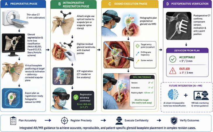
The tracking modalities are: (1) Fiducial marker-based tracking by detecting printed or physical markers attached to instruments and anatomy with the HMD camera array, which provides deterministic entry-point deviation of <2 mm and requires sterile placement of these markers; (2) Marker-less based tracking by applying the iterative closest point (ICP) algorithm to a digitized point cloud of the exposed glenoid, which is flexible but sensitive to a loss of digitized point cloud of the partially destroyed surfaces of the glenoid; and (3) Hybrid systems, which are a combination of both modalities, to provide redundancy. The Pixee Medical system uses a single-use optical tracker, mounted on a scapular reference pin, which allows continuous registration and minimises holographic drift [12,32].
Preoperative CT segmentation, virtual planning, intra-operative registration, hologram overlay, guidance
AR/MR HMD hardware platforms relevant to shoulder arthroplasty navigation are given in Table 4. There are four main steps in the AR/MR HMD workflow for revision glenoid baseplate positioning.
AR/MR HMD hardware platforms relevant to shoulder arthroplasty navigation
| Device | Manufacturer | Type | FOV | Display latency | Tracking modality | Regulatory status |
|---|---|---|---|---|---|---|
| HoloLens 2 [30] | Microsoft | MR binocular | 52° diagonal | ~20 ms | Spatial mapping+IMU+fiducial | Platform only; not medically cleared |
| Magic Leap 2 [29] | Magic Leap | MR binocular | 70° diagonal | ~7 ms | Spatial mapping+depth sensor | Platform only; not medically cleared |
| NextAR Shoulder [22] | Medacta | Dedicated surgical AR | Custom | N/A | Optical fiducial+real-time tracking | CE mark (EU); FDA general arthroplasty |
| Pixee Medical HMD [12,32] | Pixee Medical | Dedicated surgical AR | Custom | N/A | Single-use scapular tracker, optical | CE mark (EU); FDA Knee+ cleared |
| Blue MR Guidance [32] | Stryker | Dedicated surgical MR | Custom | N/A | Optical+spatial mapping | CE mark (EU); US investigational |
| VisAR [30] | Novarad | See-through visor AR | Immersive | Sub-2 mm accuracy | CT fiducial markers | FDA 510(k) cleared (spine, 2022) |
| xvision X2 [26] | Augmedics | Spine-specific HMD AR | 120° immersive | Sub-mm | CT-based optical tracking | FDA 510(k) cleared (spine) |
MR: Mixed reality, AR: Augmented reality, HMD: Head-mounted display, FOV: Field of view, IMU: Inertial measurement unit, CE: Conformité Européenne, FDA: Food and drug administration, CT: Computed tomography
A thin slice CT scan (1 mm collimation) of the entire scapula and proximal humerus is preoperatively obtained and loaded into dedicated planning software (Blueprint™, NextAR Planning, or Materialise Mimics). 3D segmentation of the scapula identifies the pre-deformity scapular body axis, glenoid vault geometry and defect margins. The surgeon virtually places the baseplate at the desired version and inclination, including corrective reaming vectors or augmented baseplate orientation. This planning step is integrated into the revision setting, which also includes bone defect mapping following the Sears/Antuña scheme and can involve virtual bone-graft sizing and/or custom implant design. The final plan, which includes the target entry point, axis trajectory and screw vectors, is then exported as a target registration dataset for the HMD software [11, 14].
In the beach-chair position, after hardware removal, a scapular reference tracker is applied to the patient through a dedicated percutaneous pin or scapular spine clamp. A tracked digitization pointer is used to collect any remaining glenoid landmarks, which then allows the surgeon to make a point-based ICP registration between the CT model and the live anatomy. Registration quality is checked by both an overlay inspection and quantitative error feedback in the HMD visor. After verification, the planned entry point, drilling direction and screw path are directly projected on the glenoid face via the HMD visor. Real-time instrument tracking gives the surgeon immediate feedback on the angular deviation of the instrument (version, inclination, reaming depth) in the surgeon’s FOV, without redirection of gaze. The drilling, reaming and implantation of the baseplate are done under holographic control. Post-operative CT or fluoroscopic verification proves execution fidelity [21, 22].
Phantom, cadaveric, and clinical validation studies
The first reliable quantitative accuracy evidence for AR/MR HMD systems is available for shoulder arthroplasty. Kriechling et al. performed a cadaveric feasibility study in a base study that used an HMD-based AR system (mean trajectory deviation 2.7° ± 1.3° and mean entry-point deviation 2.3 mm ± 1.1 mm for ten cadaveric scapulae), similar to other optical navigation systems [31]. In a proof-of-concept clinical series of 17 shoulders, the system NextAR Shoulder (Medacta) showed postoperative correspondence of 1.0° ± 0.7° for inclination, 1.8° ± 1.3° for retroversion and 1.1 ± 0.4 mm for entry point with the preoperative plan [21, 22, 33]. In a ten-cadaver study, Pixee Medical AR-HMD was able to significantly and widely decrease the absolute inclination deviation from 5.1° ± 3.7° (freehand) to 0.9° ± 1.6° (P = 0.007), significantly reducing numbers of outliers from 7 to 1 [12]. A recent study in the context of revision surgery for severe posterior glenoid erosion (bony-augmented offset RSA [BIO-RSA]) found that navigated AR (NextAR) resulted in a mean version deviation of 4.1° ± 3.4° and inclination deviation of 1.9° ± 2.4° at 6-month follow-up, with complete incorporation of the graft in all 15 shoulders [34].
In a systematic review and meta-analysis of rTSA glenoid positioning using PSI, optical navigation, and MR systems in 2025, 14 studies were identified, with both PSI and NAV achieving significantly fewer deviations from conventional instrumentation, and similar or greater accuracy for MR systems, but with a smaller body of clinical evidence for MR (P = 0.030) [20]. The Stryker Blue MR Guidance System was evaluated on 3D-printed Walch A, B and Favard E scapular models. It was found to be the critical step of reducing surgical errors markedly, as median version deviation was 17° ± 12° for both senior and junior surgeons without MR and 17° ± 10° with MR. The results overall indicate that the accuracy of AR/MR HMD systems is clinically acceptable for all levels of operator experience, which is not true for PSIs or navigation comparators (Table 1) [32].
Clinical outcomes: Early series in revision RSA, complications, operative time, radiation and costs
There are limited clinical outcome data for AR/MR HMD guidance in revision shoulder arthroplasty with severe glenoid bone loss. The pilot study of NextAR in 17 successive rTSAs showed no intraoperative complications due to the HMD system and no cases of hologram-induced distraction error [21]. The setup time (data transfer of CT, software preparation, and intraoperative registration) was 1–26 min for tracker placement, and a learning-curve effect was seen as operators gained experience. In experienced hands, the operative time was not significantly extended beyond the registration time. Optical fiducial tracking allowed systems to avoid a higher dose of radiation than standard pre-operative CT protocols, which was an advantage over fluoroscopy-driven navigation systems [22,33].
Single-use AR instrumentation can cost anywhere from USD 300 to 800, while PSI block fabrication can cost as much as USD 1,500–5,000, and conventional navigation infrastructure can cost as much as USD 150,000–300,000/system. No specific formal cost-utility analysis of revision shoulder surgery is available. From the larger perspective of the unmet clinical need, a 2026 systematic review of surgical treatment options for severe glenoid structural deficiencies in revision rTSA identified that across 13 articles and 556 shoulders, the failure rate of the allograft baseplate was 33.3%, compared with 6.3% for the autograft baseplate. The overall functional improvement achieved after these procedures met MCID thresholds despite significant complication rates [27].
Human factors and ergonomics: Cognitive load, holographic drift, FOV constraints, sterility, and training curve
There are many human-factors issues that arise with the use of HMDs in the operating theatre that require solutions before their routine use. A major issue is cognitive load, which is the overall information-processing load placed on the surgeon. AR interfaces augment the amount of information in the surgical FOV, but too many overlays can actually reduce situational awareness and decision-making [35]. NASA Task Load Index scores have been measured in NASA Task Load Index during AR-assisted procedure and have found that increased perceived workload during initial use of HMD decreased following structured familiarization [36]. A 66% increase in cognitive load in the immersive simulation literature compared to conventional simulation has been shown with HMDs, showing direct relevance to the HMD-AR contexts [37].
In shoulder arthroplasty, progressive misalignment of the virtual overlay with respect to the physical anatomy, called holographic anchoring drift, can be a clinically relevant issue caused by either sensor noise, IMU drift or patient motion after initial registration, and may require soft-tissue retraction and repositioning of the scapula. Systems with continuous optical fiducial tracking reduce the effects of drift by continuously updating the registration, but there is still a possibility of marker displacement or contamination by surgical drapes being a failure mode. The FOV constraint of current HMDs (52–70° diagonal) decreases peripheral awareness, the number of instruments and the number of monitoring devices, which presents patient-safety considerations in regulatory submissions [38]. HMDs are not autoclavable; sterility is achieved by using sterile sleeve draping/hanging above the surgical field, and may cause performance degradation over time due to heating during longer revision surgeries (generally 120–240 min). Data from the training-curve portion of the Stryker Blue MR Guidance study showed that MR provided the biggest performance gains to junior surgeons, and that pre-operative MR training using 3D-printed Walch B2 glenoid models enhanced guidewire placement accuracy in trainees, suggesting that MR should be utilized in residency training and in competency-based learning programmes [39].
Regulatory landscape, the integration of robotics, and future directions
In the United States, the regulatory process for AR/MR surgical navigation devices is like the Food and Drug Administration (FDA) premarketing notification process for surgical navigation devices (Class II medical devices), with Class II surgical navigation accessories being the usual medical device classification for non-implanted navigation accessories. The clearances include the Novarad VisAR (510(k) clearance for spine surgery, 2022) [30]; the Augmedics xvision X2 (510(k) clearance for spine and the use of 2024–2025 for brain surgery); the Medivis Spine and Cranial Navigation (2025) 510(k) clearance for orthopedic guidance); and the SKIA HEAD (510(k) clearance for orthopaedic guidance, 2026). The total knee arthroplasty clearance for Pixee Medical’s Knee+ NexSight AR system is a first for joint arthroplasty clearances and came before the shoulder. CE mark approvals are more widely available in Europe; the Pixee Medical AR-HMD and NextAR Shoulder are CE-marked for shoulder arthroplasty in the European Economic Area [12, 22]. In India, AR/MR HMDs for surgical navigation belong to the Class C medical device category as per the Central Drugs Standard Control Organisation (CDSCO) and till mid-2026, there is no specific approval for AR/MR HMDs for shoulder arthroplasty navigation, and as per the Medical Devices Rules 2017, a surgeon using a CE-marked device is required to ensure compliance with Rule 101.
Intraoperative bone-loss reclassification using artificial intelligence (AI) algorithms to process data in real time from a depth camera during surgery, modifying the Sears or Antuña classification, is being developed and has been peer-reviewed as a frontier technology. In a proof-of-concept experiment in multiple surgical specialities, the lecturer has been shown to be able to annotate the holographic overlay in the surgeon’s FOV remotely, a technique that has been coined “mixed-reality” tele-mentoring, and that is particularly promising for centres in low-resource regions or emerging economies where fellowship training for the shoulders and elbows may be unavailable [40].
Discussion
The available evidence from recent studies on glenoid baseplate positioning accuracy for AR/MR HMD guidance is summarized in the present narrative review, which shows that positioning of the glenoid baseplate is consistently more accurate than freehand positioning, with an accuracy range of 0.9–4.1° for the glenoid inclination and 0.9–2.5° for version, reported in cadaveric and early clinical studies [12, 22, 32, 34]. This accuracy profile is within 5°, an acceptable standard for glenoid accuracy that is conventionally considered clinically acceptable for the glenoid component. It represents an important improvement over freehand accuracy, which has reported deviations ranging from 5.1° to 17°. The current synthesis extends previous syntheses on PSI and navigation in two ways: It includes an important dimension that has never been demonstrated previously in these types of reviews: The level of equalization of performance across experience levels with MR guidance [32]. The results of this study—showing that MR resulted in the version deviation being reduced from 17° to within acceptable tolerance when the surgeon was a junior—are directly clinically relevant to revision shoulder arthroplasty, which is presently performed at specialist centres in high volume. For a surgeon with a limited number of years in practice, MR may prove to be a valuable adjunct to expand access to the entire spectrum of revision reconstruction procedures, such as angled augmented baseplates, structural bone grafting and custom implants [27]. A conceptual algorithm for AR/MR HMD-guided glenoid baseplate positioning in complex RSA is given in Fig. 1.
Several issues and controversies need to be explicitly recognized. The first is the evidence, which is still restricted to cadaveric studies and small primary series of rTSA, with the revision-specific application and significant bone loss encountered only in a single prospective clinical series (BIO-RSA, n = 15) [34]. There is no literature on the AR/MR revision shoulder literature for patient-reported outcome measures (PROMs), implant survivorship, or cost-utility analyses (CUAs). Second, a direct comparison between HMD platforms (HoloLens 2 vs. Magic Leap 2 vs. dedicated surgical HMDs) in a standardized shoulder arthroplasty task has not yet been undertaken, as the choice of platform is determined by commercial availability rather than comparative efficacy. Third, the characteristics of available systems in terms of latency and drift have been defined mostly through spine and phantom studies, but the unique challenges of revision shoulder surgery, such as a significant soft-tissue contracture, poor exposure, and unpredictable intraoperative changes in the glenoid due to cement removal, may compromise the accuracy of the registration [30]. Fourth, the cost-benefit of AR/MR navigation to PSI is still not clear in the revision setting, where the cost of implant failure and re-revision is significantly higher than the cost of the guidance technology.
The regulatory environment is a structural obstacle. As of mid-2026, there is no regulatory clearance specifically for the shoulder arthroplasty navigation AR/MR HMD system in major jurisdictions outside of Pixee Medical and NextAR Shoulder. This gap reduces reimbursement through current procedural coding systems and introduces medico-legal confusion for implementing surgeons. In India, the CDSCO framework has not yet guided devices for AR surgical navigation. International regulatory harmonization among the FDA, the European MDR and the CDSCO will be key to allowing multicentre trials worldwide. Future research priorities include: (1) Multicentric randomized controlled trials with co-primary endpoints of PROMs and implant survivorship comparing AR/MR HMD guidance with PSI and optical navigation in revision rTSA with Walch B3/Favard E3 bone loss; (2) prospective CUAs from health-economic perspectives; (3) validation studies that incorporate AI in the operating room to reclassify bone loss; and (4) clinical evaluation of tele-mentoring protocols in resource-limited settings.
Conclusion
The adoption of mixed and AR HMDs represents a paradigm shift in intraoperative navigation for revision shoulder arthroplasty with complex glenoid bone loss. Future priorities include regulatory expansion, revision-specific validation, and AI-assisted intraoperative to freehand and comparable to optical navigation or PSI. These systems uniquely provide real-time holographic overlays without line-of-sight limitations, transfer surgical precision across experience levels, and avoid additional radiation. However, evidence gaps remain: prospective randomized trials, patient-reported outcomes, implant survivorship, and CUAs. Future priorities include regulatory expansion, revision-specific validation, and AI-assisted intraoperative bone-loss reclassification.
Clinical Message
AR/MR head-mounted displays consistently achieve sub 2° angular deviation in glenoid baseplate positioning, markedly outperforming freehand and patient specific instrumentation, especially in revision settings with distorted anatomy
Mixed reality guidance reduces variability between junior and senior surgeons, democratising surgical skill and offering strong potential for integration into residency training and competency based learning programs
Compared to PSI and conventional navigation systems, AR/MR HMDs provide real time intraoperative feedback with lower setup costs and fewer ergonomic limitations, making them an accessible solution for precision in revision shoulder arthroplasty.
Conflict of Interest:
Nil
Source of Support:
Nil
Consent:
The authors confirm that informed consent was obtained from the patient for publication of this article
How to Cite this Article
Bharadwaj S, Jeyaraman N, Balasubramanian S, Sridhar AS, Muthu S, Jeyaraman M. Mixed Reality and Augmented Reality in Complex Revision Shoulder Arthroplasty: Head-Mounted Displays for Glenoid Baseplate Positioning. J Orthop Case Rep. 2026 November;16(11):574-584. doi: 10.13107/jocr.2026.v16.i11.8556.
References
- Chalmers PN, Boileau P, Romeo AA, Tashjian RZ. Revision reverse shoulder arthroplasty. J Am Acad Orthop Surg 2019;27:426-36. [Google Scholar] | [PubMed]
- Werner BC, Wong AC, Mahony GT, Craig EV, Dines DM, Warren RF. Causes of poor postoperative improvement after reverse total shoulder arthroplasty. J Shoulder Elbow Surg 2016;25:e217-22. [Google Scholar] | [PubMed]
- Walch G, Badet R, Boulahia A, Khoury A. Morphologic study of the glenoid in primary glenohumeral osteoarthritis. J Arthroplasty 1999;14:756-60. [Google Scholar] | [PubMed]
- Sears BW, Johnston PS, Ramsey ML, Williams GR. Glenoid bone loss in primary total shoulder arthroplasty: Evaluation and management. J Am Acad Orthop Surg 2012;20:604-13. [Google Scholar] | [PubMed]
- Antuna SA, Sperling JW, Cofield RH, Rowland CM. Glenoid revision surgery after total shoulder arthroplasty. J Shoulder Elbow Surg 2001;10:217-24. [Google Scholar] | [PubMed]
- Favard L, Levigne C, Nerot C, Gerber C, De Wilde L, Mole D. Reverse prostheses in arthropathies with cuff tear: Are survivorship and function maintained over time?. Clin Orthop Relat Res 2011;469:2469-75. [Google Scholar] | [PubMed]
- Jacquot A, Gauci MO, Chaoui J, Baba M, Deransart P, Boileau P. Proper benefit of a three dimensional pre-operative planning software for glenoid component positioning in total shoulder arthroplasty. Int Orthop 2018;42:2897-906. [Google Scholar] | [PubMed]
- Cabarcas BC, Cvetanovich GL, Gowd AK, Liu JN, Manderle BJ, Verma NN. Accuracy of patient-specific instrumentation in shoulder arthroplasty: A systematic review and meta-analysis. JSES Open Access 2019;3:117-29. [Google Scholar] | [PubMed]
- Verborgt O, De Smedt T, Vanhees M, Clockaerts S, Parizel PM, Van Glabbeek F. Accuracy of placement of the glenoid component in reversed shoulder arthroplasty with and without navigation. J Shoulder Elbow Surg 2011;20:21-6. [Google Scholar] | [PubMed]
- Tantone RP, Al-Humadi S, VanHelmond T, Kim M, Komatsu DE, Wang ED. Outcomes of reverse shoulder arthroplasty in patients with previous rotator cuff repair: A systematic review and meta-analysis. JSES Rev Rep Tech 2023;3:267-73. [Google Scholar] | [PubMed]
- Can Kolac U, Paksoy A, Akgün D. Three-dimensional planning, navigation, patient-specific instrumentation and mixed reality in shoulder arthroplasty: A digital orthopedic renaissance. EFORT Open Rev 2024;9:517-27. [Google Scholar] | [PubMed]
- Dordain F, Nourrissat G, Van Rooij F, Ferrand M, Petroff E, Antoni M. Augmented reality significantly reduces the absolute error between achieved and planned inclination and version of the glenoid baseplate for reversed shoulder arthroplasty. JSES Int 2025;9:1215-9. [Google Scholar] | [PubMed]
- Baethge C, Goldbeck-Wood S, Mertens S. SANRA-a scale for the quality assessment of narrative review articles. Res Integr Peer Rev 2019;4:5. [Google Scholar] | [PubMed]
- Iannotti JP, Weiner S, Rodriguez E, Subhas N, Patterson TE, Jun BJ. Three-dimensional imaging and templating improve glenoid implant positioning. J Bone Joint Surg Am 2015;97:651-8. [Google Scholar] | [PubMed]
- Klein A, Nové-Josserand L, Collotte P, Gunst S. Incidence and risk factors for aseptic glenoid baseplate loosening in primary reverse shoulder arthroplasty using the Aramis implant baseplate with a minimum of 2 years follow-up. J Shoulder Elbow Surg 2026;35:978-88. [Google Scholar] | [PubMed]
- Lädermann A, Schwitzguebel AJ, Edwards TB, Godeneche A, Favard L, Walch G. Glenoid loosening and migration in reverse shoulder arthroplasty. Bone Joint J 2019;101-B:461-9. [Google Scholar] | [PubMed]
- Bartels DW, Marigi E, Sperling JW, Sanchez-Sotelo J. Revision reverse shoulder arthroplasty for anatomical glenoid component loosening was not universally successful: A detailed analysis of 127 consecutive shoulders. J Bone Joint Surg Am 2021;103:879-86. [Google Scholar] | [PubMed]
- Hao KA, Simcox TG, Hones KM, Wright JO, Wright TW, LaMonica T. Conversion of failed anatomic total shoulder arthroplasty to reverse shoulder arthroplasty yields similar pain and functional outcomes to primary reverse shoulder arthroplasty but has a higher baseplate failure rate: A matched cohort study. J Am Acad Orthop Surg 2025;33:e1215-25. [Google Scholar] | [PubMed]
- Al-Omairi S, Albadran A, Dagher D, Leroux T, Khan M. Glenoid bone loss in shoulder arthroplasty: A narrative review. Ann Jt 2024;9:8. [Google Scholar] | [PubMed]
- Lee D, Yoo J, Yoon JP, Oh KS, Chung SW. Comparison of patient-specific instrumentation, navigation, and mixed reality technologies for accurate glenoid positioning in reverse total shoulder arthroplasty: A systematic review and meta-analysis. J Shoulder Elbow Surg 2026;35:849-63. [Google Scholar] | [PubMed]
- Rojas JT, Lädermann A, Ho SW, Rashid MS, Zumstein MA. Glenoid component placement assisted by augmented reality through a head-mounted display during reverse shoulder arthroplasty. Arthrosc Tech 2022;11:e863-74. [Google Scholar] | [PubMed]
- Rojas JT, Menzemer J, Rashid MS, Hayoz A, Lädermann A, Zumstein MA. Navigated augmented reality through a head-mounted display leads to low deviation between planned, intra- and postoperative parameters during glenoid component placement of reverse shoulder arthroplasty: A proof-of-concept case series. J Shoulder Elbow Surg 2025;34:567-76. [Google Scholar] | [PubMed]
- Kircher J, Wiedemann M, Magosch P, Lichtenberg S, Habermeyer P. Improved accuracy of glenoid positioning in total shoulder arthroplasty with intraoperative navigation: A prospective-randomized clinical study. J Shoulder Elbow Surg 2009;18:515-20. [Google Scholar] | [PubMed]
- Nguyen D, Ferreira LM, Brownhill JR, King GJ, Drosdowech DS, Faber KJ. Improved accuracy of computer assisted glenoid implantation in total shoulder arthroplasty: An in-vitro randomized controlled trial. J Shoulder Elbow Surg 2009;18:907-14. [Google Scholar] | [PubMed]
- Larose G, Greene AT, Jung A, Polakovic SV, Davis NZ, Zuckerman JD. High intraoperative accuracy and low complication rate of computer-assisted navigation of the glenoid in total shoulder arthroplasty. J Shoulder Elbow Surg 2023;32:S39-45. [Google Scholar] | [PubMed]
- Furman AA, Hsu WK. Augmented reality (AR) in orthopedics: Current applications and future directions. Curr Rev Musculoskelet Med 2021;14:397-405. [Google Scholar] | [PubMed]
- Henke RT, Resis RJ, Tobin JG, Rogalski BL, Friedman RJ, Li X. Surgical options for the management of severe glenoid structural deficiencies in the setting of revision reverse shoulder arthroplasty: A systematic review. J Shoulder Elbow Surg 2026;35:2735-45. [Google Scholar] | [PubMed]
- Thabit A, Benmahdjoub M, Jinabade R, Salim HS, Van Veelen MC, Van Vledder MG. SurgNavAR: An augmented reality surgical navigation framework for optical see-through head mounted displays. IEEE Trans Vis Comput Graph 2026;32:7175-87. [Google Scholar] | [PubMed]
- Iribar-Zabala A, Ruiz-Olalla-Del-Fresno J, Rubio-Pérez I, Moreta-Martínez R, Beristain-Iraola A, Pascau J. A comparative study of MR-guided needle insertion for surgical procedures: Insights from HoloLens 2, magic leap 2 and apple vision pro. Healthc Technol Lett 2026;13:e70037. [Google Scholar] | [PubMed]
- . Augmented Reality Surgical Navigation System FDA Cleared. Available from: https://blog.novarad.net/en/newsfeed/first/fully/immersive/3d/augmented/reality/surgical-navigation-system-achieves-fda-approval-for-precision-spine-surgery. [Last accessed on 2026 Jul 24]. [Google Scholar] | [PubMed]
- Kriechling P, Roner S, Liebmann F, Casari F, Fürnstahl P, Wieser K. Augmented reality for base plate component placement in reverse total shoulder arthroplasty: A feasibility study. Arch Orthop Trauma Surg 2021;141:1447-53. [Google Scholar] | [PubMed]
- Imbert C, Samargandi R, Facque A, Cariou D, Berhouet J. Mixed reality guidance enhances and levels the accuracy of glenoid preparation pin positioning in shoulder arthroplasty between experienced and junior surgeons. Orthop Traumatol Surg Res 2026;112:104350. [Google Scholar] | [PubMed]
- Rojas JT, Jost B, Zipeto C, Budassi P, Zumstein MA. Glenoid component placement in reverse shoulder arthroplasty assisted with augmented reality through a head-mounted display leads to low deviation between planned and postoperative parameters. J Shoulder Elbow Surg 2023;32:e587-96. [Google Scholar] | [PubMed]
- Segers T, Beckers J, Van Isacker T, Pierreux P, Berghs B. Navigated augmented reality is a valuable tool for precise glenoid component placement in angled bony augmentation reverse shoulder arthroplasty: A case series. Shoulder Elb 2026;17585732261423892. [Google Scholar] | [PubMed]
- Ursin F, Timmermann C, Benzinger L, Salloch S, Tietze FA. Intraoperative application of mixed and augmented reality for digital surgery: A systematic review of ethical issues. Front Surg 2024;11:1287218. [Google Scholar] | [PubMed]
- Liao SC, Shao SC, Gao SY, Lai EC. Augmented reality visualization for ultrasound-guided interventions: A pilot randomized crossover trial to assess trainee performance and cognitive load. BMC Med Educ 2024;24:1058. [Google Scholar] | [PubMed]
- Frederiksen JG, Sørensen SM, Konge L, Svendsen MB, Nobel-Jørgensen M, Bjerrum F. Cognitive load and performance in immersive virtual reality versus conventional virtual reality simulation training of laparoscopic surgery: A randomized trial. Surg Endosc 2020;34:1244-52. [Google Scholar] | [PubMed]
- Brown EJ, Fujimoto K, Blumenkopf B, Kim AS, Kontson KL, Benz HL. Usability assessments for augmented reality head-mounted displays in open surgery and interventional procedures: A systematic review. Multimodal Technol Interact 2023;7:49. [Google Scholar] | [PubMed]
- Erickson J, Biron D, Berg A, Mattern P, Rai R, Genovese N. Preoperative mixed reality training improves trainee performance of glenoid guidewire positioning in shoulder arthroplasty in Walch B2 glenoid model. Semin Arthroplasty JSES 2024;34:171-5. [Google Scholar] | [PubMed]
- Luigi-Martínez HE, Layuno-Matos JG, Fernández-Vélez NA, Fernández-Soltero R, Señeriz-Ortiz R. Artificial intelligence and augmented reality in orthopedic surgery: A narrative review of current applications and future directions. Cureus 2025;17:e100177. [Google Scholar] | [PubMed]
© 2026 Journal of Orthopaedic Case Reports - Published by Indian Orthopaedic Research Group
About the Authors
-

Sanjeevi Bharadwaj
-

Naveen Jeyaraman
-

Subasri Balasubramanian
-

Aadithya Siddarth Sridhar
-

Sathish Muthu
-

Madahan Jeyaraman
Related Articles in Journal of Orthopaedic Case Reports
 October 1, 2026 Digital Transformation in Orthopedics: Can India Leapfrog with Artificial Intelligence, Augmented Reality, and Robotics?
 September 1, 2026 The Evolution of CT-Based Robotic Systems in Total Knee Arthroplasty: From Precision Tools to Intelligent Surgical Partners
 February 1, 2025 Revision Reverse Total Shoulder Arthroplasty Combined With Secondary Pectoralis Major Transfer for Tray-Taper Breakage and Internal Rotational Dysfunction: A Case Report
 October 10, 2023 Common Peroneal Nerve Palsy Due to Chronic Exertional Compartment Syndrome after Military Training: A Case Report