

Severe C6–C7 translational injuries with bilateral facet dislocation may rarely remain neurologically preserved despite catastrophic radiological findings, and timely surgical stabilization can result in excellent recovery.
Dr. Tejas Vinayak Vatkar, Department of Orthopaedics, Government Medical College and Hospital, Nagpur, Maharashtra, India. E-mail: tejas.vhatkar@gmail.com
Abstract
Introduction: Subaxial cervical translational injuries with bilateral facet dislocation are among the most unstable spinal injuries and are generally associated with severe neurological compromise.
Case Report: A 30-year-old male sustained a high-velocity road traffic accident and presented with bilateral upper limb weakness, yet remarkably remained ambulatory with preserved bowel-bladder function. Radiological evaluation revealed a C6–C7 translational injury with bilateral facet dislocation, burst fracture of C7, traumatic disc disruption, posterior ligamentous complex injury, spinal cord contusion, and complete thrombosis of the left vertebral artery. The patient underwent posterior cervical decompression and stabilization using lateral mass screw fixation. Progressive neurological recovery occurred, with complete motor recovery documented at 6-month follow-up.
Conclusion: This case highlights the striking discordance between radiological severity and neurological status. Early diagnosis, meticulous clinical evaluation, prompt surgical stabilization, and structured rehabilitation are essential in such rare presentations to prevent secondary neurological deterioration and optimize outcomes.
Keywords: Cervical spine trauma, translational injury, bilateral facet dislocation, vertebral artery thrombosis, subaxial cervical injury, cervical fracture-dislocation.

Subaxial cervical spine injuries constitute some of the most unstable traumatic lesions encountered in orthopedic and spine practice, carrying substantial morbidity and mortality [1]. Translational injuries of the lower cervical spine are typically associated with profound neurological impairment, stemming from widespread disruption of osseoligamentous stabilizing structures and resultant spinal cord compression [1,2]. Bilateral facet dislocations arise following high-energy flexion-distraction mechanisms and frequently result in catastrophic neurological deficits, including complete quadriplegia [2].
Vertebral artery injury is a recognized complication of severe cervical trauma, particularly in injuries involving facet dislocation or transverse foramen fractures [3,4]. Such vascular injuries range from intimal tears to complete occlusion and may lead to posterior circulation ischemia if not promptly identified and managed [4].
We present an exceptionally rare case of a C6–C7 translational injury with bilateral facet dislocation, burst fracture, posterior ligamentous complex (PLC) disruption, cord contusion, and complete left vertebral artery thrombosis, in a patient who remained fully ambulatory at presentation. This discordance between imaging severity and clinical status underscores the importance of systematic neurological examination and urgent surgical intervention in such cases. (Table 1).
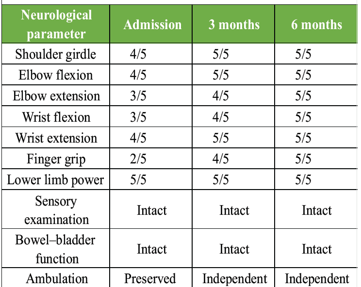
Table 1 : Neurological examination during follow-up
A 30-year-old male presented to our emergency department following a high-velocity road traffic accident in which he was struck by a car while riding a motorcycle. The patient complained of severe neck pain and bilateral upper limb weakness. Notably, he was ambulatory at presentation, with intact lower limb motor function and preserved bowel-bladder function. There was no loss of consciousness reported.
Neurological examination revealed bilateral upper limb weakness predominantly involving distal muscle groups. Grip strength and fine motor function were reduced bilaterally. Lower limb power, tone, reflexes, and sensation were within normal limits. No Horner syndrome or cranial nerve deficit was identified.
Plain radiographs of the cervical spine demonstrated translational malalignment at the C6–C7 level. Computed tomography (CT) of the cervical spine confirmed bilateral facet dislocation at C6–C7, a burst fracture of the C7 vertebral body with involvement of the posterior elements, and fractures extending into the transverse foramen bilaterally (Fig. 1 and 2).
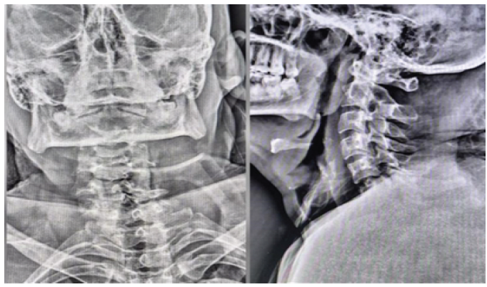
Figure 1: Pre-operative plain radiographs of the cervical spine. Anteroposterior (left) and lateral (right) plain radiographs demonstrating translational malalignment at C6–C7 with loss of normal cervical lordosis, consistent with a high-energy flexion-distraction injury.
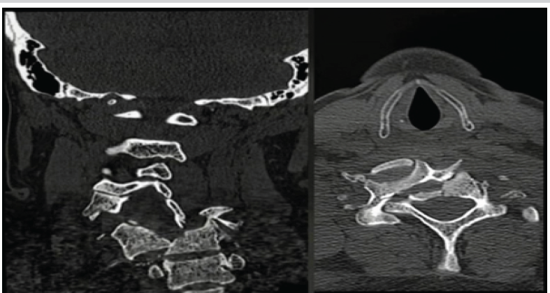
Figure 2: Pre-operative computed tomography (CT) cervical spine – coronal and axial views. Coronal (left) and axial (right) CT images demonstrating bilateral facet dislocation at C6–C7, burst fracture of C7 with posterior element comminution, and fractures extending into the transverse foramen bilaterally. Findings are consistent with arbeitsgemeinschaft für osteosynthesefragen Spine Type C/F4 injury.
CT angiography demonstrated complete thrombosis of the left vertebral artery at the level of injury. Magnetic resonance imaging (MRI) of the cervical spine revealed traumatic disc disruption at C6–C7, severe cord contusion with intrinsic signal change, PLC injury, and significant canal compromise (Fig. 3). According to the Arbeitsgemeinschaft für Osteosynthesefragen (AO) spine classification, the injury was formally designated as Type C/F4/N2/M1/M2/M4.
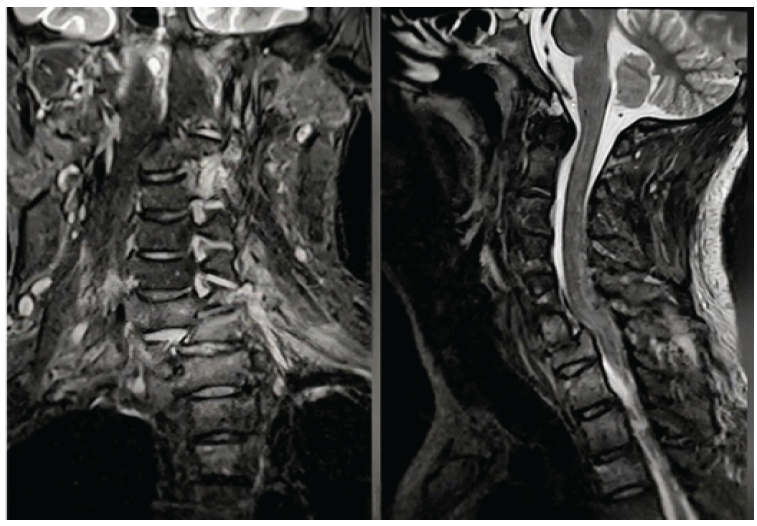
Figure 3: Pre-operative magnetic resonance imaging (MRI) cervical spine – coronal and sagittal T2-weighted images. Coronal (left) and sagittal (right) T2-weighted MRI images demonstrating severe cord contusion with intrinsic hyperintense signal change at C6–C7, traumatic disc disruption, posterior ligamentous complex injury, and significant canal compromise (Arbeitsgemeinschaft für Osteosynthesefragen modifiers M1 and M2).
In view of the radiological instability, the patient was immobilized in a cervical collar and evaluated by the neurovascular team. After interdisciplinary discussion and patient counseling, the team decided to proceed with urgent surgical stabilization.
The patient was positioned prone under general anesthesia with Gardner–Wells cervical tongs applied using 10–12 kg of axial traction to facilitate reduction. A standard posterior midline cervical incision was made, followed by careful subperiosteal dissection of the paraspinal musculature. Bilateral facet dislocation at C6–C7 was directly visualized (Fig. 4).
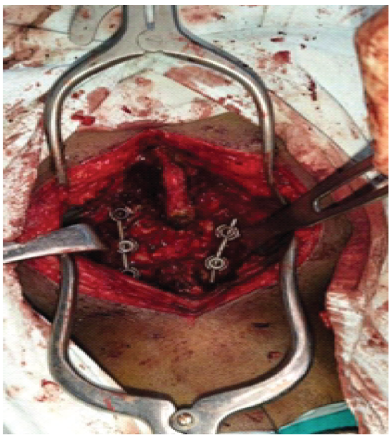
Figure 4: Intraoperative photograph – lateral mass screw placement at C5–C7. Posterior cervical surgical field following laminectomy and decompression at C6–C7. Lateral mass screws were placed bilaterally at C5 and C6, with a right-sided screw at C7 (left C7 omitted due to comminuted fracture). Self-retaining retractors maintain posterior cervical exposure.
Reduction of the dislocation was achieved intraoperatively using a combination of cervical traction and a controlled lever mechanism. Posterior stabilization was accomplished using lateral mass screws: Two screws were placed bilaterally at C5 and C6. At C7, a lateral mass screw was placed only on the right side, as the left C7 lateral mass was fractured and deemed unsuitable for screw purchase. One longitudinal rod was contoured and inserted on the right side, connecting the screw heads from C5 to C7. Laminectomy at C6–C7 was then performed to achieve adequate neural decompression. The left-side longitudinal rod was contoured and inserted, and all set screws were tightened to achieve rigid fixation.
Postoperatively, the patient was nursed in a semi-recumbent position and commenced early structured physiotherapy with a four-post collar. He demonstrated progressive neurological recovery over subsequent weeks. At 6-month follow-up, complete motor recovery was achieved in both upper and lower limbs, with full restoration of bowel-bladder function (Fig. 5 and 6). Serial imaging confirmed maintained reduction and solid arthrodesis at the operative levels.
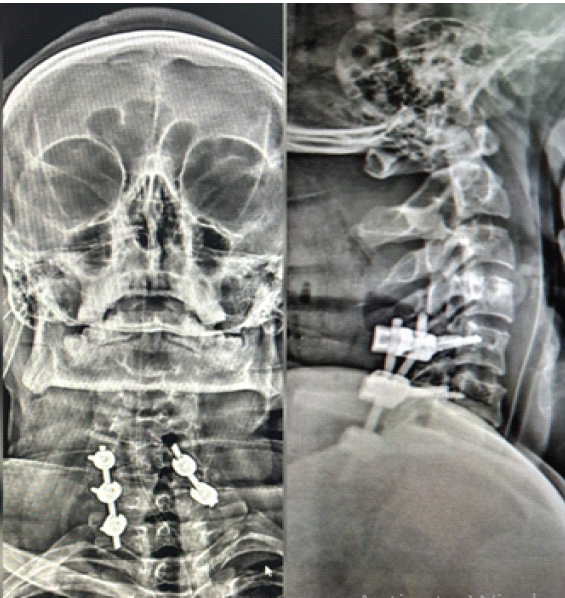
Figure 5: Post-operative plain radiographs – anteroposterior (AP) and lateral views. AP (left) and lateral (right) post-operative radiographs confirming satisfactory reduction of the C6–C7 deformity. Lateral mass screws at C5 and C6 bilaterally and at right C7, connected by two longitudinal rods, provide rigid internal fixation with restored cervical alignment.
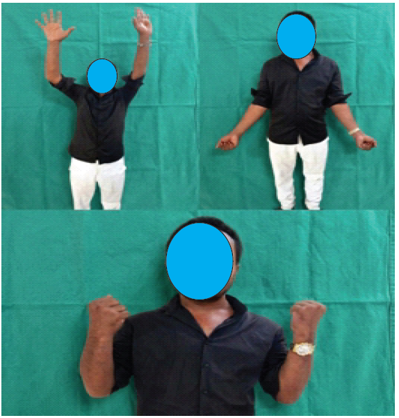
Figure 6: Clinical photographs at 6-month follow-up – Complete neurological recovery. Full shoulder abduction with hands raised overhead (top left), neutral standing posture (top right), and bilateral grip formation (bottom), confirming 5/5 power in all upper limb muscle groups at 6-month follow-up. Faces pixelated to preserve patient anonymity per JOCR editorial policy.
Subaxial cervical translational injuries with bilateral facet dislocation are among the most mechanically unstable injuries of the cervical spine. These injuries typically occur following high-energy flexion-distraction mechanisms and are generally accompanied by catastrophic neurological deficits, including complete quadriplegia [1,2]. The constellation of bilateral facet dislocation, burst fracture, disc disruption, cord contusion, and PLC injury observed in our patient represents the most severe end of the subaxial cervical injury spectrum.
The defining feature of this case was the striking discordance between the severity of radiological findings and the clinical neurological status. Despite catastrophic imaging findings, the patient remained ambulatory with intact lower limb function and preserved bowel–bladder control. Such presentations, though exceedingly rare, have been documented in limited case reports and are postulated to result from a capacious spinal canal, cord plasticity, or partial preservation of white matter tracts despite substantial compressive force [2]. The exact anatomical substrate enabling neurological preservation in the face of such structural disruption warrants further study.
Vertebral artery injury complicates approximately 19–46% of cervical facet fracture-dislocations and carries a risk of posterior circulation ischemia if undiagnosed [3,4]. In our patient, CT angiography demonstrated complete thrombosis of the left vertebral artery. Given adequate collateral circulation through the contralateral vertebral artery and the circle of Willis, the patient did not develop signs of vertebrobasilar ischemia. This underscores the importance of routine vascular screening in all patients with cervical facet dislocation and transverse foramen involvement.
Surgical stabilization remains the definitive treatment for unstable translational cervical injuries [5]. The choice of posterior decompression and lateral mass screw fixation was guided by the predominant posterior column disruption and bilateral facet injury pattern. Posterior fixation affords excellent three-column stabilization and has been associated with satisfactory clinical outcomes in similar injury patterns [6]. Clinical outcome studies based on the subaxial injury classification and severity score have further validated structured management strategies in subaxial cervical trauma [7]. The AO Spine Subaxial Cervical Spine Injury Classification System provides a comprehensive framework for injury characterization and surgical decision-making; the injury in our patient was designated Type C (translational)/F4 (bilateral facet dislocation)/N2 (incomplete deficit)/M1 (PLC injury)/M2 (disc herniation)/M4 (vertebral artery injury) – representing the most severe designation within this system [8].
Timing of surgical decompression is an important prognostic determinant in traumatic cervical spinal cord injury. Emerging evidence suggests that early decompression, ideally within 24 h of injury, is associated with improved neurological outcomes [9]. In our case, prompt surgical intervention likely contributed to the excellent neurological recovery observed at follow-up. Structured post-operative rehabilitation, including physiotherapy focused on motor re-education and functional training, further supported recovery [10].
Predictors of neurological recovery following traumatic cervical spinal cord injury include initial neurological grade, extent of cord signal change on MRI, and the timing of surgical intervention [11,12,13]. Our patient’s favorable outcome may reflect the relative preservation of cord integrity despite the severe structural injury, combined with the benefits of timely surgery.
Limitations
This report has limitations inherent to a single-patient case study, including limited generalizability and reliance on one institution’s protocols. The mechanism behind the neurological preservation remains speculative, as advanced neurophysiological or diffusion tensor imaging was not performed. Additionally, the 6-month follow-up did not allow assessment of long-term fusion durability, chronic pain, or adjacent segment degeneration; post-operative vascular imaging was not obtained to track the thrombosed artery or evolution of collateral circulation; standardized outcome scores (e.g., neck disability index, SF-36, EQ-5D, Nurick score) were not used; and alternative surgical approaches were not compared.
Severe C6–C7 translational injuries with bilateral facet dislocation and associated vertebral artery thrombosis are typically accompanied by devastating neurological deficits. Nonetheless, rare patients may remain ambulatory with preserved bowel–bladder function and intact lower limb neurological function despite catastrophic radiological findings. This case reaffirms the necessity of systematic and meticulous neurological assessment in all trauma patients, irrespective of radiological severity. Early diagnosis, prompt surgical stabilization, routine vascular screening, and structured rehabilitation collectively contribute to excellent neurological and functional recovery in such unusual presentations.
Severe subaxial cervical translational injuries can occasionally present with preserved ambulation and minimal neurological deficit despite devastating radiological abnormalities. Clinicians must not underestimate injury severity based on initial neurological status alone. Detailed neurological examination, urgent radiological evaluation including vascular imaging, and prompt surgical stabilization are essential to prevent secondary neurological deterioration and to optimize recovery.
References
- 1. Vaccaro AR, Hulbert RJ, Patel AA, Fisher C, Dvorak M, Lehman RA Jr., et al. The subaxial cervical spine injury classification system: A novel approach to recognize the importance of morphology, neurology, and integrity of the disco-ligamentous complex. Spine (Phila Pa 1976) 2007;32:2365-74. [Google Scholar] [PubMed]
- 2. Allen BL Jr., Ferguson RL, Lehmann TR, O’Brien RP. A mechanistic classification of closed, indirect fractures and dislocations of the lower cervical spine. Spine (Phila Pa 1976) 1982;7:1-27. [Google Scholar] [PubMed]
- 3. Mueller CA, Peters I, Podlogar M, Kovacs A, Urbach H, Schaller K, et al. Vertebral artery injuries following cervical spine trauma: A prospective observational study. Eur Spine J 2011;20:2202-9. [Google Scholar] [PubMed]
- 4. Biffl WL, Moore EE, Elliott JP, Ray C, Offner PJ, Franciose RJ, et al. The devastating potential of blunt vertebral arterial injuries. Ann Surg 2000;231:672-81. [Google Scholar] [PubMed]
- 5. Joaquim AF, Patel AA. Subaxial cervical spine trauma: Evaluation and surgical decision-making. Global Spine J 2014;4:63-70. [Google Scholar] [PubMed]
- 6. Aarabi B, Alexander M, Mirvis SE, Shanmuganathan K, Chesler D, Maulucci C, et al. Predictors of outcome in acute traumatic central cord syndrome due to spinal stenosis. J Neurosurg Spine 2011;14:122-30. [Google Scholar] [PubMed]
- 7. Fehlings MG, Arun R, Vaccaro AR, Arnold PM, Chapman JR, Kopjar B. Predictors of treatment outcomes in geriatric patients with odontoid fractures: AOSpine North America multi-centre prospective GOF study. Spine (Phila Pa 1976) 2013;38:881-6. [Google Scholar] [PubMed]
- 8. Badhiwala JH, Wilson JR, Witiw CD, Harrop JS, Vaccaro AR, Aarabi B, et al. The influence of timing of surgical decompression for acute spinal cord injury: A pooled analysis of individual patient data. Lancet Neurol 2021;20:117-26. [Google Scholar] [PubMed]
- 9. Schleicher P, Kobbe P, Kandziora F, Scholz M, Badke A, Brakopp F, et al. Treatment of injuries to the subaxial cervical spine: Recommendations of the Spine section of the German society for orthopaedics and trauma (DGOU). Global Spine J 2018;8 2 Suppl:25S-33. [Google Scholar] [PubMed]
- 10. Joaquim AF, Ghizoni E, Tedeschi H, Da Cruz HY, Patel AA. Clinical results of patients with subaxial cervical spine trauma treated according to the SLIC score. J Spinal Cord Med 2014;37:420-7. [Google Scholar] [PubMed]
- 11. Vaccaro AR, Koerner JD, Radcliff KE, Oner FC, Reinhold M, Schnake KJ, et al. AOSpine subaxial cervical spine injury classification system. Eur Spine J 2016;25:2173-84. [Google Scholar] [PubMed]
- 12. Wilson JR, Cadotte DW, Fehlings MG. Clinical predictors of neurological outcome, functional status, and survival after traumatic spinal cord injury: A systematic review. J Neurosurg Spine 2012;17 Suppl 1:11-26. [Google Scholar] [PubMed]
- 13. Stenimahitis V, Gharios M, Fletcher-Sandersjöö A, El-Hajj VG, Singh A, Buwaider A, et al. Long-term outcome and predictors of neurological recovery in cervical spinal cord injury: A population-based cohort study. Sci Rep 2024;14:20864. [Google Scholar] [PubMed]






Related Articles in Journal of Orthopaedic Case Reports
 October 1, 2026 Bilateral Medial Medullary Syndrome Following Anterior Odontoid Screw Fixation for a Type II Fracture: A Case Report
 September 1, 2026 Blunt Traumatic Posterior Cord Syndrome Following a Hangman Fracture: A Case Report
 April 1, 2026 A Rare Case of C5–C6 Bilateral Facetal Dislocation with Central Cord Syndrome Managed by Staged Stabilization
 July 1, 2025 Case Report – Surgical Management and Neurological Recovery in an 18-Year-Old Male with C4-C5 Cervical Spine Fracture