


Introduction
Calcific tendinitis is characterized by calcium crystal deposition in or around tendons, typically near their insertion sites, causing acute severe pain and swelling. The shoulder, particularly the supraspinatus tendon, is the most commonly affected site [1]. Other locations, such as the hip and wrist are rarely involved. Within the wrist, the Flexor Carpi Ulnaris tendon is most frequently affected [2]. Acute calcific tendinitis is often misdiagnosed as infection or inflammatory conditions, leading to inappropriate antibiotic use. Diabetes is a significant risk factor for this [3].
Case Reports
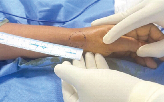
A 53-year-old diabetic female presented with abrupt-onset, throbbing pain over the left wrist lasting 2 weeks, exacerbated by movement. There was no history of trauma. Examination revealed erythema, edema, and severe tenderness over the first extensor compartment of the wrist with a positive Finkelstein test (Fig. 1).
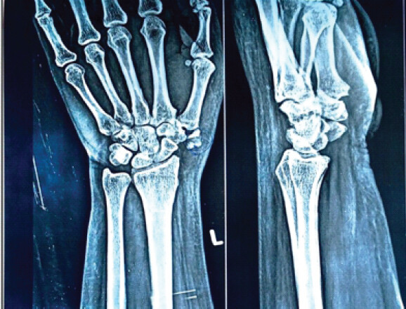
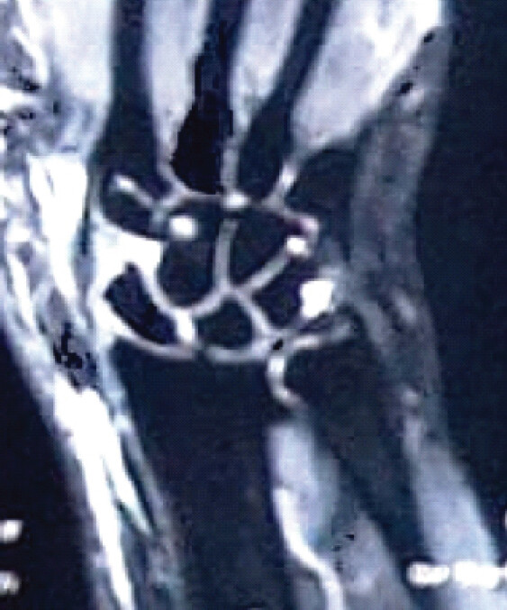
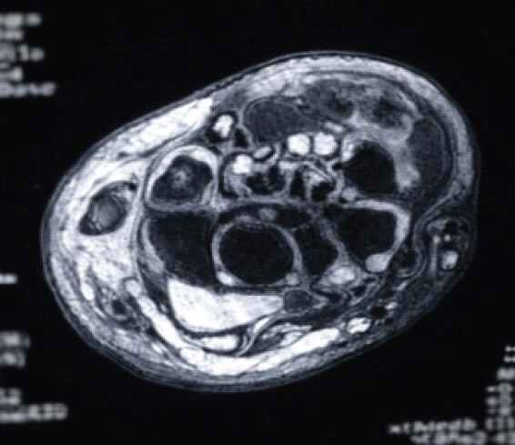
Conservative treatment with nonsteroidal anti-inflammatory drugs (NSAIDs) failed to relieve symptoms for 1 week. Radiographs showed calcification proximal to the base of the first metacarpal. Laboratory tests indicated leukocytosis with neutrophilia, elevated C-reactive protein, and high HbA1C; uric acid was normal. Magnetic resonance imaging confirmed calcific tendinitis in the first extensor compartment (Fig. 2, 3, 4).
Due to failed conservative management, surgical decompression was performed. A curvilinear incision was made over the first extensor compartment, and subcutaneous tissue was dissected to identify the compartment. Thick chalky white calcium deposit was found on releasing of the compartment. A thorough wash was given and all tendons were separately isolated (Figs. 5 and 6).
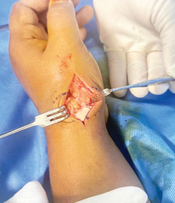
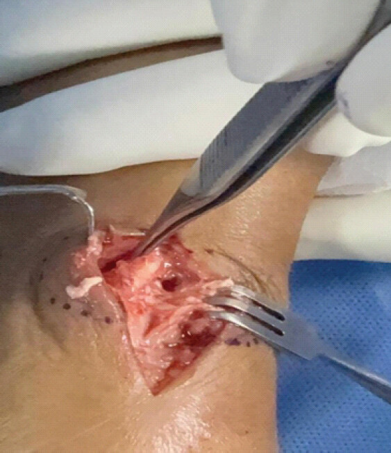
Post-operatively, the patient was immobilized with a thumb spica splint, administered NSAIDs, and underwent strict glycemic control. Gradual thumb ranges of motion exercises were initiated. At 1-week follow-up, inflammation resolved with significant pain relief, and by 2 weeks, the patient resumed normal activities without pain. At 3 months follow-up patient is fully asymptomatic.
Discussion
Acute calcific tendinitis of the wrist is exceedingly rare. A systematic review by Draghi et al. identified only 13 hand and 7 wrist cases among 184 extra-shoulder calcific tendinitis cases [4]. It predominantly affects females aged 30–60 years. The Flexor Carpi Ulnaris tendon is most commonly involved, usually near its insertion at the pisiform bone. The etiology is not fully understood but may involve tendon degeneration, reactive calcification with fibrocartilaginous metaplasia, tissue hypoxia, mechanical strain, and failed healing [5]. Uhthoff’s model describes stages from pre-calcific fibrocartilaginous metaplasia through calcific phases to tendon remodeling [6].
Clinically, patients present with acute localized pain, erythema, swelling, and tenderness, often mimicking infection [7]. Fever is usually absent or low-grade, and inflammatory markers may be normal or mildly elevated. A history of significant trauma is present in only a minority of patients, although repetitive wrist activities – such as racket sports or manual work demanding repeated flexion – may contribute, and the dominant hand is more frequently affected [8].
Plain radiographs are the initial modality of choice and typically show calcifications measuring 2–10 mm, with fluffy, ill-defined, inhomogeneous deposits in acute stages and denser, homogeneous foci in chronic stages. Ultrasonography further aids evaluation by precisely localizing calcifications, characterizing their consistency, and assessing vascularity and activity with Doppler; it also facilitates interventional procedures, such as ultrasound-guided barbotage [9].
Treatment lacks consensus, but most cases resolve with conservative management and strict diabetes control. Those cases that are refractory to conservative management have been treated by a myriad of different methods, including open resection, arthroscopic resection, steroid injection, ultrasound-guided needle lavage (barbotage), extracorporeal shockwave therapy, and platelet-rich plasma injection [10]. Refractory cases benefit from surgical or other interventional treatments. Surgical debridement provides early and definitive relief when conservative measures fail. There are no reports of recurrence at the same site after surgical debridement.
Conclusion
Acute calcific tendinitis of the first extensor compartment of the wrist is a rare entity that can mimic De Quervain’s Tenosynovitis. Radiographic evaluation is essential when patients do not respond to conservative treatment. Surgical intervention offers prompt symptom resolution and prevents prolonged morbidity.
Clinical Message
In cases of persistent wrist pain resembling De Quervain’s Tenosynovitis, especially in diabetic patients, clinicians should consider acute calcific tendinitis and perform appropriate imaging. Early diagnosis and timely surgical management can significantly improve outcomes.
Conflict of Interest:
Nil
Source of Support:
Nil
Consent
The authors confirm that informed consent was obtained from the patient for publication of this article
How to Cite this Article
Hameed H, Gupta VK, Subramanian JNK, Bapu NKS, Subramanian KN. Not All Dequervains are Dequervains – A Rare Case Report. J Orthop Case Rep. 2026 November;16(11):208-211. doi: 10.13107/jocr.2026.v16.i11.8442.
References
- Moyer RA, Bush DC, Harrington TM. Acute calcific tendinitis of the hand and wrist: A report of 12 cases and a review of the literature. J Rheumatol 1989;16:198-202. [Google Scholar] | [PubMed]
- Torbati SS, Bral D, Geiderman JM. Acute calcific tendinitis of the wrist. J Emerg Med 2013;44:352-4. [Google Scholar] | [PubMed]
- Su YC, Chung CH, Ke MJ, Chen LC, Chien WC, Wu YT. Increased risk of shoulder calcific tendinopathy in diabetes mellitus: A nationwide, population-based, matched cohort study. Int J Clin Pract 2021;75:e14549. [Google Scholar] | [PubMed]
- Draghi F, Cocco G, Lomoro P, Bortolotto C, Schiavone C. Non-rotator cuff calcific tendinopathy: Ultrasonographic diagnosis and treatment. J Ultrasound 2020;23:301-15. [Google Scholar] | [PubMed]
- Oliva F, Via AG, Maffulli N. Physiopathology of intratendinous calcific deposition. BMC Med 2012;10:95. [Google Scholar] | [PubMed]
- Uhthoff HK, Loehr JW. Calcific tendinopathy of the rotator cuff: Pathogenesis, diagnosis, and management. J Am Acad Ortho Surg 1997;5:183-91. [Google Scholar] | [PubMed]
- Ejnisman B, Andreoli CV, Monteiro GC, Pocchini Ade C, Cohen C, Tortato S. Calcifying tendinopathy: A local or a systemic condition?. Rev Bras Ortop 2012;47:479-82. [Google Scholar] | [PubMed]
- Dilley DF, Tonkin MA. Acute calcific tendinitis in the hand and wrist. J Hand Surg Br 1991;16:215-6. [Google Scholar] | [PubMed]
- Henderson RE, Walker BF, Young KJ. The accuracy of diagnostic ultrasound imaging for musculoskeletal soft tissue pathology of the extremities: A comprehensive review of the literature. Chiropr Man Therap 2015;23:31. [Google Scholar] | [PubMed]
- Kim MS, Kim IW, Lee S, Shin SJ. Diagnosis and treatment of calcific tendinitis of the shoulder. Clin Shoulder Elb 2020;23:210-6. [Google Scholar] | [PubMed]
© 2026 Journal of Orthopaedic Case Reports - Published by Indian Orthopaedic Research Group
About the Authors
-

Hany Hameed
-

Vivek Kumar Gupta
-

Jeash Narayan Kanthalu Subramanian
-

Nehasri Kuppa Srinivas Bapu
-

Kanthalu Narayanan Subramanian
Related Articles in Journal of Orthopaedic Case Reports
 August 1, 2026 Calcific Tendinitis of the Linea Aspera with Bilateral Deposits and Inflammatory Asymmetry Presenting as Unilateral Pain: A Case Report and Review of Literature
 June 1, 2026 Kienbock’s Disease in a Young Female without Classical Risk Factors: A Case Report with 1-year Functional Outcome Following Proximal Row Carpectomy
 May 1, 2026 Ultrasound and Fluoroscopy-Guided Percutaneous Hydrotenotomy with Open Conversion for Symptomatic Periligamentous Calcification of the Medial Collateral Ligament: A Case Report
 March 1, 2026 A Rare Case of Calcific Tendinitis of the Elbow Mimicking Lateral Epicondylitis: Diagnostic Challenges and Surgical Management