

Calcific tendinitis of the linea aspera may present with an aggressive-appearing cortical reaction and marrow edema and can be bilateral despite unilateral symptoms; careful CT correlation is essential to confirm intratendinous calcification and avoid misdiagnosis as infection or malignancy.
Dr. Aravindharaj Tamilselvan, Department of Radiodiagnosis, Madha Medical College and Research Institute, Chennai, Tamil Nadu, India. E-mail: aravindharajt@gmail.com
Abstract
Introduction: Calcific tendinitis (hydroxyapatite deposition disease) most commonly affects the shoulder and peri-trochanteric hip region. Involvement of the linea aspera is uncommon and may mimic aggressive pathology due to associated cortical reactions and marrow edema.
Case Report: A 53-year-old female presented with predominantly right-sided upper thigh pain. Radiographs demonstrated amorphous calcification along the lateral proximal femur at the linea aspera. Magnetic resonance imaging showed a calcific focus with marked adjacent marrow and soft-tissue edema on the right, while the contralateral side demonstrated minimal muscular hyperintensity without a definite calcific focus. Computed tomography (CT) confirmed bilateral intratendinous calcific deposits, with a larger deposit on the symptomatic right and a tiny 2.5 mm deposit on the asymptomatic left. Conservative management resulted in complete symptom resolution.
Conclusion: Calcific tendinitis of the linea aspera may be bilateral despite unilateral symptoms and can exhibit marked inflammatory asymmetry. CT is essential for confirming intratendinous calcification and avoiding misdiagnosis as infection or malignancy.
Keywords: Calcific tendinitis, linea aspera, hydroxyapatite deposition disease, bilateral involvement, magnetic resonance imaging, computed tomography.

Calcific tendinitis is a crystal-induced inflammatory disorder characterized by deposition of calcium hydroxyapatite crystals within tendons and peri-tendinous soft tissues [1]. It is distinct from degenerative enthesopathy and typically follows a self-limiting clinical course. The condition most commonly affects the rotator cuff, followed by the hip region, where deposits usually involve peri-trochanteric tendons and gluteal insertions [1-5]. Although calcific tendinitis may occur near virtually any tendinous insertion, involvement of the linea aspera is uncommon and sparsely reported in the literature [2,3]. Lesions at this site pose a diagnostic challenge due to their deep location, frequent cortical reaction, and extensive surrounding inflammatory changes, which may closely mimic infection or surface malignancy [3,6,7]. Consequently, unnecessary biopsy or surgical intervention has been reported in several cases. Awareness of this entity and its characteristic multimodality imaging features is therefore essential. We present a case of bilateral calcific tendinitis of the linea aspera with unilateral symptomatic predominance and discuss its imaging appearance, diagnostic pitfalls, and clinical significance in the context of existing literature.
A 53-year-old female presented with a 3-month history of predominantly right-sided upper thigh pain. The pain was continuous, dull, aching in nature, and aggravated by activity. There was no history of trauma, fever, weight loss, or other systemic symptoms. Routine laboratory investigations, including complete blood count, erythrocyte sedimentation rate, and C-reactive protein, were within normal limits. She had no known metabolic, endocrine, or rheumatological disorders. Clinical examination revealed localized tenderness over the proximal posterior aspect of the right thigh, while hip joint movements were full and unrestricted. The left thigh was asymptomatic. Anteroposterior radiographs of both femurs, including the hip joints, demonstrated an amorphous calcific density along the lateral aspect of the proximal right femur at the level of the linea aspera (Fig. 1).
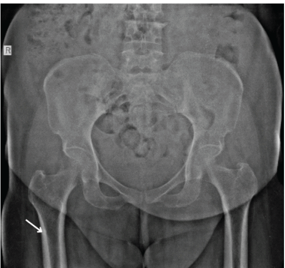
Figure 1: Anteroposterior radiograph of the pelvis demonstrates a small, well-defined amorphous calcific focus (arrow) along the lateral aspect of the proximal femoral shaft at the level of the linea aspera on the right side, without cortical destruction or aggressive periosteal reaction. The left side appears unremarkable.
No definite calcific focus was appreciable on the left side on plain radiography. There was no fracture line or discrete soft-tissue mass. Ultrasonography of the right proximal thigh (Fig. 2) showed an echogenic focus with posterior acoustic shadowing adjacent to the femoral cortex, along with a few cortical irregularities, surrounding muscular edema, and minimal fluid.
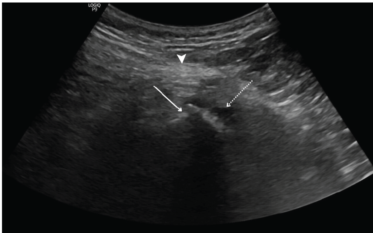
Figure 2: Ultrasonography of the proximal right thigh shows a hyperechoic focus (solid arrow) with posterior acoustic shadowing along the linea aspera with adjacent cortical irregularities. Minimal surrounding free fluid (dashed arrow) noted. Adjacent muscles appear hyperechoic, suggestive of inflammatory changes (arrow head).
No hypervascular soft-tissue mass was identified. Magnetic resonance imaging (MRI) of the thighs demonstrated a focal hypointense lesion along the posterior aspect of the proximal right femur at the linea aspera, consistent with a calcific deposit (Fig. 3).
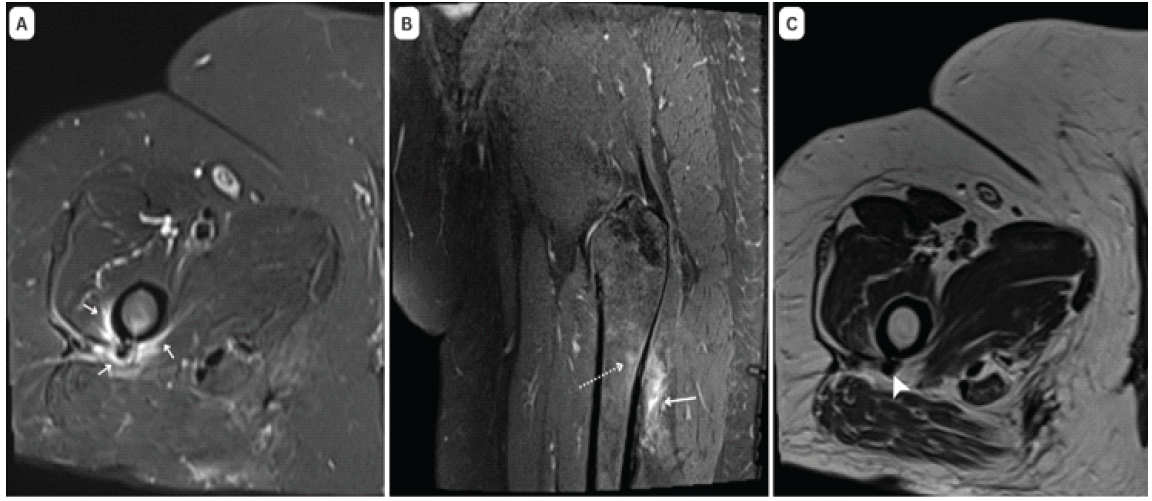
Figure 3: Magnetic resonance imaging axial T2-weighted fat-suppressed (a) and coronal T2-weighted fat-suppressed (b) images of the proximal right thigh demonstrate peri-osseous edema, subtle marrow edema (dotted arrow), and edema within the muscles attaching to the linea aspera (solid arrow) – gluteus maximus, adductors, and vastus lateralis. Axial T2-weighted image (c) reveals a corresponding hypointense focus (arrowhead), suggestive of calcification.
There was marked adjacent marrow edema, cortical reaction, and periosteal soft-tissue edema extending into the gluteus maximus, adductor musculature, and vastus lateralis. No discrete soft-tissue mass was seen. On careful contralateral assessment, MRI demonstrated minimal edema within the left vastus lateralis at the level of the linea aspera without a definite corresponding hypointense calcific focus (Fig. 4).
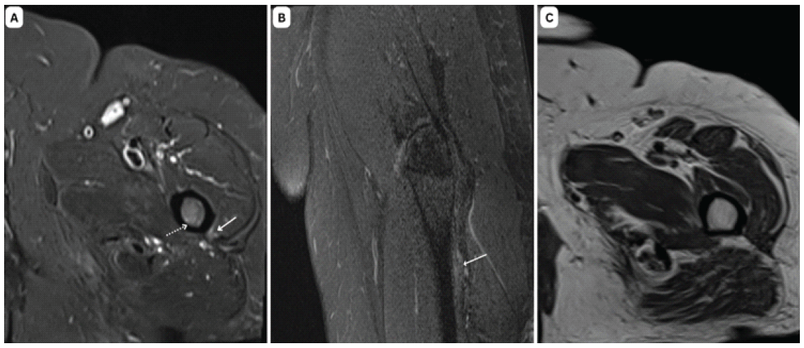
Figure 4: Magnetic resonance imaging axial T2-weighted fat-suppressed (a) and coronal T2-weighted fat-suppressed (b) images of the proximal left thigh demonstrate subtle marrow edema (dotted arrow), and minimal edema (solid arrow) within the vastus lateralis. Axial T2-weighted image (c) reveals no grossly evident corresponding hypointense focus when compared to the right side.
Given the subtle asymmetry, further evaluation with computed tomography (CT) was performed. CT confirmed bilateral intratendinous calcific deposits at the tendon insertion along the linea aspera (Fig. 5).
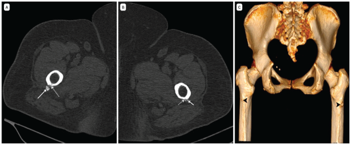
Figure 5: Axial computed tomography images of the right (a) and left (b) proximal femurs demonstrate amorphous calcific deposits (solid arrows) adjacent to the postero-lateral cortex at the level of the linea aspera, with mild associated cortical irregularity. Three-dimensional volume-rendered reconstruction (c) highlights bilateral calcific foci (arrowheads) at the linea aspera. The left-sided calcific deposit is significantly smaller than the one on the right side.
The right-sided deposit was larger and associated with cortical thickening and irregularity. On the left side, a tiny calcific focus was identified with mild adjacent cortical thickening and irregularity, corresponding to the region of minimal MRI hyperintensity. No aggressive periosteal reaction or soft-tissue mass was demonstrated on either side. Based on the multimodality imaging findings, a diagnosis of bilateral calcific tendinitis of the linea aspera with right-sided symptomatic and inflammatory predominance was established. The patient was managed conservatively with non-steroidal anti-inflammatory drugs, rest, and local cold compresses. At follow-up, she reported being symptom-free but was advised image-guided steroid injection if symptoms recur.
Calcific tendinitis, or hydroxyapatite deposition disease (HADD), is an immune and cell-mediated response rather than a strictly degenerative disorder [1,4]. Multiple mechanisms have been proposed, including fibrocartilaginous transformation under hypoxic conditions and aberrant differentiation of tendon-derived stem cells [4,7]. The course of calcific tendinitis involves the pre-calcification, calcification, and post-calcification stages, where the calcification period is further divided into the formative, resting, and resorption phases based on the classification system established by Uhthoff [1,9]. The resorptive phase is typically the most symptomatic, characterized by macrophage-mediated breakdown of the calcium deposit and extrusion of crystals into surrounding tissues, producing an intense inflammatory response and edema [1,4]. This staging model explains the asymmetry observed in the present case. The symptomatic right side demonstrated extensive periosseous and marrow edema with a conspicuous calcific focus, consistent with an active resorptive phase. In contrast, the contralateral side showed only minimal muscular hyperintensity on MRI without a definite hypointense focus. CT subsequently revealed a tiny calcific deposit at the left linea aspera. This likely represents a resting or early formative stage with limited inflammatory response. Such discordance highlights that disease burden may exceed clinical symptomatology and that bilateral involvement may be present despite unilateral symptoms. Although the differing imaging appearances likely reflect different stages of HADD, this interpretation remains inferential because serial imaging was not available to document the temporal progression of the lesions. Furthermore, the degree of marrow and soft-tissue edema observed on MRI may vary depending on the phase of disease at the time of imaging acquisition, and therefore, imaging findings should always be interpreted in conjunction with CT correlation and clinical presentation. The linea aspera is a mechanically stressed posterior femoral enthesis serving as the insertion site for the gluteus maximus, vastus lateralis, and adductor musculature [2]. Lesions at this location demonstrate a higher propensity for osseous involvement compared with more superficial sites [3,6]. Ramon et al. described three cases of calcific tendinitis at the vastus lateralis insertion along the linea aspera, all demonstrating cortical changes best appreciated on CT [10]. Similarly, Fritz et al. reported four cases of paradiaphyseal calcific tendinitis involving the linea aspera, all with cortical bone erosion, several of which were initially suspected to represent malignancy [3]. In pooled data from Low and Toms, cortical abnormalities were identified in 75% of cases evaluated with CT, underscoring that osseous reaction at this site is common rather than exceptional [8]. While there have been reports of calcific tendinitis at the linea aspera, the majority of reported cases have been unilateral in nature, including a recently published case series describing hydroxyapatite deposition disease at unusual musculoskeletal sites that included a single case involving the linea aspera [11]. Reports of bilateral linea aspera calcific tendinitis with marked inflammatory asymmetry are exceedingly rare in the literature. This report demonstrates that contralateral calcific deposits may remain clinically asymptomatic and radiographically occult, becoming apparent only on cross-sectional imaging. Imaging plays a central diagnostic role. Plain radiographs typically demonstrate amorphous, globular, or cloud-like calcifications adjacent to the posterior femoral cortex [1,8]. During the resorptive phase, deposits may appear fluffy and ill-defined, correlating with acute pain [1,9]. MRI is highly sensitive for inflammatory changes but may be misleading when interpreted in isolation. Calcifications appear as low-signal foci on all sequences; however, small deposits may be inconspicuous due to limited spatial resolution or partial volume averaging [7,9]. Marked T2/STIR hyperintensity in adjacent muscles and bone marrow may simulate infection or surface malignancy, particularly in the presence of cortical irregularity [2,7]. CT is the most reliable modality for confirming intratendinous calcification and characterizing cortical involvement [3,7,11]. CT accurately localizes the deposit to the tendon insertion, demonstrates comet-tail or flame-like morphology, and excludes an associated discrete soft-tissue mass [7,8]. Importantly, cortical erosion in HADD is reactive and may regress following resolution of the calcific deposit, as documented in prior series [3]. Approximately nine case reports of calcific tendinitis of the linea aspera have been published, which, together with some small case series, have added up to a total of 55 cases in a recently conducted narrative review [8]. Most patients present with acute or subacute posterior thigh pain and demonstrate favorable outcomes with conservative management [2,3]. Although interventions like CT-guided barbotage help alleviate symptoms more quickly, there is no difference in long-term results between interventions and conservative treatment methods [5]. The self-limiting nature of the condition underscores the importance of accurate imaging diagnosis. Histological features such as chondroid metaplasia, inflammatory cell infiltration, multinucleated giant cells, and reactive bone formation can easily be mistaken for malignancy, especially when performing a biopsy without proper radiologic correlation [3,6,11]. Presence of specific imaging signs, such as intratendinous calcification at the linea aspera, reactive bone changes without an identifiable mass lesion, and spontaneous resolution of symptoms, ensures that a clear diagnosis is made, thus preventing unnecessary invasive procedures. Histopathological confirmation was not obtained in the present case because the imaging findings were characteristic and the patient demonstrated complete clinical resolution following conservative treatment. Recognition of the typical multimodality imaging features is important to avoid unnecessary invasive diagnostic procedures. However, this report has a few limitations. As a single-patient observation, the findings may not be generalizable to the broader population of patients with calcific tendinitis of the linea aspera. Long-term clinical and imaging follow-up was not available to assess complete radiological resolution, recurrence, or the future evolution of the asymptomatic contralateral deposit. Standardized pain or functional outcome measures were not recorded, and treatment response was assessed clinically based on symptom resolution. Finally, although bilateral deposits were identified in this patient, no conclusions can be drawn regarding the prevalence of bilateral involvement in calcific tendinitis of the linea aspera. The present case expands the recognized spectrum of linea aspera HADD by demonstrating bilateral deposits with marked inflammatory and clinical asymmetry. Careful contralateral evaluation and multimodality imaging, particularly CT correlation, are essential for establishing diagnostic confidence and preventing overinterpretation of aggressive imaging features. From an orthopedic standpoint, the presence of cortical irregularity and marrow edema along the femoral diaphysis can be indicative of infection or surface malignancy and may warrant further investigation, such as a biopsy or oncological consultation. The detection of intra-tendinous calcification on a CT scan is definitive and justifies conservative treatment.
Calcific tendinitis of the linea aspera is a rare manifestation of HADD, which can be confused with aggressive osseous pathology owing to the associated cortical reaction and bone marrow edema. It must be remembered that bilateral involvement is possible, even when there are symptoms only on one side, with differences in disease severity being related to the stage of calcification. While MRI is exquisitely sensitive in the demonstration of edema, it may overestimate the disease severity or miss small calcifications, while CT is the most reliable modality for the detection of intratendinous calcification and associated changes in the cortex. Recognition of this entity and appropriate multimodality imaging correlation are essential to avoid misdiagnosis, unnecessary biopsy, and overtreatment, given the typically self-limiting course of the condition.
Calcific tendinitis of the linea aspera should be considered in patients presenting with posterior thigh pain and aggressive-appearing cortical changes on imaging. The condition may be bilateral despite unilateral symptoms, and MRI findings of marrow and soft-tissue edema can exaggerate lesion severity. CT confirmation of intratendinous calcification is essential to establish the diagnosis, prevent misinterpretation as malignancy or infection, and guide appropriate conservative management.
References
- 1. Siegal DS, Wu JS, Newman JS, Del Cura JL, Hochman MG. Calcific tendinitis: A pictorial review. Can Assoc Radiol J 2009;60:263-72. [Google Scholar] [PubMed]
- 2. Patil AR, Samson M. Imaging in calcific tendinitis of linea aspera: A case report and review of literature. Indian J Musculoskelet Radiol 2021;3:129-32. [Google Scholar] [PubMed]
- 3. Fritz P, Bardin T, Laredo JD, Ziza JM, D’Anglejan G, Lansaman J, et al. Paradiaphyseal calcific tendinitis with cortical bone erosion. Arthritis Rheum 1994;37:718-23. [Google Scholar] [PubMed]
- 4. Hegazi T. Hydroxyapatite deposition disease: A comprehensive review of pathogenesis, radiological findings, and treatment strategies. Diagnostics (Basel) 2023;13:2678. [Google Scholar] [PubMed]
- 5. Klontzas ME, Vassalou EE, Zibis AH, Karantanas AH. Hydroxyapatite deposition disease around the hip: Outcomes of CT-guided treatment. Diagn Interv Radiol 2016;22:466-70. [Google Scholar] [PubMed]
- 6. Tamangani J, Davies AM, James SL, Christie-Large M. Calcific tendonitis of the adductor brevis insertion. Clin Radiol 2009;64:940-3. [Google Scholar] [PubMed]
- 7. Hongsmatip P, Cheng KY, Kim C, Lawrence DA, Rivera R, Smitaman E. Calcium hydroxyapatite deposition disease: Imaging features and presentations mimicking other pathologies. Eur J Radiol 2019;120:108653. [Google Scholar] [PubMed]
- 8. Low SB, Toms AP. Calcification of the linea aspera: A systematic narrative review. Eur J Radiol Open 2019;6:101-5. [Google Scholar] [PubMed]
- 9. Albano D, Coppola A, Gitto S, Rapisarda S, Messina C, Sconfienza LM. Imaging of calcific tendinopathy around the shoulder: Usual and unusual presentations and common pitfalls. Radiol Med 2021;126:608-19. [Google Scholar] [PubMed]
- 10. Ramon FA, Degryse HR, De Schepper AM, Van Marck EA. Calcific tendinitis of the vastus lateralis muscle. A report of three cases. Skeletal Radiol 1991;20:21-3. [Google Scholar] [PubMed]
- 11. Natarajan M, Cannane S, Vasu L. Hydroxyapatite deposition disease beyond the shoulder: A case series on unusual musculoskeletal locations. Case Rep Clin Radiol 2026;4:23-6. [Google Scholar] [PubMed]




Related Articles in Journal of Orthopaedic Case Reports
 October 1, 2026 Acute Cauda Equina Syndrome in Rapid Succession: A Case Series of Eight Consecutive Patients Undergoing Emergency Surgical Decompression Over One Week
 October 1, 2026 Magnetic Resonance Imaging-Arthroscopy Correlation in Traumatic Knee Injuries: A Retrospective Diagnostic Accuracy Study
 October 1, 2026 Incidence, Arthroscopic Detection, and Clinical Outcomes of Ramp Lesions in Patients Undergoing Anterior Cruciate Ligament Reconstruction: A Prospective Observational Study
 September 1, 2026 Diagnostic Accuracy of Magnetic Resonance Imaging Signs for Detecting Meniscal Root Tears: A Prospective Comparison with Arthroscopic Findings