


Introduction
Congenital coxa vara is a rare deformity of the proximal femur characterized by a decreased femoral neck-shaft angle and increased vertical orientation of the proximal femoral physis. It presents as a limp after the onset of walking and may be associated with a mild limb-length discrepancy in unilateral cases [1]. In contrast, slipped capital femoral epiphysis (SCFE) most commonly occurs during adolescence and has been reported to be associated with chronic conditions, such as endocrine and metabolic disorders and renal disease [2,3].
We report a case of chronic SCFE in a lean pre-adolescent girl with bilateral congenital coxa vara.
Case Report
A girl with no notable medical or family history presented with an out-toeing gait at 8 years and 2 months of age. She was observed, but her symptoms fluctuated over time; therefore, she first visited a local clinic at 9 years and 9 months of age. She was diagnosed with SCFE and was referred to our department at 9 years and 10 months of age for further evaluation and treatment. Hip range of motion showed mild limitation of internal rotation. There was no obvious limb-length discrepancy, and no abnormal findings were noted in other joints.
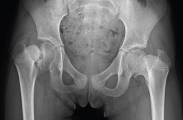
Plain radiographs showed bilateral coxa vara, and the right proximal femoral physis was widened, with posteromedial displacement of the epiphysis and a triangular metaphyseal fragment (Fig. 1). The Hilgenreiner-epiphyseal angle [4] was 76° on the right and 59° on the left. The posterior tilt angle [5] was 68° on the right and 32° on the left.
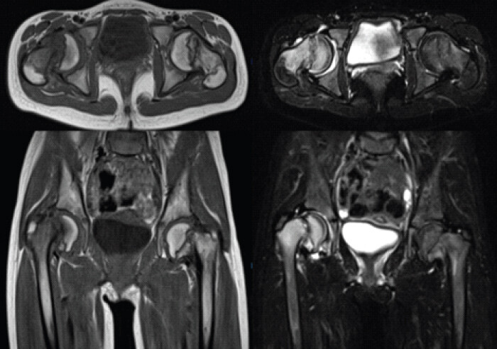
Magnetic resonance imaging showed low signal intensity on T1-weighted images and high signal intensity on short tau inversion recovery images around the right proximal femoral physis, consistent with chronic SCFE. No obvious signal change was observed on the left (Fig. 2).
Prophylactic internal fixation was also planned for the asymptomatic left side, and bilateral in situ pinning of the proximal femoral physes was performed at 9 years and 10 months of age.
Post-operatively, the affected right hip was kept non-weight-bearing using an ischial weight-bearing orthosis, whereas full weight-bearing was permitted on the left side. Evaluation for underlying disease was performed, but no skeletal abnormalities other than those involving the hips were identified, and skeletal dysplasia was considered unlikely. Blood tests showed Blood Urea Nitrogen 10 mg/dL, Creatinine 0.33 mg/dL, and Thyroid-stimulating hormone 2.710 μIU/mL, which did not suggest renal or thyroid dysfunction. Although Parathyroid Hormone was not measured, the only abnormal finding was a mildly decreased 25-hydroxyvitamin D level at 15.0 ng/mL; Calcium 10.0 mg/dL, Inorganic Phosphorus 4.2 mg/dL, and Bone-specific Alkaline Phosphatase 48.3 μg/L were within normal ranges, and metabolic bone disease was considered unlikely. After consultation with pediatricians, skeletal deformity secondary to metabolic bone disease or endocrine disease was also considered unlikely.
At 3 months post-operatively, union of the right triangular metaphyseal fragment was observed, and partial weight bearing on the right side was started. At 6 months post-operatively, radiographic stability on the left side was confirmed, and full weight bearing on both sides was allowed (Fig. 3).
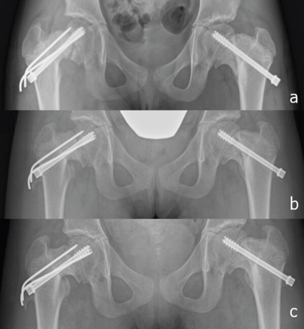
Because of skeletal growth, bilateral screws were exchanged 1 year and 3 months post-operatively. After physeal closure associated with skeletal maturity, implant removal was performed at 14 years and 3 months of age (4 years after the initial surgery).
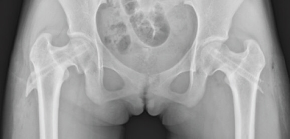
At the final follow-up, she had no hip pain or gait disturbance and was able to participate in dance activities without restrictions (Fig. 4).
Discussion
Congenital coxa vara is rare and is estimated to occur in approximately 1 in 25,000 births [1]. It may be unilateral or bilateral, and some cases are associated with skeletal dysplasias, such as osteogenesis imperfecta, osteopetrosis, epiphyseal dysplasia, and metaphyseal dysplasia [6].
Radiographically, the proximal femoral physis becomes more vertical, and varus deformity may progress as shear force acts on the physis. The Hilgenreiner-epiphyseal angle is used to quantify the deformity and determine the treatment strategy [4].
Weinstein et al. introduced the Hilgenreiner-epiphyseal angle and proposed that valgus osteotomy should be considered when this angle exceeds 60°, that 45–59° represents a gray zone requiring observation, and that angles <45° may improve without surgery [4]. Trigui et al. retrospectively evaluated osteotomy for coxa vara associated with skeletal dysplasia and reported a high rate of recurrence after osteotomy [7]. Similarly, the possibility of recurrence after valgus osteotomy and the importance of sufficient correction have also been mentioned in other reports of long-term outcomes [8,9,10].
To our knowledge, only one case report has described SCFE associated with congenital coxa vara, similar to the present case; that patient was a 5-year-old girl. In that case, treatment consisted of in situ pinning combined with valgus osteotomy [11]. To our knowledge, there have been no previous reports describing favorable outcomes with in situ pinning alone in patients with both congenital coxa vara and SCFE. Although long-term follow-up is required, this case suggests that some patients may achieve satisfactory clinical outcomes without valgus osteotomy.
SCFE in children younger than 10 years is rare and has been reported to be associated with underlying diseases, such as thyroid dysfunction, growth hormone-related disorders, and renal osteodystrophy [2,3,11]. In situ pinning is generally performed for SCFE; however, poor long-term outcomes have been reported in severe slips, and open reduction may be an option in selected cases [12,13]. Osteotomy for SCFE generally involves flexion correction, whereas osteotomy for coxa vara involves valgus correction; therefore, osteotomic correction of the combined deformity may require three-dimensional correction and increases surgical complexity.
In the present case, chronic SCFE of the right hip was observed in the setting of bilateral congenital coxa vara. No underlying disease was identified for either congenital coxa vara or SCFE. At the time of diagnosis, the pathophysiology could not be clearly determined, and internal fixation with bilateral in situ pinning alone was selected, taking into account the possibility that staged osteotomy might be required in the future. As a result of carefully advancing weight bearing on the affected side, a favorable clinical outcome was achieved at mid-term follow-up without osteotomy.
Conclusion
We report a case of SCFE associated with congenital coxa vara. Given the chronic clinical course and careful post-operative weight-bearing management was used, a favorable mid-term outcome was achieved with in situ pinning alone.
Clinical Message
SCFE may occur in children with congenital coxa vara. Depending on the clinical course and post-operative management, favorable outcomes may be achieved with in situ pinning alone.
Conflict of Interest:
Nil
Source of Support:
Nil
Consent
The authors confirm that informed consent was obtained from the patient for publication of this article
How to Cite this Article
Ikezawa S, Kamada H, Tsukagoshi Y, Tomaru Y, Nishino T, Homma Y. Slipped Capital Femoral Epiphysis Associated with Congenital Coxa Vara: A Case Report. J Orthop Case Rep. 2026 November;16(11): 212-215. doi: 10.13107/jocr.2026.v16.i11.8444.
References
- Johanning K. Coxa vara infantum I. Clinical appearance and aetiological problems. Acta Orthop Scand 1951;21:273-99. [Google Scholar] | [PubMed]
- Novais EN, Millis MB. Slipped capital femoral epiphysis: Prevalence, pathogenesis, and natural history. Clin Orthop Relat Res 2012;470:3432-8. [Google Scholar] | [PubMed]
- Witbreuk M, Van Kemenade FJ, Van der Sluijs JA, Jansma EP, Rotteveel J, Van Royen BJ. Slipped capital femoral epiphysis and its association with endocrine, metabolic and chronic diseases: A systematic review of the literature. J Child Orthop 2013;7:213-23. [Google Scholar] | [PubMed]
- Weinstein JN, Kuo KN, Millar EA. Congenital coxa vara: A retrospective review. J Pediatr Orthop 1984;4:70-7. [Google Scholar] | [PubMed]
- Rennie AM. The pathology of slipped upper femoral epiphysis. A new concept. J Bone Joint Surg Br 1960;42-B:273-9. [Google Scholar] | [PubMed]
- Say B, Tunçbilek E, Pirnar T, Tokgözoglu N. Hereditary congenital coxa vara with dominant inheritance?. Humangenetik 1971;11:266-8. [Google Scholar] | [PubMed]
- Trigui M, Pannier S, Finidori G, Padovani JP, Glorion C. Coxa vara in chondrodysplasia: Prognosis study of 35 hips in 19 children. J Pediatr Orthop 2008;28:599-606. [Google Scholar] | [PubMed]
- Carroll K, Coleman S, Stevens PM. Coxa vara: Surgical outcomes of valgus osteotomies. J Pediatr Orthop 1997;17:220-4. [Google Scholar] | [PubMed]
- Cordes S, Dickens DR, Cole WG. Correction of coxa vara in childhood. The use of Pauwels' Y-shaped osteotomy. J Bone Joint Surg Br 1991;73:3-6. [Google Scholar] | [PubMed]
- Desai SS, Johnson LO. Long-term results of valgus osteotomy for congenital coxa vara. Clin Orthop Relat Res 1993;294:204-10. [Google Scholar] | [PubMed]
- Kushare I, Wiltfong RE, Klingele KE. Acute, unstable slipped capital femoral epiphysis with associated congenital coxa vara. J Pediatr Orthop B 2015;24:511-4. [Google Scholar] | [PubMed]
- Ziebarth K, Zilkens C, Spencer S, Leunig M, Ganz R, Kim YJ. Capital realignment for moderate and severe SCFE using a modified Dunn procedure. Clin Orthop Relat Res 2009;467:704-16. [Google Scholar] | [PubMed]
- De Poorter JJ, Beunder TJ, Gareb B, Oostenbroek HJ, Bessems GH, Van der Lugt JC. Long-term outcomes of slipped capital femoral epiphysis treated with in-situ pinning. J Child Orthop 2016;10:371-9. [Google Scholar] | [PubMed]
© 2026 Journal of Orthopaedic Case Reports - Published by Indian Orthopaedic Research Group
About the Authors
-

Shunsuke Ikezawa
-

Hiroshi Kamada
-

Yuta Tsukagoshi
-

Yohei Tomaru
-
Tomofumi Nishino
-

Yasuhiro Homma
Related Articles in Journal of Orthopaedic Case Reports
 November 1, 2026 Identical Twins with Legg-Calve-Perthes Disease: A Report of Two Cases
 August 1, 2026 Surgical Excision of a Rare Ventral Scapular Osteochondroma using the Prone “Chicken-Wing” Position: A Case Report
 August 1, 2026 Serendipitous Healing of a Congenital Pseudoarthrosis of the Clavicle Following Acute Injury: A Case Report
 July 1, 2026 Step-cut and Butterfly Femoral Osteotomies Following Intraoperative Fracture Propagation during Distraction Osteogenesis: Report of Two Cases