


Introduction
The bone graft is the second most commonly implanted material in the body, after blood Transfusion. Taylor et al., in 1975 [1], first described the use of a two-free microvascular fibula graft for reconstructing tibial bone gaps. The vascularized free fibula flap, popularized by Hidalgo in 1989 [2], is currently used to reconstruct bone defects, particularly during limb reconstruction. The fibula has been used as a bone graft in different reconstructive orthopedic procedure, namely core decompression, limb salvage surgery in giant cell tumor of distal end radius using proximal fibula [3,4,5], like a strut graft for bone defects, bridging defects in long bones resulting from tumor or trauma, etc., [6,7,8,9].
The fibula bone graft efforts many advantages: Long length of donor bone (>25 cm), adequate bone stock, bone strength permits good screw fixation and solid reconstruction, bony reconstruction can be shaped with multiple segmental osteotomies, thin, pliable overlying skin, very little soft tissue bulk, resemblance in shape to the distal end of radius [10].
However, harvesting fibula is not without complications. Some complications, such as donor site pain, hematoma, transient peroneal nerve palsy, ankle instability, and sensory deficit, were reported in previous studies.
Despite the widespread use of fibula as a bone graft, there is very limited literature available on the donor site morbidity.
The purpose of our study is to assess the complications related to bone grafting site of the fibula that we have encountered after this graft had been obtained and to evaluate morbidity following free fibular bone grafting by evaluating patient satisfaction using the ankle-hind-foot scale (Kitaoka et al) and radiographic analysis [11].
Material and Methods
Fifty-four patients (60 cases) were included in this study protocol was approved by the Institutional Ethics Committee, and written informed consent was obtained from all patients prior to inclusion in the study after explaining the necessity of the surgery, complications likely to occur and prognosis about the bone grafting outcome.
It is a prospective and retrospective study, with a duration period from 2015 to 2019. Patients aged >18 years and <60 years were included in the study (mean age 34.83). Patients had a follow-up of at least 6 months. A significant number of patients had long-term follow-up. In 27 (45%) cases, a fibular graft was taken from the right side, and in 33 (55%) cases, a fibular graft was taken from left side. Middle third of the fibula was harvested in 42 cases (70%) while only in 18 cases (30%) had proximal third fibular resection. None of the patients had distal third fibular resection. In all the cases, the length of distal fibular remnant was more than 7.5 cm. All patients were thoroughly investigated and underwent free fibula (non-vascularized) grafting surgery. No patient had any motor or sensory impairment in the leg before harvesting the graft.
The reasons for harvesting a fibular graft were core decompression for avascular necrosis of the head of femur, limb salvage surgery in giant cell tumor of the distal end of the radius using proximal fibula, like a strut graft for bone defects, bridging defects in long bones resulting from tumor or trauma, etc.
The standard posterolateral approach of Henry was used in all cases [12]. The fibula was approached in the plane between peroneus longus and soleus. Medium-sized right-angled retractors were used for retracting the peronei anteriorly and soleus posteriorly. The peroneal and extensor muscles were reflected gently from the fibula by subperiosteal dissection using a periosteal elevator starting at the distal end. The length of fibula required was measured and small multiple drill holes were made at the proximal and distal ends of the graft. Bone-cutting forceps were used to complete the osteotomy. Neither a saw, osteotome, cautery, nor tourniquet was used. Direct subcuticular and skin sutures were applied to prevent compartment syndrome.
The mean length of the fibula harvested was 6.25 cm. In all cases, the proximal and middle third of the fibula were excised, preserving the most proximal and distal portions. In the immediate post-operative period, a detailed neurological examination of the donor and opposite limb was carried out in this study, and follow-up was recorded at immediate post-operative, at 1st month and 6th month.
We used the Kitaoka ankle-hind-foot score to assess functional outcome. With 100 available points, this score incorporates both subjective and objective factors into numerical scale to design pain (40 points), function (50 points), and alignment (10 points).
Radiological parameters were assessed on X-ray films of anteroposterior and lateral views, including both the knee and ankle of the same side and contralateral side; radiological signs are seen at donor-site regeneration and osteoporosis at the graft site were observed, and varus and valgus instability of both knees and ankles were assessed. The length of fibula resected and the length of the proximal and distal fibular remnants were measured.
Statistical analyses were described with its mean±SD/percentage (95% confidence levels), as applicable. The data were analyzed using the Statistical Package for the Social Sciences 16.0 version. The Pearson correlation coefficient was calculated as well as the t-test was carried out with P < 0.05 considered significant.
Results
In our study, the maximum number of patients was in the age group of 18-30 years (43.3%) whereas the least number of cases (10%) were found in 51–60-year age group. Out of the 60 cases, 78.3 % were men and 21.7% were women; thus, in this study, men dominated with an M:F ratio of 3.6:1 (Table 1).
Age-wise distribution of study cases
| S. No. | Age Group | Frequency | Percentage |
|---|---|---|---|
| 1. | 18–30 yrs | 26 | 43.3 |
| 2. | 31–40 yrs | 15 | 25 |
| 3. | 41–50 yrs | 13 | 21.7 |
| 4. | 51–60 yrs | 6 | 10 |
| Total | 60 | 100 |
Kitaoka ankle-hind-foot score
In our study, there was a significant rise in total ankle-hind-foot-scale scores during follow-up period mean 74.07 at 1st month and 92.33 at 6th month (t-test applied and P-value found to be <0.0001 and hence statistically significant). 57 (95%) patients had excellent and 3 (5%) good results (Table 2).
Number of patients according to grade of Kitaoka score
| Kitaoka score (Points) | Number (%) of donor site | |
|---|---|---|
| At 1st month (%) | At 6th month (%) | |
| • Excellent (90–100) | 2 (3.3) | 57 (95) |
| • Good (75–89) | 22 (36.6) | 3 (5) |
| • Fair (60–74) | 35 (58.3) | 0 |
| • Poor(<60) | 1 (1.6) | 0 |
Radiographic follow-up
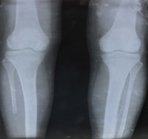
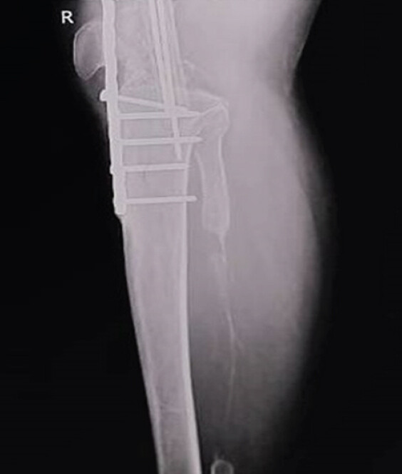
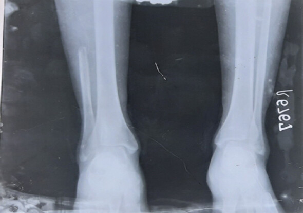
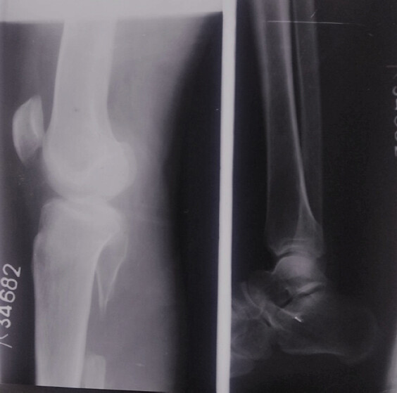
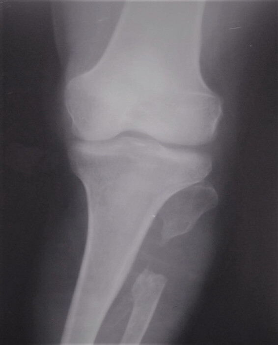
The most significant finding was osteoporosis of the distal fibular remnant (Fig. 1) in patients with fibular resection in 17 patients (28.3%). The radiograph showed rounding of the cut ends of fibula (Fig. 2 & 3). There was a thin bridged bone formation in the fibular bed in 9 cases (10.58%). None of the cases had varus and valgus instability of the knee and ankle. None of the cases had proximal migration of the fibula. In our study, 11 (18.3%) cases were found to have proximal fibula regeneration (Fig. 4 & 5).
Donor site related complication
In our series of 60 cases, mainly minor complication was found like cosmesis linear scar in 36 cases, minor pain in 11 cases, wound healing minor complication in 22 cases, and neuroma and sensory deficit in 14 cases. The most frequent complication after the fibular grafts had been obtained was cosmesis related (Table 3).
Frequency of complication at donor site
| Complications | No. of patients (%) | |
|---|---|---|
| Moderate (%) | Minor (%) | |
| • Cosmesis | 2 (3.3) | 36 (60) |
| • pain | – | 11 (19) |
| • Wound healing | – | 22 (36.7) |
| • Iatrogenic injury | – | 14 (23.3) |
Discussion
The donor site morbidity is considered to be low in the existing published studies. However, most of these studies did only a subjective evaluation of the functional consequences [7,8,9,10], so the incidence of donor site complications has been variable, ranging from 10% to 40%. The paucity of Indian literature regarding donor site morbidity following free fibular bone graft compelled us to investigate it. We used a non-vascularized fibular graft to fill bone defect.
The results of the Kitaoka ankle hindfoot score showed about 95% excellent and 5% poor results were with 6th month follow up. Results were compared to Garrett et al., (2006) studies showed the mean average score to be 84.82 in 14 patients [13]. Peter Sieg et al., (2009) studies showed mean average score to be 93 in 62 cases [14]. Catalá-Lehnen et al. (2012) reported a mean average score of 85.7 in 42 patients [15] and Feuvrier et al., (2015) reported 78 in 11 patients [16].
The most significant radiological finding in our series was osteoporosis of the distal fibular remnant in 17 patients (28.3%) out of 60 and is probably due to reduced load transmission.
Lee et al., (1990) studies showed that 90% of patients had radiological osteoporosis of the distal fibular remnant which is a direct result of a decrease in load transmission [17].
The resected part of fibula may be regenerated completely or in part from the periosteum, especially in younger patients. In our study, 11 (18.3%) cases were found to have proximal fibula regeneration.
Many authors have suggested that all attempts should be made to preserve the distal 6–8 cm of the fibula to maintain the lateral stability of the ankle [1,18,19,20,21,22]. Distal fibular remnant, in our series, was at least 7.5 cm or more, and none of the cases evaluated had ankle instability.
Conclusion
On the basis of our clinical observation, a non-vascularized fibular graft is associated with minor clinical morbidity related to pain, numbness, range of motion, osteoporosis of the distal fibular remnant, and muscular weakness.
Careful soft-tissue dissection may decrease the morbidity, and a distal fibular remnant of at least 8–10 cm should be retained for ankle stability. Attention to wound closure and meticulous wound care are required to further reduce donor leg complications.
Clinical Message
Free fibular graft harvesting is a reliable reconstructive option with good functional outcomes. Meticulous surgical technique and preservation of ankle stability are essential to minimize donor-site morbidity and ensure optimal post-operative recovery.
Conflict of Interest:
Nil
Source of Support:
Nil
Consent
The authors confirm that informed consent was obtained from the patient for publication of this article
How to Cite this Article
Sen KK, Ahirwar PC, Prajapati G, Chourasia A, Gupta DP, Ahirwar R. Retrospective Assessment of Donor Site Morbidity after Partial Fibula Resection. J Orthop Case Rep. 2026 November;16(11): 562-567. doi: 10.13107/jocr.2026.v16.i11.8552.
References
- Taylor GI, Miller GD, Ham FJ. The free vascularized bone graft. A clinical extension of microvascular techniques. Plast Reconstr Surg 1975;55:533-44. [Google Scholar] | [PubMed]
- Hidalgo DA. Vascularized fibular graft: A new method of mandible reconstruction. Plast Reconstr Surg 1989;84:71-9. [Google Scholar] | [PubMed]
- Murray RA, Brindley HH. The proximal fibula: Indications for excision and use. South Med J 1957;50:297-302. [Google Scholar] | [PubMed]
- Murray JA, Schlafly B. Giant-cell tumors in the distal end of the radius. Treatment by resection and fibular autograft interpositional arthrodesis. J Bone Joint Surg Am 1986;68:687-94. [Google Scholar] | [PubMed]
- Bassiony AA. Giant cell tumour of the distal radius: Wide resection and reconstruction by non-vascularised proximal fibular autograft. Ann Acad Med Singap 2009;38:900-4. [Google Scholar] | [PubMed]
- Wilson PDJr. A clinical study of the biomechanical behavior of massive bone transplants used to reconstruct large bone defects. Clin Orthop Relat Res 1972;87:81-109. [Google Scholar] | [PubMed]
- Dell PC, Burchardt H, Glowczewskie FPJr. A roentgenographic, biomechanical, and histological evaluation of vascularized and non-vascularized segmental fibular canine autografts. J Bone Joint Surg 1985;67-A:105-12. [Google Scholar] | [PubMed]
- Enneking WF, Eady JL, Burchardt H. Autogenous cortical bone grafts in the reconstruction of segmental skeletal defects. J Bone Joint Surg Am 1980;62:1039-58. [Google Scholar] | [PubMed]
- Yadav SS. Dual-fibular grafting for massive bone gaps in the lower extremity. J Bone Joint Surg 1990;72-A:486-94. [Google Scholar] | [PubMed]
- Chen ZW, Yan W. The study and clinical application of the osteocutaneous flap of fibula. Microsurgery 1983;4:11-6. [Google Scholar] | [PubMed]
- Kitaoka HB, Alexander IJ, Adelaar RS, Nunley JA, Myerson MS, Sanders M. Clinical rating systems for the ankle-hindfoot, midfoot, hallux, and lesser toes. Foot Ankle Int 1994;15:349-53. [Google Scholar] | [PubMed]
- Crenshaw AHJr. Surgical techniques and approaches Canale ST. Campbell's Operative Orthopaedics.;1 9thed. St Louis. Mosby 1998:42-3. [Google Scholar] | [PubMed]
- Garrett A, Ducic Y, Athre RS, Motley T, Carpenter B. Evaluation of fibula free flap donor site morbidity. Am J Otolaryngol 2006;27:29-32. [Google Scholar] | [PubMed]
- Sieg P, Taner C, Hakim SG, Jacobsen HC. Long-term evaluation of donor site morbidity after free fibula transfer. Br J Oral Maxillofac Surg 2010;48:267-70. [Google Scholar] | [PubMed]
- Catalá-Lehnen P, Rendenbach C, Heiland M, Khakpour P, Rueger JM, Schmelzle R. Long-term donor-site morbidity after microsurgical fibular graft: Is there a difference between the medial approach and the lateral approach?. J Oral Maxillofac Surg 2012;70:2198-204. [Google Scholar] | [PubMed]
- Feuvrier D, Sagawa YJr, Béliard S, Pauchot J, Decavel P. Long-term donor-site morbidity after vascularized free fibula flap harvesting: Clinical and gait analysis. J Plast Reconstr Aesthet Surg 2016;69:262-9. [Google Scholar] | [PubMed]
- Lee EH, Goh JC, Helm R, Pho RW. Donor site morbidity following resection of the fibula. J Bone Joint Surg Br 1990;72-B:129-31. [Google Scholar] | [PubMed]
- Gore DR, Gardner GM, Sepic SB, Mollinger LA, Murray MP. Function following partial fibulectomy. Clin Orthop Relat Res 1987;220:206-10. [Google Scholar] | [PubMed]
- Babhulkar SS, Pande KC, Babhulkar S. Ankle instability after fibular resection. J Bone Joint Surg Br 1995;77:258-61. [Google Scholar] | [PubMed]
- Anthony JP, Rawnsley JD, Benhaim P, Ritter EF, Sadowsky SH, Singer MI. Donor leg morbidity and function after fibula free flap mandible reconstruction. Plast Reconstr Surg 1995;96:146-52. [Google Scholar] | [PubMed]
- Vail TP, Urbaniak JR. Donor-site morbidity with use of vascularized autogenous fibular grafts. J Bone Joint Surg Am 1996;78:204-11. [Google Scholar] | [PubMed]
- Shpitzer T, Neligan P, Boyd B, Gullane P, Gur E, Freeman J. Leg morbidity and function following fibular free flap harvest. Ann Plast Surg 1997;38:460-4. [Google Scholar] | [PubMed]
© 2026 Journal of Orthopaedic Case Reports - Published by Indian Orthopaedic Research Group
About the Authors
-

Keshav Kumar Sen
-

Prem Chand Ahirwar
-

Gourav Prajapati
-

Atul Chourasia
-

Dharm Prakash Gupta
-

Rahul Ahirwar
Related Articles in Journal of Orthopaedic Case Reports
 September 1, 2026 Posterior Cruciate Ligament Avulsion Fracture with Fibular Avulsion of Lateral Collateral Ligament-biceps Femoris Complex Avulsion Injury in Ipsilateral Diaphyseal Tibia Fracture – A Rare Complex Injury
 September 1, 2026 Acetabular Giant Cell Tumor Presenting with Protrusio Acetabuli: A Rare Case Report
 June 1, 2026 Bone Graft Harvesting Site Morbidity in Donor Area (Anterior Iliac Crest): A Retrospective Study
 April 1, 2026 A Rare Case of Chronic Pan-Diaphyseal Osteomyelitis of the Fibula in a Child Treated by Near Total Fibulectomy: A Case Report