


Introduction
Synovial chondromatosis is a rare, benign disorder of the synovium that leads to loose-body formation due to metaplastic transformation. It manifests as multiple cartilaginous or osteochondral bodies within synovial joints, bursae, and tendon sheaths [1,2]. The knee is the most commonly affected joint, followed by the hip, elbow, shoulder and ankle. It is more frequently seen in males and usually occurs during the third to fifth decades of life [1, 2].
Synovial chondromatosis of the hip presents with pain, stiffness and difficulty in walking. If untreated, progressive mechanical effects and synovial disease may result in articular cartilage damage and secondary osteoarthritis [1,2]. Symptoms may become apparent with the development of synovitis, effusion, mechanical symptoms, or restriction of movement.
Radiographs may be normal in the early stages when loose bodies are not mineralized. Magnetic resonance imaging (MRI) is useful for demonstrating non-mineralized loose bodies, synovial disease and joint effusion, while computed tomography (CT) can better demonstrate mineralized or ossified loose bodies [2]. Milgram described three stages: Active intrasynovial disease without loose bodies, a transitional stage with both synovial lesions and loose bodies, and a late stage with loose bodies without active synovial disease [3].
Treatment of symptomatic disease involves removal of loose bodies with or without synovectomy. Arthroscopy, mini-arthrotomy, and open procedures, including safe surgical dislocation (SSD) have all been described [4,5,6,7].
We present a case of synovial chondromatosis of the hip in a young adult treated with staged hip arthroscopy followed by SSD, extensive removal of loose bodies, and synovectomy, with preservation of the native hip at 4-year follow-up.
Case Report
A 28-year-old male presented with a history of groin pain localized to the right hip for 6 weeks. His pain was worse on standing and walking. On clinical assessment, the patient had a normal gait pattern and was tender over the anterior aspect of the right hip. Internal and external rotation of the right hip in flexion were terminally restricted and painful.
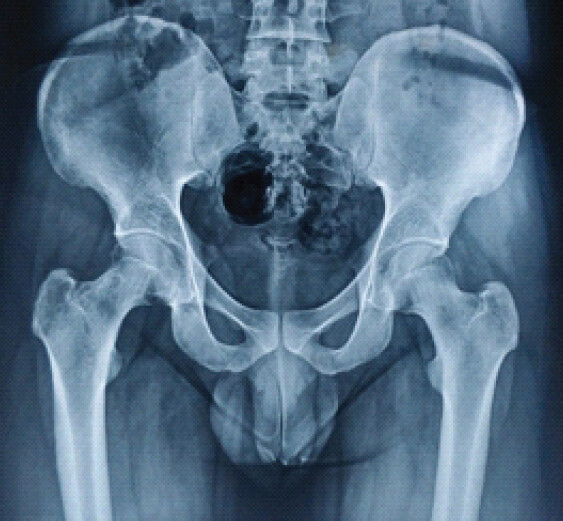
Plain radiographs of the pelvis with both hips revealed two radiolucent areas in the neck of the femur with femoral-neck erosion, narrowing and lytic lesions (Fig. 1). Blood investigations, including inflammatory markers, were normal.
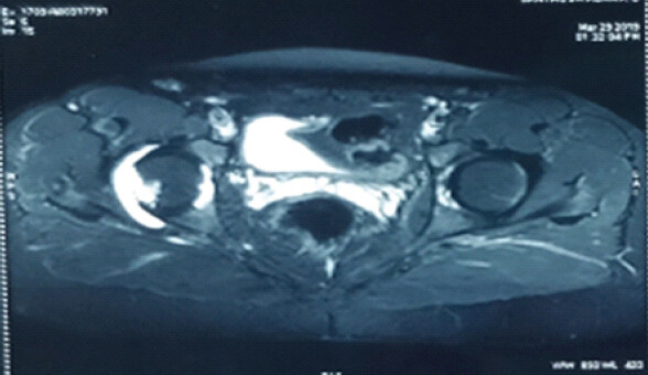
Symptomatic treatment was advised for 6 weeks. As the patient’s symptoms did not improve, MRI of the pelvis with both hips was performed. MRI demonstrated hyperintense signal around the right hip joint with effusion and synovitis (Fig. 2). The differential diagnosis included non-specific synovitis, Koch’s synovitis, synovial chondromatosis and pigmented villonodular synovitis.
After pre-operative counseling and evaluation, right hip arthroscopy and synovial biopsy were performed under general anesthesia on a traction table. Standard anterolateral and mid-anterior portals were utilized.
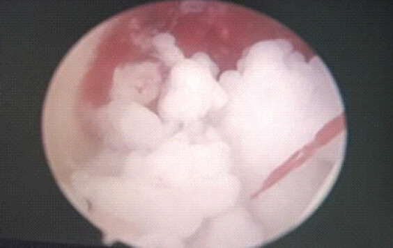
Arthroscopy demonstrated synovitis, effusion and multiple chondral loose bodies, suggestive of synovial chondromatosis (Fig. 3). The articular cartilage of the femoral head and acetabulum was carefully inspected and was noted to be intact and pristine.
Histopathological examination confirmed synovial chondromatosis.
As there were numerous loose bodies with extensive synovial involvement, SSD was planned for extensive removal of loose bodies and near-total synovectomy. The decision was based on the pre-operative MRI and intraoperative findings. In this setting, complete arthroscopic removal was considered potentially prolonged and technically demanding, with a possibility of incomplete removal of loose bodies or inadequate synovectomy.
Six weeks after the initial arthroscopy, open SSD was performed under combined spinal and epidural anesthesia in the left lateral position.
Operative technique
SSD with Ganz trochanteric flip osteotomy
Under combined spinal and epidural anesthesia, with intravenous cefuroxime (1.5 g) as prophylaxis, a posterior approach with a Kocher-Langenbeck incision was performed until the split in the fascia lata. The leg was internally rotated, and the posterior border of gluteus medius was identified.
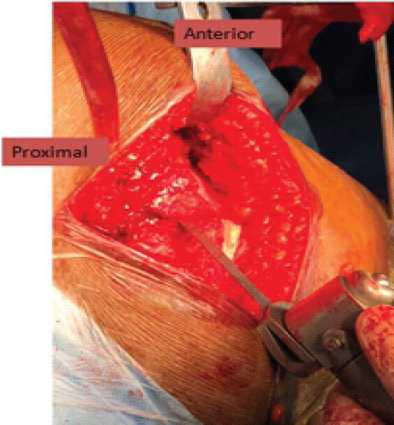
An incision was made from the posterosuperior edge of the greater trochanter extending distally to the posterior border of the ridge of vastus lateralis. A trochanteric osteotomy with a maximal thickness of approximately 1.5 cm was made along this line using an oscillating saw (Fig. 4). At its proximal limit, the osteotomy exited just anterior to the most posterior insertion of gluteus medius.
The greater trochanteric fragment was mobilized anteriorly with its attached vastus lateralis after releasing it along its posterior border to approximately the middle of the tendon of gluteus maximus. The most posterior fibers of gluteus medius were also released from the remaining trochanteric base.
With the leg flexed and slightly externally rotated, vastus lateralis and vastus intermedius were elevated from the lateral and anterior aspects of the proximal femur. The inferior border of gluteus minimus was separated from the relaxed piriformis and underlying capsule. The entire flap, including gluteus minimus, was retracted anteriorly and superiorly to expose the superior capsule.
The Z capsulotomy was performed as follows (Fig. 5). First, an anterolateral incision was made along the long axis of the femoral neck. Second, an anteroinferior capsular incision was made. The first capsular incision was then extended toward the acetabular rim and sharply turned posteriorly parallel to the labrum, reaching the retracted tendon of piriformis.
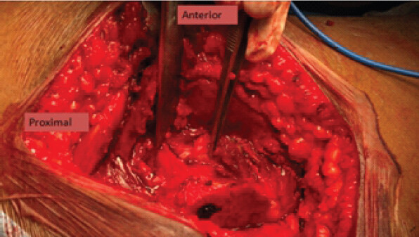
The hip was dislocated, and the leg was flexed, externally rotated, brought over the front of the operating table, and placed in a sterile bag.
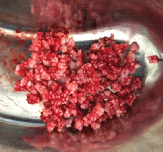
The intra-articular findings included synovitis, two erosions in the femoral neck, and multiple chondral loose bodies (Fig. 6). The articular cartilage of the femoral head and acetabulum was again inspected and was noted to be intact and pristine.
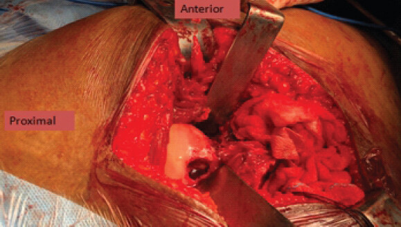
All loose bodies were carefully removed, and synovectomy was performed (Fig. 7 and 8). The femoral head was relocated after thorough irrigation with normal saline. The capsule was closed with No. 1 Vicryl suture.
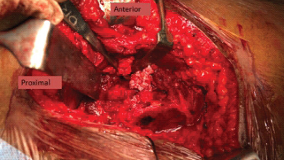
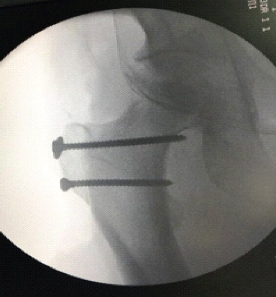
The trochanteric osteotomy was fixed with two 3.5-mm cortical screws and one washer. Good screw position, reduction, and compression of the osteotomy were confirmed using an image intensifier (Fig. 9).
Post-operatively, the patient was allowed active exercises in bed. He was mobilized non-weight bearing on the 1st post-operative day. Active range-of-motion exercises, quadriceps strengthening, and hip abductor strengthening exercises were allowed. Non-weight bearing was continued for 6 weeks.
A single post-operative dose of radiotherapy was administered within 48 h as an adjunct intended to reduce the risk of recurrence.
Follow-up and outcome
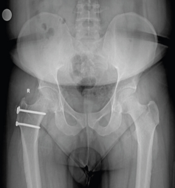
The patient was followed at 2 weeks, 6 weeks, 6 months, and subsequently for 4 years (Fig. 10).
The operative wound healed well, and the patient’s pain improved significantly by 6 weeks. At 6 months, he was mobilizing well and had no pain or stiffness around the hip.
At 4-year follow-up, the patient remained asymptomatic in the right hip. Follow-up plain radiographs did not demonstrate further progression of the femoral-neck erosive changes or narrowing of the joint space. There was no radiographic evidence of femoral-head collapse, avascular necrosis, or secondary osteoarthritis. The most recent radiograph has been included to demonstrate the long-term radiographic outcome.
There was no clinical evidence of recurrence during the 4-year follow-up period. As the patient remained asymptomatic and follow-up radiographs did not demonstrate new abnormalities, routine MRI was not performed. MRI would be considered if recurrent symptoms developed.
Discussion
Synovial chondromatosis is a benign osteochondral disorder characterized by the formation of multiple cartilaginous or osteochondral nodules within the synovium [1,2]. It may be primary or secondary, with secondary disease occurring in association with trauma, osteochondritis dissecans, and arthropathies [1].
Although hip involvement is rare, synovial chondromatosis should be considered in the differential diagnosis of persistent hip pain, particularly in young adults. A high index of suspicion and appropriate imaging are important for diagnosis.
The present patient had symptoms for only 6 weeks, and therefore this case does not demonstrate delayed diagnosis. Symptoms may become clinically apparent when synovitis, effusion, mechanical symptoms, or restriction of movement develops. The duration of the underlying disease before the onset of symptoms cannot be established in this patient.
The important message from this case is early recognition and treatment. Progressive disease can damage articular cartilage and contribute to secondary osteoarthritis. In this patient, despite multiple loose bodies and femoral-neck erosions, the articular cartilage of the femoral head and acetabulum remained intact and pristine.
Arthroscopic removal of loose bodies with synovectomy is an established treatment for hip synovial chondromatosis. A systematic review of 197 patients treated arthroscopically reported a recurrence rate of 7.1%, although the available evidence was predominantly Level IV [4]. Other reports have demonstrated favorable clinical outcomes following arthroscopic treatment [5].
In the present case, arthroscopy was initially performed for diagnosis, synovial biopsy, and assessment of the extent of disease. Multiple loose bodies and extensive synovitis were identified. Because of the number and distribution of the loose bodies, SSD was subsequently chosen to facilitate extensive removal and near-total synovectomy.
The purpose of SSD in this case was therefore not to establish superiority over arthroscopy. Rather, it provided extensive access to the hip joint when numerous loose bodies were present. Attempting complete arthroscopic removal in this particular patient could have required prolonged anesthetic and surgical time, with the possibility of incomplete removal or inadequate synovectomy.
Ganz et al. described SSD as providing extensive access to the femoral head and acetabulum while preserving the blood supply to the femoral head when performed appropriately [6]. Surgical dislocation has also been reported specifically for extensive synovial chondromatosis of the hip [7].
Thus, this case illustrates an individualized treatment strategy for extensive disease. It does not establish that staged arthroscopy and SSD are superior to arthroscopy, mini-arthrotomy, or other open procedures.
Role of radiotherapy
A single post-operative dose of radiotherapy was administered as an adjunct intended to reduce recurrence. The evidence supporting radiotherapy in synovial chondromatosis is limited. Chong et al. reported radiotherapy in a patient with recurrent, extensive synovial chondromatosis of the knee that had been refractory to multiple operations, with no progression during 5 years of follow-up [8].
Therefore, radiotherapy should not be considered a routine treatment for synovial chondromatosis. In this case, it was used as an adjunctive, case-specific measure. Potential long-term risks of ionizing radiation, including tissue effects and the theoretical risk of radiation-induced malignancy, should be considered, particularly in a young patient.
Recurrence and malignant transformation
Recurrence can occur following treatment of synovial chondromatosis, particularly when residual synovial disease or loose bodies remain [3,4,9]. In the present case, there was no clinical or radiographic evidence of recurrence during 4 years of follow-up. However, because routine serial MRI was not performed, clinically occult non-mineralized recurrence cannot be completely excluded.
Malignant transformation to secondary synovial chondrosarcoma is extremely rare [10]. Progressive symptoms, rapid recurrence, and aggressive features, such as muscle or medullary involvement should raise suspicion [10]. In the present case, histopathology confirmed synovial chondromatosis, and there was no clinical or radiographic evidence of recurrence during 4 years of follow-up.
This is a single case report, and therefore the findings cannot be generalized to all patients with hip synovial chondromatosis.
There was no control or comparison group, and the case cannot establish superiority of staged arthroscopy followed by SSD over arthroscopy alone, mini-arthrotomy, or conventional open excision.
The patient had symptoms for only 6 weeks, so this case cannot establish delayed diagnosis or the natural history of the disease. CT was not performed because plain radiographs did not demonstrate a clear calcified or ossified component, and MRI provided adequate information for diagnosis and operative planning.
Follow-up was based on clinical assessment and plain radiographs rather than a standardized serial MRI protocol. Therefore, non-mineralized or clinically occult recurrence cannot be completely excluded.
The use of post-operative radiotherapy was based on limited evidence and should be regarded as a case-specific adjunct rather than a standard treatment.
Conclusion
A high index of suspicion is important when evaluating persistent hip pain in a young adult, as synovial chondromatosis is rare and may present with non-specific symptoms.
Early recognition and treatment may help preserve the native hip before significant articular cartilage damage develops. In this patient, staged hip arthroscopy followed by SSD allowed extensive removal of multiple loose bodies and near-total synovectomy, while preserving the articular cartilage and native hip.
At 4-year follow-up, the patient remained asymptomatic with no clinical or radiographic evidence of recurrence, avascular necrosis, or secondary osteoarthritis.
This case demonstrates successful native hip preservation in an individual patient but does not establish superiority of staged arthroscopy and SSD over other contemporary treatment options.
Clinical Message
Synovial chondromatosis of the hip is rare but should be considered in young patients presenting with persistent hip pain. Early recognition and treatment may help preserve the native hip. In patients with extensive disease and multiple loose bodies, safe surgical dislocation can provide extensive access for loose-body removal and synovectomy when arthroscopic access may be technically demanding.
Conflict of Interest:
Nil
Source of Support:
Nil
Consent
The authors confirm that informed consent was obtained from the patient for publication of this article
How to Cite this Article
Mereddy P, Mazhar CSMS, Kanth GS, Prakash GV, Reddy RY, Reddy VG. Synovial Chondromatosis of Hip: A Rare Cause of Hip Pain in a Young Adult. J Orthop Case Rep 2026 November;16(11): 43-49. doi: 10.13107/jocr.2026.v16.i11.8382.
References
- Murphey MD, Vidal JA, Fanburg-Smith JC, Gajewski DA. Imaging of synovial chondromatosis with radiologic-pathologic correlation. Radiographics 2007;27:1465-88. [Google Scholar] | [PubMed]
- McCarthy C, Anderson WJ, Vlychou M, Inagaki Y, Whitwell D, Gibbons CL. Primary synovial chondromatosis: A reassessment of malignant potential in 155 cases. Skeletal Radiol 2016;45:755-62. [Google Scholar] | [PubMed]
- Milgram JW. Synovial osteochondromatosis: A histopathological study of thirty cases. J Bone Joint Surg Am 1977;59:792-801. [Google Scholar] | [PubMed]
- De Sa D, Horner NS, MacDonald A, Simunovic N, Ghert M, Philippon MJ. Arthroscopic surgery for synovial chondromatosis of the hip: A systematic review of rates and predisposing factors for recurrence. Arthroscopy 2014;30:1499-504.e2. [Google Scholar] | [PubMed]
- Boyer T, Dorfmann H. Arthroscopy in primary synovial chondromatosis of the hip: description and outcome of treatment. J Bone Joint Surg Br 2008;90 3:314-8. doi:10.1302/0301-620X.90B3.19664.. [Google Scholar] | [PubMed] | [CrossRef]
- Ganz R, Gill TJ, Gautier E, Ganz K, Krügel N, Berlemann U. Surgical dislocation of the adult hip a technique with full access to the femoral head and acetabulum without the risk of avascular necrosis. J Bone Joint Surg Br 2001;83:1119-24. [Google Scholar] | [PubMed]
- Fang S, Li H, Wang Y, Xu P, Sun H, Li S. Surgical hip dislocation for treatment of synovial chondromatosis of the hip. Int Orthop 2021;45:2819-24. [Google Scholar] | [PubMed]
- Chong CC, Kneebone A, Kirsh G. Radiotherapy in the management of recurrent synovial chondromatosis. Australas Radiol 2007;51:95-8. [Google Scholar] | [PubMed]
- van der Valk MR, Veltman ES, Assink J, Veen MR. Synovial chondromatosis of the hip, a case report and literature review. Journal of Orthopaedics. 2019;16 3:249-253. doi:10.1016/j.jor.2019.02.010.. [Google Scholar] | [PubMed] | [CrossRef]
- Ng VY, Louie P, Punt S, Conrad EU. Malignant transformation of synovial chondromatosis: A systematic review. Open Orthop J 2017;11:517-24. [Google Scholar] | [PubMed]
© 2026 Journal of Orthopaedic Case Reports - Published by Indian Orthopaedic Research Group
About the Authors
-

Praveen Mereddy
-

Chennur S M S Mazhar
-

G Shashi Kanth
-

G Veda Prakash
-

Raghuram Yeshwanth Reddy
-

Venu Gopal Reddy
Related Articles in Journal of Orthopaedic Case Reports
 November 1, 2025 Primary Synovial Chondromatosis of the Hip Joint: A Case Report
 August 1, 2026 The Boba Tea Sign: A Novel Magnetic Resonance Imaging Finding in Synovial Chondromatosis of the Knee – A Case Report
 June 1, 2026 Secondary Synovial Chondromatosis of the Shoulder Associated with Rotator Cuff Tear: Diagnostic Challenge and Arthroscopic Management – A Case Report
 January 1, 2026 Primary Synovial Chondromatosis of the Elbow Joint Presenting with Ulnar Nerve Compression and Restricted Range of Motion: A Case Report