


Introduction
Congenital insensitivity to pain with anhidrosis (CIPA) is a rare genetic disorder—with an estimated incidence of 1 in 125 million—defined by a complete absence of nociception alongside a systemic inability to sweat. This sensory deficit frequently results in repetitive microtrauma, culminating in progressive joint destruction and complex deformities secondary to untreated and neglected fractures [1,2,3]. They often present with multiple musculoskeletal deformities following fractures and dislocations. Neurological disorders of this nature often manifest in the lower extremities as severe instability and profound functional impairment. From the surgeon’s perspective, managing these cases is exceptionally demanding due to the combination of distorted anatomy, poor bone mineral density, accelerated bone resorption, and gross instability [4,5]. While hindfoot arthrodesis is the present established gold standard treatment for irreducible or non-braceable neuropathic deformities, standard implants often result in construct failure because of the underlying biological and mechanical compromises [6,7]. This report outlines the successful management of a uniquely complex neuropathic ankle using an extended fusion “superconstruct” strategy—a technique that, to our knowledge, is yet to be documented in the medical literature.
Case Report
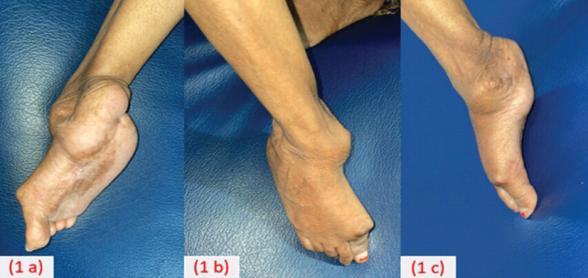
The patient, a 24-year-old female who had been wheelchair-bound for 7 years, presented to a tertiary care orthopedic center, with a grossly deformed and highly unstable flail ankle (Fig. 1). Her clinical history was significant for recurrent localized infections and a series of multiple fractures in the past that resulted in deformities of the upper limbs. Upon physical examination, she demonstrated a complete absence of pain during joint manipulation, a hallmark finding that led to a detailed clinical evaluation and the subsequent diagnosis of CIPA. On examination, the patient exhibited severely dry skin and a Tendo-Achilles contracture alongside an unstable flail ankle and flexion contractures of the toes. The combination of a biologically compromised limb and a mechanically unstable joint presented a unique therapeutic hurdle to the treating orthopedic surgeon.
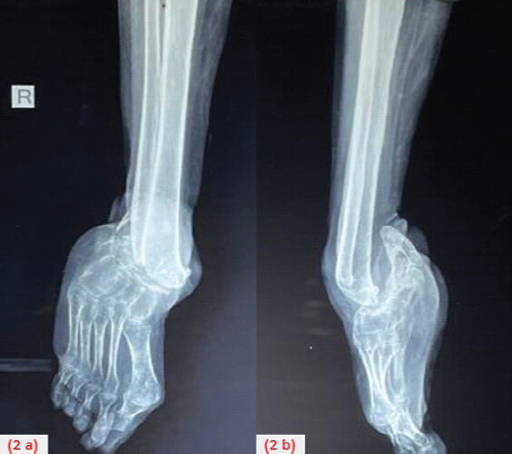
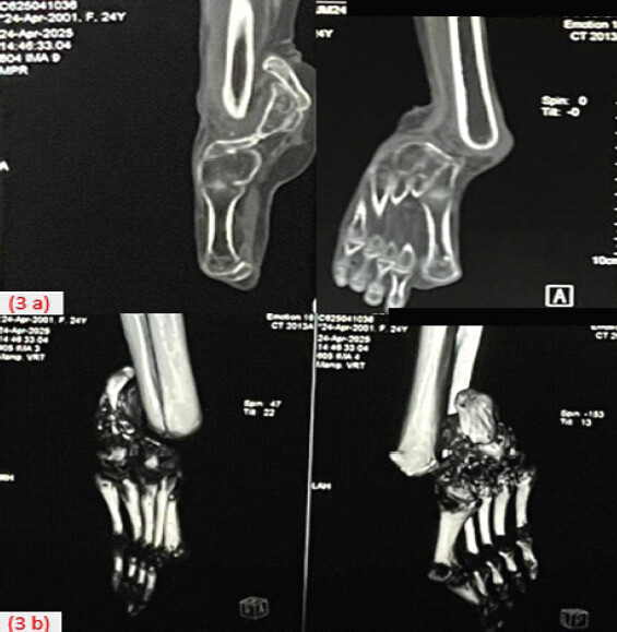
Radiographic evaluation revealed severe regional osteopenia, grossly distorted ankle anatomy with an atrophic boat-shaped calcaneum, and complete resorption of the talus, consistent with advanced Charcot neuroarthropathy (Fig. 2). Computed tomography with 3D reconstruction (Fig. 3) and 3D printing was done to aid in pre-operative planning.
The primary surgical objective was limb salvage through stable hindfoot arthrodesis, aimed at achieving a plantigrade foot capable of functional weight-bearing. The surgical challenges were multifaceted: A profound neuropathic state predisposing to delayed union; compromised bone stock increasing the risk of hardware pull-out and construct failure; severely distorted anatomy; a compromised soft-tissue envelope; and a significant Tendo-Achilles contracture necessitating a formal release.
Pre-operative planning focused on the surgical approach, implant selection, and optimal hardware positioning. Several internal fixation modalities were evaluated during the planning phase. Tibiotalocalcaneal intramedullary nailing was precluded by an exceptionally narrow medullary canal and a severely deformed calcaneus, compounded by the complete absence of the talus. Consequently, an open tibiocalcaneal arthrodesis was performed using a modified Mann’s technique. Stability was ensured through a rigid “superconstruct” comprising beam screws and a fusion plate, providing the necessary mechanical environment to facilitate fusion in a neuropathic limb.
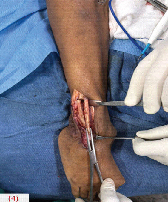
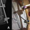
Surgical intervention commenced with a Hoke triple hemisection of the Achilles tendon to correct the equinus contracture. A lateral transfibular approach was employed, as described in the modified Mann’s technique, utilizing a fibular osteotomy performed approximately 5 cm proximal to the ankle joint. After osteotomizing its medial one-third (Fig. 4) to freshen the surface, the distal fibular segment was reflected posteriorly, with careful attention paid to preserving its vascular supply for subsequent use as an onlay graft. This provided direct visualization and allowed for meticulous preparation of the tibiocalcaneal arthrodesis site (Fig. 5). To promote osteogenesis, autologous corticocancellous bone graft was harvested from the ipsilateral iliac crest and packed into the fusion site. Initial stabilization and compression were achieved using headless, variable-pitch compression screws directed cephalad from the calcaneus into the tibia (Fig. 6).
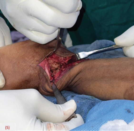
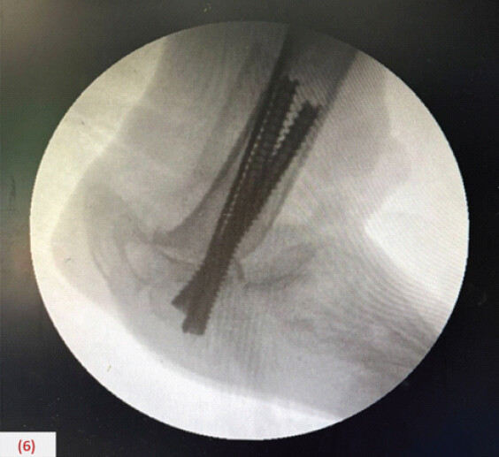
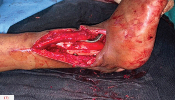
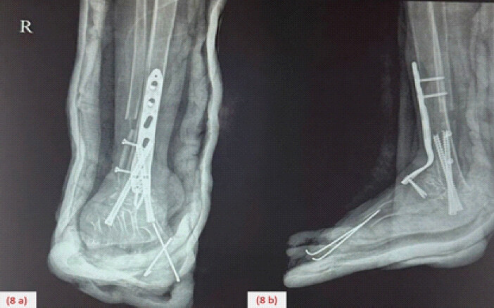
The “superconstruct” principle was implemented through extended fusion, aggressive bone resection, and the use of high-strength implants in optimal alignment. This screw fixation was augmented with an anterior ankle fusion plate to establish a rigid, load-sharing construct. Furthermore, the reflected distal fibula was utilized as a vascularized onlay graft bridging the lateral cortices of the tibia and calcaneus, secured with two cortical screws (Fig. 7). Concurrently, a symptomatic hallux flexus deformity was addressed through a flexor hallucis longus tenotomy and K-wire arthrodesis of the first metatarsophalangeal joint. Post-operative radiography confirmed optimal implant positioning and the successful restoration of plantigrade alignment (Fig. 8).
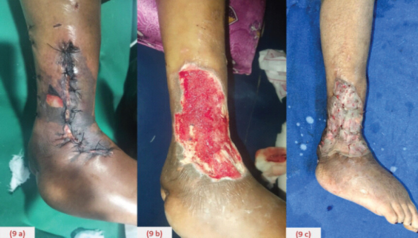
On post-operative day 5, the patient developed marginal skin necrosis. This necessitated surgical debridement, during which one exposed screw was removed. The wound was managed successfully with a vacuum-assisted closure (VAC) device and subsequently reconstructed with a split-thickness skin graft (STSG) (Fig. 9).
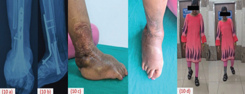
Post-operatively, the limb was immobilized in a below-knee cast for 3 months. A strict weight-bearing protocol was enforced: Absolute non-weight-bearing for the initial 6 weeks, progressing to toe-touch weight-bearing, and gradually advancing to full weight-bearing by 3 months. A custom orthosis was prescribed for long-term wear to protect against future neuropathic breakdown and limb length variation of 1.5 cm. At the 3-, 6-, and 12-month follow-ups, solid bony consolidation was evident both clinically and radiographically (Fig. 10). After 7 years of wheelchair dependency, the patient successfully regained the ability to ambulate independently without assistance. American Orthopaedic Foot and Ankle Society improved from 30 pre-operatively to 81 at 1 year follow-up.
Discussion
CIPA is an extremely rare genetic entity [1,4], characterized by a systemic lack of nociception frequently manifesting as a series of unrecognized musculoskeletal injuries, including recurrent fractures and dislocations that inevitably progress to severe deformities and neuropathic arthropathy. On review of the literature, clinical reports of this condition remain sparse; consequently, the management of the resulting joint destruction is often highly empiric, relying heavily on localized institutional experience rather than standardized protocols [2]. The absolute absence of protective sensation leads to repeated microtrauma, unchecked joint destruction, and classic Charcot-like neuroarthropathy. In advanced stages, this presents as a grossly deformed, non-braceable, and flail limb necessitating amputation [3,5].
Traditional treatment methods for patients with severe bony resorption, compromised soft tissue envelopes, and gross instability often made amputation the most predictable – and sometimes the only – viable treatment option for the resultant neuropathic joint [7]. While hindfoot arthrodesis is the gold standard for limb salvage, traditional fusion techniques in neuropathic conditions are fraught with unacceptably high complication rates, including non-union, implant failure, and surgical site infection [6,8]. Indeed, non-union rates have been reported as high as 41% in the literature [9]. In patients with CIPA, the native bone quality is frequently characterized by profound osteopenia. This makes standard isolated screw fixation or intramedullary nailing insufficient to withstand the unopposed, abnormal mechanical stresses generated during weight-bearing. The challenge is further exacerbated by the patient’s underlying biological compromise, which significantly impairs the healing response.
To counter this compromised biomechanical environment, we applied the “superconstruct” principles popularized by Sammarco [10]. This strategy mandates extending the fusion beyond the immediate zone of injury, performing aggressive bone resection down to healthy, bleeding bone to correct the deformity, and utilizing the strongest possible hardware. In our case, the combination of headless compression screws for dynamic interfragmentary compression, augmented by a dedicated rigid fusion plate, created a robust load-sharing construct. This construct effectively neutralized deforming forces while maximizing the surface area for bony consolidation.
Furthermore, our choice of surgical approach provided a dual mechanical and biological advantage. We utilized a lateral transfibular approach employing a modified Mann’s technique [11]. This not only offers extensive, direct visualization for thorough joint debridement and deformity correction but also yields the resected distal fibula for use as an onlay vascularized fibular graft. In neuropathic joints where the biologic healing cascade is often severely blunted, supplementing a rigid mechanical superconstruct with a robust local bone graft is a critical step in achieving the solid bony union observed at our patient’s 1-year follow-up.
Finally, the surgical management of CIPA-associated neuropathic ankles extends well beyond osseous fixation; it is equally a soft-tissue challenge. The soft tissue envelope in these patients is notoriously fragile, and the patient’s lack of pain eliminates the primary early warning sign of wound ischemia or impending breakdown. Our patient’s complication on post-operative day 5 underscores this vulnerability. The successful salvage of the limb and the underlying hardware was entirely dependent on prompt recognition, aggressive surgical debridement, and the integration of VAC and a STSG. This highlights that achieving a union in these complex deformities requires a vigilant, multidisciplinary post-operative protocol.
Conclusion
Neuropathic ankles secondary to CIPA are exceedingly rare and present formidable reconstructive challenges. Nevertheless, this case demonstrates that limb salvage is achievable through hindfoot arthrodesis using a modified Mann’s technique. By integrating compression screws with a plate superconstruct, surgeons can achieve the rigid stability necessary for successful fusion. Such an approach underscores that amputation is not an inevitability; rather, through robust reconstruction, it is possible to restore ambulation and dramatically improve functional outcomes. In the management of complex neuropathic deformities, amputation should be the final chapter, not the first option.
Clinical Message
The primary clinical message is that limb salvage and functional restoration remain achievable, even in severe flail neuropathic ankles. This case demonstrates that hindfoot arthrodesis using a “superconstruct” can overcome challenges in congenital insensitivity to pain with anhidrosis, yielding excellent outcomes and restoring independent ambulation after years of wheelchair dependency.
Conflict of Interest:
Nil
Source of Support:
Nil
Consent
The authors confirm that informed consent was obtained from the patient for publication of this article
How to Cite this Article
Tholgapiyan T, Senthilkumar S, Madhan A, Kumar TD. Limb Salvage of a Flail Neuropathic Ankle in Congenital Insensitivity to Pain with Anhidrosis Using a Hindfoot Superconstruct. J Orthop Case Rep 2026 November;16(11):37-42. doi: 10.13107/jocr.2026.v16.i11.8380.
References
- Nagasako EM, Oaklander AL, Dworkin RH. Congenital insensitivity to pain: An update. Pain 2003;101:213-9. [Google Scholar] | [PubMed]
- Udayashankar C, Oudeacoumar P, Nath AK. Congenital insensitivity to pain and anhidrosis: A case report from South India. Indian J Dermatol 2012;57:503. [Google Scholar] | [PubMed]
- Batouk OA, Almutairi MM, Saemaldahar MA, Ambon BZ. Congenital insensitivity to pain with anhidrosis and multiple Charcot joints in a child: A case report. J Musculoskelet Surg Res 2021;5:298-302. [Google Scholar] | [PubMed]
- Greider TD. Orthopedic aspects of congenital insensitivity to pain. Clin Orthop Relat Res 1983;172:177-85. [Google Scholar] | [PubMed]
- Mifsud M, Spiteri M, Camilleri K, Bonello M, Azzopardi T, Abela M. The orthopedic manifestations of congenital insensitivity to pain: A population-based study. Indian J Orthop 2019;53:665-73. [Google Scholar] | [PubMed]
- Huang PJ, Fu YC, Lu CC, Wu WL, Cheng YM. Hindfoot arthrodesis for neuropathic deformity. Kaohsiung J Med Sci 2007;23:120-7. [Google Scholar] | [PubMed]
- Alvarez RG, Barbour TM, Perkins TD. Tibiocalcaneal arthrodesis for nonbraceable neuropathic ankle deformity. Foot Ankle Int 1994;15:354-9. [Google Scholar] | [PubMed]
- Uehara K, Tsuchiya H, Kabata T, Sakurakichi K, Shimozaki E, Tomita K. Ankle arthrodesis and tibial lengthening for congenital sensory neuropathy with anhidrosis. J Orthop Sci 2001;6:430-4. [Google Scholar] | [PubMed]
- Allport J, Ramaskandhan J, Siddique MS. Nonunion rates in hind- and midfoot arthrodesis in current, ex-, and nonsmokers. Foot Ankle Int 2021;42:582-8. [Google Scholar] | [PubMed]
- Sammarco VJ. Superconstructs in the treatment of Charcot foot deformity: Plantar plating, locked plating, and axial screw fixation. Foot Ankle Clin 2009;14:393-407. [Google Scholar] | [PubMed]
- Napiontek M, Jaszczak T. Ankle arthrodesis from lateral transfibular approach: Analysis of treatment results of 23 feet treated by the modified Mann's technique. Eur J Orthop Surg Traumatol 2015;25:1195-9. [Google Scholar] | [PubMed]
© 2026 Journal of Orthopaedic Case Reports - Published by Indian Orthopaedic Research Group
About the Authors
-

T Tholgapiyan
-

Senthilkumar Sivakumar
-

Madhan Athmanathan
-

T Dilip Kumar
Related Articles in Journal of Orthopaedic Case Reports
 November 1, 2026 Compression of the Diseased Segment and Regeneration through Healthy Bone for Salvage of Infected Recurrent Congenital Pseudarthrosis of the Tibia in an Adult: A Case Report
 November 1, 2026 Limb Salvage in Near-Total Traumatic Elbow Amputation with Bone Loss Using Spanning Titanium Elastic Nails: A Case Report
 November 1, 2026 Immediate Limb Lengthening Following Vascular Repair of Iatrogenic Arterial Lesions – A Report of Two Cases
 October 1, 2026 Soft-Tissue Closure through Acute Deformation and Gradual Correction Using Taylor Spatial Frame in a Gustilo-Anderson Type IIIB Open Tibial Pilon Fracture: A Case Report