


Introduction
Aneurysmal bone cyst (ABC) is an uncommon benign but locally aggressive osteolytic bone lesion characterized by blood-filled cystic spaces separated by connective tissue septa containing fibroblasts, multinucleated giant cells, and reactive woven bone [1]. First described by Jaffe and Lichtenstein in 1942, ABC has since been recognized as a distinct clinicopathological entity that can exhibit aggressive behavior despite its benign histological nature. Although ABC accounts for only approximately 1% of all primary bone tumors, its propensity for rapid expansion, cortical destruction, pathological fracture, and recurrence makes it a clinically significant condition in Orthopaedic oncology [2].
ABCs predominantly affect children and adolescents, with nearly 80% of cases occurring during the first two decades of life. The lesion most commonly arises in the metaphyseal regions of long bones, particularly the distal femur, proximal tibia, and proximal humerus, as well as the posterior elements of the spine [3]. However, ABCs have been reported in virtually every skeletal site, including uncommon locations such as the pelvis, clavicle, scapula, ribs, talus, calcaneum, metatarsals, craniofacial bones, and small bones of the hands and feet [4]. Lesions arising in atypical locations frequently pose diagnostic and therapeutic challenges because of their rarity and the broad range of differential diagnoses.
Historically, the pathogenesis of ABC was attributed to local circulatory disturbances resulting in increased venous pressure, vascular dilatation, and progressive bone resorption [5]. Advances in molecular pathology have substantially improved the understanding of this lesion. Recurrent chromosomal translocations involving the USP6 oncogene have been identified in primary ABCs, supporting the concept that these lesions represent true neoplasms rather than purely reactive processes [6]. In contrast, secondary ABCs develop within pre-existing osseous lesions such as giant cell tumors, chondroblastoma, osteoblastoma, fibrous dysplasia, and non-ossifying fibroma. This distinction has important implications for diagnosis, biological behavior, and treatment planning.
The clinical presentation of ABC is highly variable and depends on lesion size, anatomical location, and biological aggressiveness. Pain and swelling are the most common presenting complaints, although patients may also present with a palpable mass, deformity, restricted joint motion, neurological symptoms in spinal lesions, or pathological fractures. The nonspecific nature of these symptoms often leads to delayed diagnosis, particularly in uncommon anatomical sites.
Radiological evaluation plays a crucial role in the diagnostic workup. Conventional radiographs typically demonstrate an expansile, eccentric, osteolytic lesion with cortical thinning and the characteristic “multiloculated” appearance [2]. Computed tomography (CT) assists in assessing cortical integrity and surgical planning, whereas magnetic resonance imaging (MRI) frequently reveals multiloculated cystic cavities with fluid–fluid levels, a feature considered highly suggestive of ABC. Nevertheless, these imaging findings are not pathognomonic, and several lesions, including telangiectatic osteosarcoma, giant cell tumor (GCT), unicameral bone cyst, and chondroblastoma, may demonstrate overlapping radiological features. Consequently, histopathological examination remains the gold standard for definitive diagnosis.
Management of ABC has evolved considerably over the past few decades. Traditional treatment consisted primarily of intralesional curettage with or without bone grafting; however, recurrence rates ranging from 10% to 30% prompted the development of adjunctive techniques and alternative treatment modalities. Contemporary management options include extended curettage, high-speed burring, adjuvant local therapies, percutaneous sclerotherapy, selective arterial embolization, en bloc resection, and various reconstructive procedures. The optimal treatment strategy depends on patient age, lesion location, extent of bone destruction, proximity to neurovascular structures, and risk of recurrence [2, 7].
Given the wide spectrum of clinical presentations, anatomical locations, and available treatment options, a comprehensive understanding of the epidemiology, pathogenesis, diagnostic characteristics, and management strategies of ABC is essential for Orthopaedic surgeons, musculoskeletal radiologists, pathologists, and oncologists. This review aims to summarize the current evidence regarding the demographics, clinical presentation, radiological and histopathological features, differential diagnosis, treatment modalities, and outcomes of ABCs, while highlighting the challenges associated with atypical presentations and contemporary management approaches.
Materials and Methods
Study design
This study was conducted as a narrative review of published literature on ABCs, with particular emphasis on demographic characteristics, clinical presentation, anatomical distribution, diagnostic features, histopathology, treatment modalities, and clinical outcomes. The review aimed to provide a comprehensive overview of the clinicopathological spectrum of ABCs, including both typical and atypical presentations reported in the literature.
Literature source
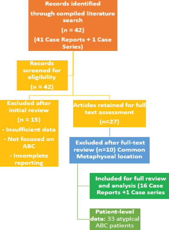
The review was based on published case reports and case series pertaining to ABCs in atypical anatomical locations in the Journal of Orthopaedic Case Reports from 2011 to 2026. The compiled literature included reports of ABCs occurring in both common (n = 10) and uncommon anatomical locations (n = 17), such as long bones, pelvis, clavicle, spine, foot, and other rare skeletal sites (Fig. 1).
Eligibility criteria
-
Studies were considered eligible for inclusion if they:
-
Reported patients with histologically confirmed ABCs
-
Described demographic characteristics, clinical presentation, imaging findings, pathology, management, or outcomes
-
Included primary or secondary ABCs affecting atypical skeletal location
-
Were available in full text and published in the English language.
Studies were excluded if they:
-
Lacked histopathological confirmation of diagnosis
-
Contained insufficient clinical or radiological information
-
Described the long bone metaphyseal ABC.
Study-selection clarification
The apparent discrepancy between 42, 27, and 21 reports reflects sequential screening stages. Forty-two records were initially identified; 15 were excluded during initial screening, leaving 27 for full-text assessment. Ten of these were excluded because they described conventional metaphyseal long-bone ABCs, leaving 17 reports (16 case reports and 1 case series) focused on atypical locations. The 33 patients represent the patient-level aggregate reported within these 17 included publications; therefore, article counts and patient counts should not be interpreted interchangeably.
Data synthesis
This review was designed as a narrative review. The literature search was restricted to Journal of Orthopaedic Case Reports (JOCR), and therefore the findings should be interpreted as a focused synthesis of reports published in that journal. The original search did not use PubMed/MEDLINE, Embase, Scopus, Web of Science, or the Cochrane Library; consequently, a comprehensive database search strategy, Boolean search string, and database-specific search dates cannot be retrospectively reported as having been performed.
Extracted data were analyzed descriptively and synthesized narratively. The collected evidence was compared with established Orthopaedic oncology literature to identify common patterns, emerging trends, and areas of clinical importance.
Ethical considerations
As this study was a review of previously published literature and did not involve direct patient recruitment, intervention, or access to identifiable patient information, institutional ethical approval and informed consent were not required.
Results
ABCs occurring outside the metaphysis of long bones represent a diagnostically challenging subset of lesions. Foot bones, pelvis, spine, clavicle, ribs, and craniofacial bones were the most commonly reported atypical sites. While imaging characteristics remained similar to conventional ABCs, atypical location substantially broadened the differential diagnosis and often necessitated advanced imaging and histopathological confirmation. Surgical treatment tailored to anatomical location achieved excellent functional outcomes and low recurrence rates, particularly when complete excision was feasible.
Aggregate analysis of atypical ABCs
Number of Articles:
-
20 individual case reports
-
1 Case series
-
Total atypical ABC cases reviewed: 33 patients.
The following table synthesized the available data from the reviewed articles (Table 1).
Analysis of aneurysmal bone cysts in atypical locations
| No. | Site | Clinical presentation | Diagnostic approach | Differential diagnosis | Management | Outcome and follow-up |
|---|---|---|---|---|---|---|
| 1 | Lateral Cuneiform | Midfoot pain, swelling, difficulty walking, pathological fracture | X-ray, CT, MRI, biopsy | Chondromyxoid fibroma, GCT, osteoblastoma | En bloc excision+tricortical iliac crest graft+arthrodesis | Complete union, pain- free walking, no recurrence |
| 2 | Talus | Chronic ankle pain, swelling, gait disturbance | Radiographs, MRI, histopathology | Chondroblastoma, GCT, telangiectatic osteosarcoma | Curettage+bone grafting | Good function, no recurrence reported |
| 3 | Calcaneum | Heel pain, swelling, difficulty weight-bearing | X-ray, MRI, biopsy | Unicameral bone cyst, GCT, chondroblastoma | Curettage+grafting | Radiological healing and symptom resolution |
| 4 | Third Metatarsal | Progressive foot swelling and pain | X-ray, MRI, biopsy | Enchondroma, osteomyelitis, GCT | En bloc excision+fibular strut graft | Excellent functional outcome, no recurrence |
| 5 | Clavicle | Painful enlarging clavicular mass | X-ray, CT, MRI, biopsy | Eosinophilic granuloma, plasmacytoma, GCT | Wide/en bloc excision | Full shoulder function, no recurrence |
| 6 | Acetabulum/P elvis | Hip pain, limp, reduced range of motion | X-ray, CT, MRI, biopsy | Chondrosarcoma, metastatic lesion, GCT | Curettage±reconstruction | Preserved hip function, no recurrence reported |
| 7 | Ilium/Iliac Bone | Pelvic pain and swelling | CT, MRI, biopsy | Fibrous dysplasia, giant cell tumor, sarcoma | Surgical excision/curettage | Good local control |
| 8 | Rib | Chest wall swelling and pain | CT, MRI, histopathology | Fibrous dysplasia, chondrosarcoma, metastasis | Wide excision | No recurrence during follow-up |
| 9 | Distal Ulna | Wrist pain and swelling | X-ray, MRI, biopsy | Osteoblastoma, GCT | Wide excision | No recurrence reported |
| 10 | Sphenoid Wing/Craniofa cial Skeleton | Headache, facial asymmetry, orbital symptoms | CT, MRI, histopathology | Fibrous dysplasia, giant cell lesion, osteoblastoma | Surgical excision | Symptomatic improvement, no recurrence |
| 11 | Lumbar Spine (L3 vertebra) | Acute paraparesis, lower limb weakness, severe pain, gait inability in pregnancy | MRI showing multiloculated lesion with fluid-fluid levels, biopsy confirmation | Transverse myelitis, multiple sclerosis, metastatic lesion, Guillain–Barré syndrome, spinal epidural abscess | Staged decompression, tumor excision, pedicle screw fixation, cage reconstruction | Independent ambulation at 10 months follow-up |
| 12 | Tibial Diaphysis | Pain, swelling, cortical expansion | X-ray, MRI, histopathology | Fibrous dysplasia, adamantinoma, osteofibrous dysplasia | Wide excision+ipsilateral fibular strut graft | Excellent functional and radiological outcome at 1 year; no recurrence |
CT: Computed tomography, MRI: Magnetic resonance imaging, GCT: Giant cell tumor
Anatomical distribution of atypical ABCs
The reviewed literature demonstrated a wide anatomical distribution of atypical ABCs involving both appendicular and axial skeletal sites outside the conventional metaphyseal regions of long bones. The foot represented the most frequently affected region, with lesions reported in the talus (n = 2), calcaneum (n = 3), cuneiform bones (n = 2), navicular (n = 1), and metatarsals (n = 2). Pelvic involvement included the ilium (n = 2), acetabulum (n = 1), and pubic rami (n = 1). Lesions affecting the axial skeleton were identified in the lumbar vertebrae (n = 2), cervical vertebra (n = 1), and sacroiliac joint (n = 1). In the upper limb, atypical ABCs were reported in the clavicle (n = 2), metacarpals (n = 2), distal ulna (n = 1), radial head (n = 1), and scapular glenoid (n = 1). Additional uncommon sites included the rib (n = 1) within the thoracic cage and the sphenoid wing (n = 1) within the craniofacial skeleton. Lower-limb lesions outside the typical metaphyseal location involved the patella (n = 2), tibial diaphysis (n = 1), femoral head (n = 1), femoral shaft (n = 1), and femoral neck (n = 1). Overall, the foot and pelvic regions constituted the largest proportion of atypical ABCs, highlighting the diverse anatomical spectrum and diagnostic complexity associated with these uncommon presentations.
Common clinical presentations
Overall, the clinical presentation of atypical ABCs was largely determined by lesion location and extent of local bone involvement, although pain and swelling remained the most consistent presenting features across all reported cases. A palpable mass was observed in several cases, particularly in superficial locations such as the clavicle, rib, and foot bones. Patients with lesions involving the pelvis and foot commonly presented with limping, gait disturbances, or difficulty bearing weight due to pain and structural compromise of the affected bone. Pathological fractures were encountered occasionally, particularly in lesions associated with extensive cortical thinning or aggressive bone destruction. Neurological deficits were uncommon and were primarily restricted to spinal ABCs, where tumor expansion resulted in neural compression, manifesting as lower-limb weakness, sensory disturbances, or gait impairment.
Diagnostic strategy
Nearly all articles depicted a similar diagnostic pathway: Plain radiographs, CT scan, MRI, Histopathological confirmation.
Differential diagnosis
The diagnosis of ABCs in atypical skeletal locations is often challenging because of significant overlap with several benign and malignant bone lesions. In the foot, particularly in lesions involving the talus and calcaneum, the most common differential diagnoses include GCT and chondroblastoma, both of which may demonstrate expansile osteolytic features and similar MRI characteristics. Pelvic lesions frequently raise suspicion for GCT, metastatic disease, brown tumor of hyperparathyroidism, and telangiectatic osteosarcoma because of their aggressive radiographic appearance and extensive bone destruction. ABCs involving the clavicle may mimic GCT or eosinophilic granuloma, whereas lesions affecting the spine often necessitate differentiation from telangiectatic osteosarcoma and osteoblastoma because of their expansile nature and potential neurological manifestations. Craniofacial lesions, particularly those involving the sphenoid wing, may resemble fibrous dysplasia or other giant cell-rich lesions. Similarly, rib lesions are commonly confused with fibrous dysplasia or chondrosarcoma. The presence of fluid–fluid levels on MRI may strongly suggest ABC; however, histopathological confirmation remains essential to exclude these important differential diagnoses and establish a definitive diagnosis.
Management patterns
Treatment strategies for atypical ABCs are largely determined by the anatomical location of the lesion, extent of bone involvement, functional demands of the affected region, and risk of recurrence. Lesions involving the foot bones, including the talus, calcaneum, cuneiforms, and metatarsals, were most commonly treated with intralesional curettage and bone grafting, although en bloc excision with structural reconstruction was preferred in cases associated with extensive bone destruction. ABCs arising in relatively expendable bones such as the clavicle, rib, and distal ulna were generally managed by wide excision, which provided excellent local control with minimal functional compromise. Pelvic lesions were commonly treated with extended curettage with or without reconstruction depending on the extent of acetabular or pelvic involvement. Spinal ABCs often required a combination of neural decompression, complete or subtotal tumor excision, and instrumented stabilization to restore spinal stability and neurological function. Craniofacial lesions were managed primarily by surgical excision, whereas diaphyseal lesions frequently required wide excision followed by structural graft reconstruction to restore skeletal integrity. Overall, treatment selection was individualized according to lesion location and anticipated biomechanical consequences.
Outcomes and follow-up
Most individual reports described favorable local disease control and functional recovery after definitive treatment. However, these observations cannot be generalized to all atypical ABCs because the evidence consisted predominantly of case reports and one case series, with heterogeneous follow-up and non-standardized functional outcomes.
Discussion
ABCs remain one of the most intriguing benign skeletal lesions encountered in Orthopaedic oncology because of their variable biological behavior, diverse anatomical distribution, and potential to mimic aggressive neoplastic conditions. The findings synthesized in this review largely corroborate the established Orthopaedic oncology literature while also emphasizing the demographic profile, diagnostic and therapeutic challenges associated with atypical presentations (Tables 2 and 3).
Demographic profile of atypical ABCs in comparison to literature
| Parameter | Standard literature | Findings in this study |
|---|---|---|
| Age | Usually <20 years; peak in 2nd decade | Majority pediatric/adolescent cases; age range 5–45 years in atypical series |
| Sex | Slight female predominance or equal distribution | 11 males and 9 females in atypical series |
| Incidence | ~1% of all bone tumors | Repeatedly cited throughout articles |
| Common Sites | Distal femur, proximal tibia, proximal humerus, spine | Several uncommon sites reported: talus, calcaneum, clavicle, sphenoid wing, metatarsal, acetabulum, ulna, rib, femoral neck |
| Primary versus Secondary | Primary ~70–80%; Secondary ~20–30% | Secondary ABC associated with chondroblastoma, fibrous dysplasia, giant cell tumor, metabolic lesions |
ABCs: Aneurysmal bone cysts
Comparison of atypical ABCs in this review with standard orthopaedic oncology literature
| Parameter | Atypical ABCs from your documents | Standard orthopaedic oncology literature |
|---|---|---|
| Age | Predominantly children, adolescents, and young adults; occasional adult cases | Approximately 80% occur before 20 years of age |
| Location | Foot (talus, calcaneum, cuneiform, metatarsal), clavicle, rib, pelvis, acetabulum, sphenoid wing, distal ulna, spine | Most commonly metaphysis of long bones and posterior elements of spine; atypical sites are uncommon but recognized |
| Presentation | Pain, swelling, limp, pathological fracture, neurological deficit (spine) | Pain and swelling remain the most common symptoms; pathological fractures and neurological deficits occur in aggressive lesions |
| Diagnostic Difficulty | Frequently mistaken for GCT, chondroblastoma, sarcoma, fibrous dysplasia, metastasis | Diagnostic confusion is well recognized, especially with telangiectatic osteosarcoma and giant cell tumor |
| MRI Findings | Fluid-fluid levels reported in nearly all atypical lesions | Fluid-fluid levels considered highly suggestive but not pathognomonic |
| Histopathology | Blood-filled spaces, fibroblastic septa, giant cells, reactive bone | Identical classical histological description in standard literature |
| Treatment | Curettage, grafting, wide excision, arthrodesis, reconstruction, spinal stabilization | Curettage with grafting remains standard treatment; adjuvants increasingly used |
| Recurrence | Recurrence was infrequently reported; rates cannot be estimated reliably | Recurrence rates generally 10–30%, especially after intralesional procedures |
| Functional Outcome | Favorable outcomes were commonly described, but reporting was non-standardized | Excellent when local control achieved |
| Follow-up | Usually 10 months–5 years | Most recurrences occur within the first 2 years after treatment |
ABCs: Aneurysmal bone cysts, GCT: Giant cell tumor, MRI: magnetic resonance imaging
Site-Specific comparison in literature reveal that foot ABCs are exceptionally rare in large Orthopaedic oncology series. Most major series reported very few lesions in the tarsal bones [5]. Consequently, diagnosis is often delayed because clinicians initially suspect more common foot tumors. Our collection contains a disproportionately high number of foot ABCs compared with classical Orthopaedic oncology series, making it particularly valuable for understanding rare-site disease.
The 2023 review identifies pelvic ABCs as among the most difficult lesions to manage because of large size at presentation, difficult surgical access and significant intraoperative bleeding risk. Selective arterial embolization is often recommended for pelvic lesions and may achieve local control rates approaching 94%. None of our reviewed cases used embolization as primary treatment despite its strong support in Orthopaedic oncology literature [8].
Spinal ABCs are among the most extensively studied atypical ABCs. Standard literature recommends preoperative embolization, complete excision where possible, and instrumented stabilization when instability exists. Neurological recovery following decompression is generally favorable, mirroring our findings [9, 10, 11, 12].
The clavicle and rib are considered relatively expendable bones, making wide resection an attractive option with minimal functional compromise [13,14,15]. There was complete concordance with standard practice. Craniofacial ABCs are rare but recognized. Standard literature notes greater diagnostic difficulty, higher risk of morbidity because of adjacent neurovascular structures and frequent need for multidisciplinary management [15]. Our findings closely match these observations.
Compared with contemporary Orthopaedic oncology literature, atypical ABCs in the present review demonstrated similar demographic characteristics and histopathological features but substantially greater diagnostic complexity owing to their uncommon anatomical locations. While curettage and grafting remain the cornerstone of treatment, many lesions in atypical locations were managed by en bloc excision and reconstruction, resulting in excellent local control and functional outcomes [16,17,18,19]. Interestingly, recurrence rates appeared lower than those reported in large Orthopaedic oncology series, likely reflecting careful patient selection and the frequent use of wide excision [20,21,22,23,24,25]. Emerging minimally invasive treatments such as selective arterial embolization, polidocanol sclerotherapy, doxycycline injection, and denosumab, increasingly advocated in contemporary literature, were infrequently utilized in the reviewed reports, highlighting an evolving area of ABC management (Table 4) [26,27,28,29,30,31].
Treatment comparison
| Treatment | Our review | Orthopaedic oncology literature |
|---|---|---|
| Curettage+grafting | Frequently reported | Established conventional option |
| Wide resection | Frequently used in atypical sites | Recommended for expendable bones and recurrent lesions |
| Ethanol sclerotherapy | Reported in a proximal humerus case | Increasingly accepted modality |
| Polidocanol | Rarely reported in your cases | Healing rates 84–93% reported |
| Embolization | Not used in reviewed cases | Used selectively; evidence from broader literature |
| Doxycycline injection | Not reported | Recurrence approximately 5–6% in early series |
| Denosumab | Not reported | Emerging option for sacral and spinal ABCs |
ABCs: Aneurysmal bone cysts
Recurrence remains the most clinically relevant challenge in the management of ABC. The literature consistently identifies young age, open growth plates, aggressive radiological characteristics, secondary nature and incomplete excision as major predictors of recurrence [32,33,34]. Most recurrences occur within the first 2 postoperative years, emphasizing the importance of vigilant follow-up during this period. Encouragingly, recurrence rates appear substantially lower following wide resection and modern adjuvant therapies, although long-term comparative data remain limited [15].
The current evidence suggests that successful management of ABC requires a multidisciplinary approach integrating clinical assessment, advanced imaging, histopathological confirmation, and patient-specific treatment planning. Future multicentre studies with longer follow-up periods are required to better define the optimal role of emerging minimally invasive therapies and to establish evidence-based treatment algorithms for lesions arising in uncommon anatomical locations.
Limitations
The findings of this review are subject to important limitations. First, this was a narrative review rather than a systematic review, so author selection bias and publication bias cannot be excluded. Second, the search was restricted to JOCR from 2011 to 2026 and did not include major bibliographic databases; therefore, the evidence base is not representative of the broader international ABC literature. Third, the final evidence base comprised 17 reports (16 case reports and 1 case series) describing 33 patients, making comparisons between anatomical sites and treatment modalities exploratory rather than definitive. Fourth, the included lesions were clinically heterogeneous, including primary and secondary ABCs, different skeletal sites, age groups, fracture status, lesion aggressiveness, and treatment strategies. Primary and secondary ABCs were not sufficiently stratified to permit separate outcome estimates. Fifth, no validated formal risk-of-bias tool was applied; the methodologicalquality of case reports and the case series therefore remain uncertain. Sixth, functional outcomes were inconsistently reported and were often described qualitatively rather than with validated scores. Follow-up ranged from approximately 10 months to 5 years, which may miss late recurrence, deformity, or functional impairment. Finally, treatment comparisons are confounded by indication: curettage, wide excision, sclerotherapy, embolization, and reconstruction were selected according to lesion location and clinical circumstances. Accordingly, recurrence rates and treatment effectiveness should not be interpreted as pooled or comparative estimates.
Conclusion
ABCs represent uncommon but clinically significant benign bone lesions characterized by variable presentation and biological behavior. Atypical anatomical locations may pose substantial diagnostic and therapeutic challenges. Advances in imaging, molecular pathology, and minimally invasive treatment have expanded management options beyond traditional curettage. In the present narrative review, histopathological confirmation and individualized treatment planning were recurring themes across published case reports. However, because the evidence was restricted to a single journal and was predominantly composed of case reports, the available data do not permit reliable estimates of recurrence or comparative effectiveness of treatment modalities. Larger multicentre studies using standardized diagnostic, functional, and follow-up measures are required to establish evidence-based treatment algorithms.
Clinical Message
Aneurysmal bone cysts are benign but locally aggressive lesions that predominantly affect children and adolescents, with a predilection for the metaphyseal regions of long bones
Clinical and radiological presentations can vary considerably, particularly when ABCs occur in uncommon locations such as the pelvis, clavicle, foot, spine, or small bones, often mimicking more aggressive neoplastic conditions
MRI demonstrating fluid–fluid levels is highly suggestive of ABC, but histopathological confirmation remains essential to differentiate it from lesions such as telangiectatic osteosarcoma and giant cell tumor
Management should be individualized based on lesion size, anatomical location, skeletal maturity, and biological aggressiveness, with options ranging from sclerotherapy and curettage to en bloc resection and reconstruction
Modern minimally invasive treatments, particularly percutaneous sclerotherapy, have emerged as effective alternatives to surgery in selected patients, offering high healing rates with reduced morbidity
Recurrence remains the most important complication, especially in young patients and aggressive lesions; therefore, careful long-term follow-up is essential
Early diagnosis and multidisciplinary management can achieve excellent functional outcomes while minimizing recurrence and preserving skeletal integrity.
Conflict of Interest:
Nil
Source of Support:
Nil
Consent:
The authors confirm that informed consent was obtained from the patient for publication of this article
How to Cite this Article
Lingaiah P, Abraham S, Sai BM, Lavudi R, Shanmugham S, Uthup M. Clinicopathological Spectrum and Management of Aneurysmal Bone Cysts in Atypical Anatomical Location: A Comprehensive Review of Demographics, Presentation, Diagnosis, and Treatment Outcomes. Journal of Orthopaedic Case Reports 2026 October;16(10): 571-579.
References
- Cottalorda J, Bourelle S. Modern concepts of primary aneurysmal bone cyst. Arch Orthop Trauma Surg 2007;127:105-14. [Google Scholar] | [PubMed]
- Park HY, Yang SK, Sheppard WL, Hegde V, Zoller SD, Nelson SD. Current management of aneurysmal bone cysts. Curr Rev Musculoskelet Med 2016;9:435-44. [Google Scholar] | [PubMed]
- Mascard E, Gomez-Brouchet A, Lambot K. Bone cysts: Unicameral and aneurysmal bone cyst. Orthop Traumatol Surg Res 2015;101 Suppl 1:S119-27. [Google Scholar] | [PubMed]
- Papagelopoulos PJ, Currier BL, Shaughnessy WJ, Sim FH, Ebsersold MJ, Bond JR. Aneurysmal bone cyst of the spine. Management and outcome. Spine (Phila Pa 1976) 1998;23:621-8. [Google Scholar] | [PubMed]
- Stevens KJ, Stevens JA. Aneurysmal Bone Cysts. [Updated 2023 Aug 14]. In: StatPearls [Internet].. Treasure Island (FL): StatPearls Publishing; 2023. Jan. Available from: https://www.ncbi.nlm.nih.gov/sites/books/NBK546654/ Last Accessed: 29/09/2026.. [Last Accessed: 29/09/2026].. [Google Scholar] | [PubMed]
- Lau AW, Pringle LM, Quick L, Riquelme DN, Ye Y, Oliveira AM. TRE17/ubiquitin-specific protease 6 (USP6) oncogene translocated in aneurysmal bone cyst blocks osteoblastic maturation via an autocrine mechanism involving bone morphogenetic protein dysregulation. J Biol Chem 2010;285:37111-20. [Google Scholar] | [PubMed]
- Rossi G, Rimondi E, Bartalena T, Gerardi A, Alberghini M, Staals EL. Selective arterial embolization of 36 aneurysmal bone cysts of the skeleton with N-2-butyl cyanoacrylate. Skeletal Radiol 2010;39:161-7. [Google Scholar] | [PubMed]
- Saeed A, Rometsch E, Chong AH. Aneurysmal bone cyst of the pelvis: Systematic literature review. J Musculoskelet Surg Res 2023;7:11-23. [Google Scholar] | [PubMed]
- Prasad HR, Murthy PV, Reddy DA, Lingala H, Immaneni SR, Reddy AV. Percutaneous management of aneurysmal bone cyst of axis vertebrae in a 12-year-old child: A case report. J Orthop Case Rep 2023;13:47-51. [Google Scholar] | [PubMed]
- Iorio C, Mazza O, Tundo F, Stracuzzi A, Crostelli M. Lumbar spine osteoblastoma with secondary aneurysmal bone cyst causing severe trunk imbalance and radiculopathy: A Case report. J Orthop Case Rep 2020;10:18-22. [Google Scholar] | [PubMed]
- Guin B, Balakrishnan D, Sekar A, Ayyanar P. Spinal aneurysmal bone cyst in a pregnant patient presenting with neurological symptoms: A case report. J Orthop Case Rep 2026;16:30-3. [Google Scholar] | [PubMed]
- Nidhin TS, Thomas A, Kumar VV, Nayar PP, John D. Aneurysmal bone cyst of thoracic spine in an elderly treated by two stage 360° surgical excision and reconstruction with expandable cage - a case report. J Orthop Case Rep 2025;15:178-81. [Google Scholar] | [PubMed]
- Thanigaimani K, Kumar S, Mahendran V. Aneurysmal bone cyst of the clavicle: A rare orthopedic entity. J Orthop Case Rep 2025;15:78-83. [Google Scholar] | [PubMed]
- Rajak A, Sethia A, Limbu A, Kalawar RPS. Aneurysmal bone cyst of the clavicle: A case report. J Orthop Case Rep 2020;9:3-5. [Google Scholar] | [PubMed]
- Kancherla NR, Jagadabi S, Bodla AK, Peddamadyam SK, Cherukuri CN, Cholleti SL. Journey and roadblocks in the management of aneurysmal bone cysts in patients with unusual presentations. J Orthop Case Rep 2026;16:475-82. [Google Scholar] | [PubMed]
- Lingaiah P, Nataraj AR. Aneurysmal bone cyst of lateral cuneiform: Challenge in diagnosis and management. J Orthop Case Rep 2026;16:233-7. [Google Scholar] | [PubMed]
- Ahmad S, Harun F, Anwar A, Ansari MO, Shoaib SM, Shaan SF. Aneurysmal bone cyst of talus: Case report of a rare presentation. J Orthop Case Rep 2026;16:217-21. [Google Scholar] | [PubMed]
- Tiwari V, Sahoo S, Roy M, Dwidmuthe S, Bhavani P, Reddy H. Aneurysmal Bone cyst of the third metatarsal treated using free nonvascular fibular strut graft-a rare case report. J Orthop Case Rep 2024;14:136-40. [Google Scholar] | [PubMed]
- Chigblo SP, Tidjani IF, Hadonou AA, Takin R, Agbessi FO, Hans-Moevi AA. Aneurysmal bone cyst of talus: A case report. J Orthop Case Rep 2025;15:125-9. [Google Scholar] | [PubMed]
- Jain M, Pradhan SS, Khan S, Tripathy S, Lubaib KP, Raj KS. Aneurysmal bone cyst of the distal ulna in an elderly lady. J Orthop Case Rep 2024;14:79-83. [Google Scholar] | [PubMed]
- Kumar DN, Nagasamy S. Aneurysmal bone cyst-cloaked codman's tumor in the patella: Case report of an easily misdiagnosed entity. J Orthop Case Rep 2026;16:174-8. [Google Scholar] | [PubMed]
- Mohapatra S, Pani SK, Khandelwal C, Mishra S, Sharma A. Ipsilateral fibula grafting in a case of aneurysmal bone cyst in tibia diaphysis: A case report. J Orthop Case Rep 2024;14:36-9. [Google Scholar] | [PubMed]
- Mukherjee K, Ghorai TK, Kumar A, Basak I. Giant cell tumor of patella with secondary aneurysmal bone cyst-a rare case report of anterior knee pain in young adults. J Orthop Case Rep 2024;14:65-9. [Google Scholar] | [PubMed]
- Singh P, Kumar R. Aneurysmal bone cyst in metacarpal of a child. J Orthop Case Rep 2013;3:3-6. [Google Scholar] | [PubMed]
- Nanda SN, Tripathi S, Shiraz SM, Warrier S. Aneurysmal bone cyst of 3rd metacarpal, management and follow-up: A case report. J Orthop Case Rep 2018;8:9-12. [Google Scholar] | [PubMed]
- Naraval KY, Guerzon EJ, Robles AF, Carolino DK. Percutaneous doxycycline ablation for recurrent aneurysmal bone cyst in the sacroiliac joint: A case report. J Orthop Case Rep 2024;14:125-32. [Google Scholar] | [PubMed]
- Burdorf B, Kesarla P, MacDonald W. Young female with calcaneal chondroblastoma & secondary aneurysmal bone cyst: Case report. J Orthop Case Rep 2025;15:160-3. [Google Scholar] | [PubMed]
- Jain M, Tripathy SK, Mishra NP, Ayyanar P, Singh AK. Primary aneurysmal bone cyst in the iliac bone: A case report. J Orthop Case Rep 2024;14:109-13. [Google Scholar] | [PubMed]
- Saraf A, Kumar SK. Sclerotherapy as a primary or salvage procedure for aneurysmal bone cyst: A case report. J Orthop Case Rep 2024;14:102-6. [Google Scholar] | [PubMed]
- Bansal K, Baskaran S, Dahuja A, Gupta N. A rare case of aneurysmal bone cyst - navicular bone treated by intralesional sclerotherapy agent polidocanol. J Orthop Case Rep 2021;11:1-4. [Google Scholar] | [PubMed]
- Rao A, Samant PD, Varshneya A. Aneurysmal bone cyst of pubic ramus: A rare entity. J Orthop Case Rep 2013;3:42-5. [Google Scholar] | [PubMed]
- Chaudhary S, Dhawale R, Sawarbandhe S, Agrawal P, Kate A, Sakhare K. Aneurysmal bone cyst of femoral head: A rare case report. J Orthop Case Rep 2025;15:133-8. [Google Scholar] | [PubMed]
- Chhawra S, Jain R, Aggarawal R, Pandey A. A rare case of radius head epiphyseal aneurysmal bone cyst with predisposing factor as trauma tuberculosis of elbow apart from genetic. J Orthop Case Rep 2019;9:23-7. [Google Scholar] | [PubMed]
- Guhan TV, Raghav RV, Kumar MM, Vanchi PK. Giant cell tumor of intermediate cuneiform evolving into aneurysmal bone cyst: A rare case report. J Orthop Case Rep 2021;11:12-5. [Google Scholar] | [PubMed]
© 2026 Journal of Orthopaedic Case Reports - Published by Indian Orthopaedic Research Group
About the Authors
-

Purushotham Lingaiah
-

Showry Abraham
-

Bache Manmohan Sai
-

Rambabu Lavudi
-

Sabaricharan Shanmugham
-

Mikhael Uthup
Related Articles in Journal of Orthopaedic Case Reports
 July 1, 2026 Aneurysmal Bone Cyst of Calcaneum in a Young Adult: A Rare Case Report
 September 1, 2025 Aneurysmal Bone Cyst of Talus: A Case Report
 July 1, 2025 Aneurysmal Bone Cyst of Thoracic Spine in an Elderly Treated by Two Stage 360° Surgical Excision and Reconstruction with Expandable Cage – A Case Report
 June 10, 2024 Response of Recurrent Aggressive Aneurysmal Bone Cyst of Distal Tibia to Denosumab Treatment