


Introduction
Osteoarthritis (OA) of the knee is one of the most prevalent and debilitating musculoskeletal disorders worldwide. The prevalence in India ranges from 22% to 39%, with Indians accounting for approximately one-third of the estimated 500 million individuals affected globally [1]. The epidemiological burden has escalated significantly: The number of affected individuals in India increased 2.66-fold from 23.46 million in 1990 to 62.35 million in 2019 [2]. Total knee replacement remains the most widely practiced elective surgical procedure for advanced knee OA [3], but carries inherent surgical risks, high costs, and prolonged rehabilitation, making identification of effective conservative strategies a public health priority.
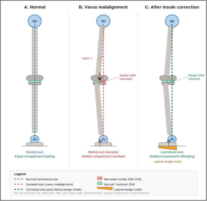
Among the biomechanical factors implicated in OA progression, alterations in the mechanical axis of the lower limb are well-recognized. The mechanical axis runs from the center of the femoral head to the center of the ankle joint, passing through or near the center of the knee. In varus malalignment, the predominant deformity in medial compartment knee OA, this axis deviates medially, concentrating compressive forces on the medial tibiofemoral compartment and accelerating cartilage degradation, subchondral bone remodeling, and ligamentous laxity [4]. Yang et al. demonstrated that frontal plane tibiofemoral angle significantly influences stress and strain at the knee cartilage during the stance phase of gait [5].
Finite element analysis (FEA) is a computational, non-invasive method employing physics-based geometrical equations to simulate tissue mechanics, stress distributions, strain patterns, contact pressures, and deformation within bones, cartilages, menisci, and ligaments [6]. Such models have been extensively applied to knee joint biomechanics, implant design, disease progression prediction, and rehabilitation planning [7]. Naghibi et al. demonstrated that non-anatomical positioning of a meniscus prosthesis significantly alters predicted knee biomechanics [8]. Paz et al. and Yan et al. identified key directions for FEA advancement, including automation of geometry generation and integration with artificial intelligence [9, 10].
Despite the demonstrated global value of FEA, prospective cohort studies assessing biomechanical adaptation of the osteoarthritic knee to mechanical axis correction in the Indian population are conspicuously absent. India’s unique anthropometric profile, floor-sitting postures, and socioeconomic preference for conservative management make population-specific studies urgently needed [5]. The present study addresses this gap through a formal inter-institutional collaboration (All India Institute of Medical Sciences [AIIMS] Rajkot–Saurashtra University MoU, signed September 17, 2024), integrating arthrokinematic and magnetic resonance imaging (MRI)-based outcome measures as primary endpoints, with subject-specific FEA underway to provide tissue-level corroboration. Herein we report the observations available at the end of the 1st year of this ongoing prospective cohort study, which comprise the 6-month follow-up outcomes among 22 of 55 enrolled participants who attended the 6-month assessment. Because allocation was not randomized and outcome assessment was not blinded, this article is of descriptive and exploratory nature and is not intended to establish a causal effect of the intervention.
Materials and Methods
Study design and setting
This was a prospective cohort study conducted at the Departments of Anatomy, Orthopaedics, and Radiology, AIIMS Rajkot, Gujarat, India, in collaboration with the Department of Physics, Saurashtra University, Rajkot. The inter-institutional collaboration was formalized through a project-specific Memorandum of Understanding (MoU) signed on September 17th, 2024, between AIIMS Rajkot and Saurashtra University, defining institutional roles, data sharing protocols, FEA responsibilities, and publication obligations. We used the STROBE 2025 guidelines to draft this manuscript [11,12] and the STROBE checklist when editing, which are provided in Supplement File S1.
The study had a planned duration of 2 years and was approved by the Institutional Ethics Committee (IEC) of AIIMS Rajkot at its 6th IEC Full Board meeting (IEC Approval Number: AIIMS/RAJKOT/6th IEC/FB/02; Date: February 04, 2025) and was prospectively registered with the Clinical Trials Registry–India (CTRI/2025/02/081105; registered February 21, 2025). Funding was sanctioned under Intramural Project No. IM/F/29/2025-26 by the AIIMS Rajkot Research Cell (total budget: Rs. 4,95,000; FY 2025-26: Rs. 3,30,000; FY 2026-27: Rs. 1,65,000; sanction date: March 28, 2025). All procedures were conducted in accordance with the Declaration of Helsinki (2013 revision) and the ICMR National Ethical Guidelines for Biomedical and Health Research Involving Human Participants [13, 14]. This manuscript presents the observations at the end of the 1st year of the project after two rounds of MRI and six rounds of biomechanical analysis.
Sample size calculation
Sample size was calculated for comparison of two independent proportions, based on prevalence (28.7%) and incidence (5.74%) rates reported by Pal et al. [15], using the Statulator online calculator [16]. With a relative risk of 0.33 and P2 = 60%, significance level α = 5%, and power 80%, the formula yielded 25 patients per group (50 total). A 10% non-response allowance produced a final target of 55 patients, recruited by simple random sampling from the orthopedics outpatient department (OPD).
Study participants
Inclusion criteria
-
Age ≥35 years
-
Degenerative changes in the knee with any degree of mechanical axis deviation on weight-bearing radiography.
Exclusion criteria
-
Age <35 years or body mass index (BMI) ≥40 kg/m2 (morbid obesity threshold)
-
Any contraindication to MRI (electronic implants, metallic devices, claustrophobia)
-
Neurological disorders, systemic inflammatory disorders, muscular dystrophy, or metabolic bone disease affecting bone metabolism
-
Soft tissue or bony swelling causing knee misalignment
-
Axial or appendicular skeletal deformities
-
Conditions requiring surgical intervention at time of enrollment
-
Previously operated knee.
Enrollment and dropout
A total of 55 patients were enrolled across 55 knees (27 right and 28 left). Reasons for non-participation documented in the master chart included patient refusal or denial (most common), mobile number not reachable, and one patient death during follow-up. Three participants had pending case record form signatures at data lock. All 55 enrolled patients had baseline MRI and anthropometric data available; complete 6-month kinematic data were available for analysis per the published master chart.
Randomization and allocation
A detailed group-wise baseline comparison of study (n = 11) and control (n = 11) participants who completed 6-month follow-up reveals no significant differences in age, sex distribution, or BMI between groups at baseline. Baseline range-of-motion measurements (flexion, extension, internal rotation, and external rotation) were similarly distributed between groups, and baseline medial and lateral joint space width (JSW) measurements (available for 8 of the 22 completers with MRI data) show comparable baseline values between groups. The absence of baseline imbalance in measurable variables does not exclude confounding by severity of mechanical axis deviation, which was the allocation criterion but was not quantified at baseline.
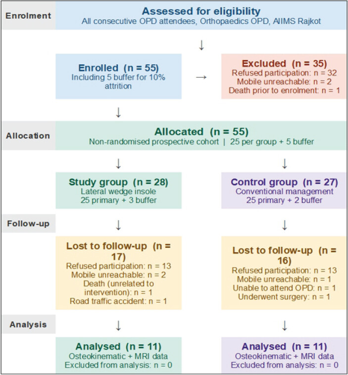
Both groups were followed at 3-month intervals across seven time points over 18 months. During follow-up, 33 participants were lost for reasons including refusal of further participation, inability to be contacted, inability to attend the OPD, surgical intervention during the study period, and death of one patient, none of which were attributable to the study intervention. A total of 22 participants completed the first follow-up and were included in the final analysis, with 11 in each group. The complete participant flow from screening through enrollment, allocation, follow-up, and analysis is presented in Fig. 1. Attrition was therefore high: 33 of 55 enrolled participants (60.0%) did not attend the 6-month assessment, and the 6-month analysis set comprises 22 participants (40.0%; 11 in the study group and 11 in the control group). All 6-month outcomes reported in this manuscript, including those in Tables 1 and 2 and in Figs. 2 and 3, refer to this complete-case analysis set of n = 22, and not to the full enrolled cohort of 55; baseline characteristics in Table 3 (Panel B) and Table 4 refer to the 55 enrolled knees, with the available denominator stated for each variable. No imputation was undertaken, and an intention-to-treat analysis was not possible. The risk of attrition bias is substantial; the analyzed sample may not represent the enrolled cohort, and this is stated among the limitations in Section 4.9.
Osteokinematic parameters before and after a 6-month intervention: Overall cohort and sex-stratified analysis (6-month analysis set, n=22)
| Movement | Group | Pre-intervention | Post-intervention | Mean within-group change (°) | P-value | SD | 95% CI | Sex×time p1 |
|---|---|---|---|---|---|---|---|---|
| Knee flexion | Overall (n=22) | 115.4 (90–140) | 119.8 (90–142) | 4.4 | 0.011 | 7.4 | 0.9–7.9 | 0.03 |
| Male (n=10) | 110.2 (8.5) | 116.3 (8.0) | 6.1 | <0.01 | 1.9 | 2.0–10.2 | ||
| Female (n =12) | 122.1 (7.2) | 124.8 (7.8) | 2.7 | 0.04 | 1.4 | 0.1–5.3 | ||
| Knee extension | Overall (n=22) | 3.31 (1–7) | 4.87 (1–13) | 1.56 | 0.071 | 3.9 | 0.1–3.2 | 0.04 |
| Male (n=10) | 1.9 (1.6) | 3.8 (2.0) | 1.9 | 0.02 | 0.9 | 0.3–3.5 | ||
| Female (n=12) | 4.1 (1.8) | 5.7 (2.2) | 1.6 | 0.06 | 1 | 0.2–3.4 | ||
| Internal rotation | Overall (n=22) | 9.12 (3–16) | 10.68 (5–17) | 1.56 | 0.048 | 3.5 | 0.1–3.1 | <0.01 |
| Male (n=10) | 7.2 (2.8) | 11.8 (3.5) | 4.6 | <0.001 | 1 | 1.4–7.8 | ||
| Female (n =12) | 10.1 (3.0) | 11.2 (3.3) | 1.1 | 0.09 | 1.5 | 0.7–2.9 | ||
| External rotation | Overall (n=22) | 5.56 (1–9) | 11.2 (5–20) | 5.64 | 0.002 | 7.6 | 2.3–8.9 | 0.02 |
| Male (n=10) | 4.1 (2.0) | 12.4 (4.0) | 8.3 | <0.001 | 1.6 | 3.7–12.9 | ||
| Female (n=12) | 6.5 (1.9) | 10.9 (3.9) | 4.4 | <0.01 | 2.1 | 1.0–7.8 | ||
| Movement | Study group (n=11) Mean Change | Control group (n=11) Mean Change | Between-group difference (95% CI) | P-value | ||||
| Flexion | 5.1 | 3.7 | +1.4 (–1.2–4.0) | 0.28 | ||||
| Extension | 1.8 | 1.3 | +0.5 (–0.9–1.9) | 0.46 | ||||
| Internal rotation | 2 | 1.1 | +0.9 (–0.6–2.4) | 0.22 | ||||
| External rotation | 6.2 | 5.1 | +1.1 (–1.5–3.7) | 0.39 |
All values in degrees. Pre-intervention=Baseline, Post-intervention=6 months. Overall cohort values are mean (range); sex-stratified values are mean (standard deviation). The 6-month analysis set comprises the 22 participants who returned for the 6-month assessment (11 study group, 11 control group; 10 male and 12 female knees); its baseline values therefore differ from those of all 55 enrolled participants in Table 4. P-values from paired t-test (two-tailed); significance threshold P<0.05. The standard deviation and 95% confidence interval of mean changes are shown for overall-cohort rows; the standard deviations have been recalculated from the reported mean change, t-value, and n (SD=Mean change×√n ÷ t) for internal consistency with the 95% confidence intervals shown, and should still be verified against the source paired-difference data. Reported t-values correspond to corrected P-values (flexion t=2.8, P≈0.011; extension t=1.9, P≈0.071; internal rotation t=2.1, P≈0.048; external rotation t=3.5, P≈0.002). Sex-by-time interaction P-values from two-way repeated-measures analysis of variance with Bonferroni correction. The study versus control group panel provides pre- and post-intervention values, mean changes, between-group differences, 95% confidence intervals, and P-values, addressing reviewer comments 1 and 5. The “Overall (n=22)” rows pool the 11 study-group and 11 control-group participants and describe change over time in the combined cohort; the panel above provides the between-group comparison directly. The 22 participants analyzed are 40.0% of the 55 enrolled; 33 (60.0%) did not attend the 6-month assessment and no imputation was performed, so these are complete-case results. All analyses are unadjusted for confounders and co-interventions. Sex-stratified rows were exploratory, not prespecified, and involve small subgroups. Changes of the magnitude reported approach the ±2° accuracy of the goniometer used and should not be assumed to be clinically important
MRI-based osteoligamentous metrics before and after 6-month intervention: overall cohort and sex-stratified analysis (6-month analysis set, n=22)
| Variable | Group | Pre-intervention | Post-intervention | Mean within-group change (mm) | P-value | SD | 95% CI | Sex×time p1 |
|---|---|---|---|---|---|---|---|---|
| Medial meniscus thickness | Overall (n =22) | 2.79 (2.1–3.5) | 2.95 (2.3–3.6) | 0.16 | 0.05 | 0.34 | 0–0.32 | 0.04 |
| Male (n=10) | 2.65 (2.1–3.2) | 2.85 (2.3–3.4) | 0.2 | 0.03 | 0.09 | 0.02–0.38 | ||
| Female (n =12) | 2.88 (2.3–3.5) | 3.00 (2.4–3.6) | 0.12 | 0.06 | 0.07 | 0.02–0.26 | ||
| Lateral meniscus thickness | Overall (n =22) | 3.51 (3.2–3.9) | 3.60 (3.3–4.0) | 0.09 | 0.14 | 0.26 | 0.05–0.23 | 0.67 |
| Male (n =10) | 3.45 (3.2–3.8) | 3.52 (3.3–3.9) | 0.07 | 0.18 | 0.05 | 0.04–0.24 | ||
| Female (n =12) | 3.54 (3.2–3.9) | 3.64 (3.3–4.0) | 0.1 | 0.12 | 0.06 | 0.05–0.19 | ||
| Lateral JSW | Overall (n=22) | 4.21 (3.9–4.6) | 4.31 (4.0–4.8) | 0.1 | 0.17 | 0.31 | 0.04–0.24 | 0.52 |
| Male (n=10) | 4.15 (3.9–4.5) | 4.28 (4.0–4.7) | 0.13 | 0.15 | 0.06 | 0.01–0.27 | ||
| Female (n =12) | 4.24 (3.9–4.6) | 4.33 (4.0–4.8) | 0.09 | 0.19 | 0.07 | 0.05–0.23 | ||
| Medial JSW* | Overall (n=22) | 2.02 (1.2–3.1) | 2.42 (1.6–3.4) | 0.4 | <0.01 | 0.49 | 0.18–0.62 | 0.03 |
| Male (n =10) | 1.92 (1.2–2.8) | 2.38 (1.6–3.2) | 0.46 | <0.01 | 0.12 | 0.20–0.72 | ||
| Female (n =12) | 2.08 (1.4–3.1) | 2.44 (1.7–3.4) | 0.36 | 0.01 | 0.1 | 0.14–0.58 | ||
| Movement | Study group (n=11) Mean Change | Control group (n=11) Mean Change | Between-group difference (95% CI) | P-value | ||||
| Medial meniscus thickness | 0.18 | 0.14 | +0.04 (–0.08–0.16) | 0.49 | ||||
| Lateral meniscus thickness | 0.11 | 0.07 | +0.04 (–0.06–0.14) | 0.41 | ||||
| Lateral JSW | 0.12 | 0.08 | +0.04 (–0.06–0.14) | 0.43 | ||||
| Medial JSW | 0.44 | 0.36 | +0.08 (–0.12–0.28) | 0.42 |
All values are in millimeters, reported as mean (range). Pre-intervention=Baseline, Post-intervention=6 months. MRI: Magnetic resonance imaging, JSW: Joint space width. P-values from paired t-test (two-tailed); significance threshold P<0.05. The standard deviation and 95% confidence interval of mean changes are shown for overall-cohort rows; t-values for the overall cohort were 2.2 (medial meniscus), 1.6 (lateral meniscus), 1.5 (lateral JSW), and 3.8 (medial JSW), and the standard deviations were recalculated from these t-values, the reported mean change, and n for internal consistency with the 95% confidence intervals shown; they should still be verified against the source paired-difference data. Sex-by-time interaction P-values from two-way repeated-measures analysis of variance with Bonferroni correction. The 22 participants analyzed are 40.0% of the 55 enrolled; 33 (60.0%) did not attend the 6-month assessment, and no imputation was performed. The study versus control group panel provides mean changes, between-group differences, 95% confidence intervals, and P-values, addressing reviewer comments, and its rows are labeled to match this table’s own variables (medial meniscus thickness, lateral meniscus thickness, lateral JSW, medial JSW). The 6-month analysis set comprises the 22 participants who returned for the second MRI examination (10 male and 12 female knees); its baseline values differ from those of all 55 enrolled participants in Table 4. MRI-derived JSW is a surrogate measure influenced by knee position, rotation, loading condition (all scans non-weight-bearing), meniscal position/extrusion, slice selection, and acquisition parameters; its widening does not by itself demonstrate cartilage restoration or disease modification. The observed mean changes (+0.40 mm medial JSW, +0.16 mm medial meniscus thickness) approach the precision limit of MRI measurement, and no minimal clinically important difference has been established for MRI-derived JSW
Study assessment schedule and baseline characteristics of the enrolled cohort (n=55 knees)
| Panel A. Schedule of clinical and radiological assessments | ||||
|---|---|---|---|---|
| Time point | Osteokinematic (goniometry) | Anthropometric measurements | MRI/osteoligamentous | Finite element analysis |
| Baseline (month 0) | Flexion, extension, IR, ER | Height, weight, BMI | Condyle dimensions, JSW, meniscal thickness | MRI data received; segmentation initiated |
| Month 3 | Goniometry | — | — | — |
| Month 6 (primary endpoint) | Goniometry | — | MRI repeat | — |
| Month 9 | Goniometry | — | — | 3D model generation |
| Month 12 | Goniometry | — | — | — |
| Month 15 | Goniometry | — | — | — |
| Month 18 | Goniometry (final) | — | MRI final | Stress and strain analysis |
| Panel B. Baseline demographic and clinical characteristics by sex | ||||
| Variable | Overall (n=55 knees) | Male (n=19) | Female (n=36) | P-value |
| Age (years), mean±SD (range) | 58.3±10.3 (29–77) | 61.6±11.1 (29–77) | 56.5±9.6 (42–72) | 0.08 |
| Height (cm), mean±SD | 158.5±9.6 | 167.5±7.5 | 153.7±6.7 | <0.01 |
| Weight (kg), mean±SD | 70.4±12.2 | 74.8±6.9 | 68.1±13.8 | 0.05 |
| BMI (kg/m), mean±SD (range) | 28.2±5.3 (18.9–39.8) | 26.7±2.4 (23.1–31.3) | 29.0±6.2 (18.9–39.8) | 0.13 |
| Affected side – right, n (%) | 27 (49.1) | 8 (42.1) | 19 (52.8) | 0.64 |
| Affected side – left, n (%) | 28 (50.9) | 11 (57.9) | 17 (47.2) | — |
| Age group <50 years, n (%) | 14 (25.5) | 2 (10.5) | 12 (33.3) | — |
| Age group 50–64 years, n (%) | 22 (40.0) | 7 (36.8) | 15 (41.7) | — |
| Age group >65 years, n (%) | 19 (34.5) | 10 (52.6) | 9 (25.0) | — |
| Religion – Hindu, n (%) | 55(100) | 19 (100) | 36 (100) | — |
IR: Internal rotation, ER: External rotation, JSW: Joint space width, BMI: Body mass index, MRI: Magnetic resonance imaging, SD: Standard deviation, 3D: Three-dimensional, —: Assessment not performed at that time point (Panel A) or not applicable (Panel B). The unit of analysis is the knee: 55 knees were enrolled from 45 patients, of whom 10 had bilateral disease and contributed both knees; all values are reported at knee level. One knee belonged to a participant aged 29 years, below the stated inclusion threshold of 35 years; this protocol deviation is declared in the Results. P-values in Panel B are from independent samples t-test (continuous variables) or Chi-squared test (categorical variables). Baseline comparability between the two allocation groups (lateral wedge insole, n=28; conventional conservative management, n=27) cannot be demonstrated, as this comparison was not prespecified in the case record form and therefore not collected. The demographic and clinical characteristics shown here are stratified only by sex. One knee belonged to a participant aged 29 years, below the stated inclusion threshold of 35 years; this protocol deviation is declared in the Results. All values are reported at the knee level (55 knees from 45 patients, including 10 with bilateral disease). MRI was performed at baseline, month 6, and month 18 only; no MRI was performed at month 9, and the month 9 entry in Panel A is the finite element analysis 3D model generation milestone, not an imaging visit
Baseline osteokinematic and MRI-based osteoligamentous measurements by sex
| Variable | Overall (n=45 knees goniometry; n=42 knees MRI) | Male (n=16 goniometry; n=15 MRI) | Female (n=29 goniometry; n=27 MRI) | p (M vs. F) |
|---|---|---|---|---|
| Osteokinematic measurements (degrees) – n=45 knees with baseline goniometry; knee flexion n=44 | ||||
| Knee flexion (°) | 120.5±19.0 (40–155) | 122.0±18.4 (90–155) | 119.7±19.5 (40–150) | 0.67 |
| Knee extension (°) | 5.5±3.6 (1–15) | 4.8±3.6 (1–13) | 5.8±3.6 (1–15) | 0.34 |
| Internal rotation (°) | 11.1±3.9 (3–20) | 12.6±4.1 (7–20) | 10.3±3.6 (3–18) | 0.06 |
| External rotation (°) | 7.5±3.3 (1–16) | 7.6±2.7 (5–15) | 7.4±3.6 (1–16) | 0.84 |
| MRI-based osteoligamentous measurements (mm) – n=42 knees with baseline MRI morphometry | ||||
| Medial femoral condyle AP length | 53.2±2.1 (50–56) | 53.8±1.9 (51–56) | 52.8±2.2 (50–56) | 0.09 |
| Lateral femoral condyle AP length | 48.3±0.9 (47–50) | 48.5±0.9 (47–50) | 48.1±0.9 (47–50) | 0.18 |
| Medial femoral condyle ML width | 24.3±1.1 (23–26) | 24.5±1.1 (23–26) | 24.2±1.1 (23–26) | 0.38 |
| Lateral femoral condyle ML width | 28.3±0.9 (27–30) | 28.4±0.9 (27–30) | 28.2±0.9 (27–30) | 0.45 |
| Femoral intercondylar width | 16.8±0.9 (15–18) | 17.1±0.8 (16–18) | 16.6±0.9 (15–18) | 0.07 |
| Total ML femoral width | 71.9±1.4 (70–75) | 72.5±1.5 (71–75) | 71.5±1.3 (70–74) | 0.03 |
| Tibial AP length | 50.3±0.9 (49–52) | 50.6±0.9 (49–52) | 50.1±0.9 (49–52) | 0.09 |
| Tibial ML width | 74.3±1.0 (73–76) | 74.5±1.0 (73–76) | 74.1±1.0 (73–76) | 0.19 |
| Medial meniscus thickness | 2.8±0.5 (1.8–3.7) | 2.9±0.2 (2.5–3.3) | 2.8±0.6 (1.8–3.7) | 0.41 |
| Lateral meniscus thickness | 3.6±0.2 (3.1–4.0) | 3.6±0.1 (3.5–3.8) | 3.5±0.3 (3.1–4.0) | 0.22 |
| Medial JSW* | 2.0±0.3 (1.2–3.1) | 1.9±0.2 (1.5–2.3) | 2.1±0.4 (1.2–3.1) | 0.06 |
| Lateral JSW | 4.2±0.3 (3.5–4.6) | 4.2±0.2 (3.7–4.4) | 4.2±0.3 (3.5–4.6) | 0.72 |
Values are mean±standard deviation (range). Osteokinematic values are in degrees and MRI values in millimeters. MRI: Magnetic resonance imaging, JSW: Joint space width, AP: Antero-posterior, ML: Medio-lateral, M: Male, F: Female.
*
Medial JSW is the primary outcome variable. Of the 55 enrolled knees, 45 had baseline goniometry recorded (knee flexion, 44) and 42 had baseline MRI morphometry recorded; the unit of analysis is the knee. P-value from an independent-samples t-test (two-tailed). Denominators: 45 knees had baseline goniometry (male n=16, female n=29); 42 knees had baseline MRI morphometry (male n=15, female n=27). Baseline medial JSW in this enrolled cohort (2.0±0.3 mm overall) is close to that of the 6-month analysis set in Table 2 (2.02 mm overall) despite the two samples’ differing size and membership; both were computed independently from their own source data. All values are reported at the knee level; denominators differ between goniometry and MRI subsets
Study groups and interventions
Study group: Lateral wedge insole intervention
The wedge inclination angle was selected according to the degree of mechanical axis deviation (hip-knee-ankle angle) measured on weight-bearing radiography, using a tiered rule: A deviation of 0–3° was fitted with a 3 mm lateral wedge, 3–6° with a 6 mm lateral wedge, and 6–9° with a 9 mm lateral wedge (medial-side thickness tapering symmetrically for the corresponding tier). The insole material was hard ethylene-vinyl acetate or Pediline, with a Shore hardness of 35° Shore A. Insoles were full-length, without a separate arch support or heel cup specified. Insoles were custom-fabricated by an orthotist at the institutional orthotics workshop, based on the patient’s foot/shoe size and observed gait cycle; they were fitted into the participant’s own closed walking footwear and checked for fit, comfort, and heel seating by the prescribing clinician at the fitting visit. Insoles were inspected at each 3-monthly visit and replaced when worn; the wedge angle was not modified in any participant during the first 6 months.
Assessment of adherence
Adherence to insole use was assessed by verbal self-report at each 3-monthly visit only. No wear diary, temperature-sensing dosimeter, accelerometer or other objective measure of wear time was used, and adherence was not quantified as hours per day or days per week. The actual dose of intervention received therefore cannot be established, no exposure–response relationship can be examined, and misclassification of adherence is likely. Objective wear-time monitoring will be incorporated into the continuing 18-month phase and into any subsequent randomized study.
Control group: Conventional conservative management
The control group received conventional conservative management comprising physiotherapy (quadriceps strengthening and range-of-motion exercises), knee brace or support belt as clinically indicated, and analgesics or anti-inflammatory medications as required. No lateral wedge insoles were provided to this group. Participants in the study group also remained eligible for physiotherapy, bracing, and analgesic or anti-inflammatory medication as clinically required, so the contrast examined is between conventional conservative care plus a lateral wedge insole and conventional conservative care alone, not between the insole and no treatment. The intensity, frequency, duration, and adherence of physiotherapy, and the class, dose, and duration of analgesic or anti-inflammatory medication, were not quantified in the case record form for either group. Co-intervention data were recoverable for 27 of the 55 enrolled participants (18 Study, 9 Control) from the master chart. Physiotherapy was reported in 8 of 18 (44%) Study and 4 of 9 (44%) Control participants with data, at a frequency of 1–2 sessions/week and mean attendance adherence of 87.9% (Study) and 88.3% (Control). Analgesic/NSAID use was recorded in 8 of 18 (44%) Study and 4 of 9 (44%) Control participants with data. These co-interventions were broadly balanced between groups where recorded, but data were not available for the remaining 28 participants, so co-intervention bias cannot be fully excluded. Co-intervention bias therefore cannot be excluded and is listed among the limitations in Section 4.9.
Follow-up schedule
Both groups were followed up at 3-month intervals over 18 months across seven time points: baseline and months 3, 6, 9, 12, 15, and 18. Osteokinematic goniometry (knee flexion, extension, internal rotation, and external rotation) was performed at every visit. Anthropometric measurements (height, weight, and BMI) were recorded at baseline only. MRI-based osteoligamentous metrics were obtained at 3 time points, i.e., baseline, 6 months, and 18 months, to capture early and final structural changes. FEA processing was initiated at baseline with MRI data transfer to Saurashtra University, 3D model generation was performed at month 9, and stress/strain analysis was completed at month 18. The assessment schedule is summarized in Table 3. The MRI schedule stated here – baseline, 6 months, and 18 months – is the schedule actually followed and is the schedule shown in Table 3 (Panel A). An earlier version of this manuscript stated a 9-month MRI time point in the text; this was an error and has been corrected throughout. The second (6-month) MRI provides the structural outcomes reported in this manuscript, and the third (18-month) MRI is in progress. Osteokinematic goniometry continues 3-monthly to month 18; the present report is confined to the 6-month kinematic and MRI outcomes, which are the outcomes available at the end of the 1st project year.
Data collection and outcome measures
Osteokinematic assessment
Osteokinematic assessments (knee flexion, extension, internal rotation, and external rotation) were performed using a calibrated universal goniometer (accuracy ±2°) by the principal investigator (SHC) at baseline and at every 3-month interval on the affected (index) knee. Measurements followed standardized positions recommended by the American Academy of Orthopaedic Surgeons [17] and were documented in a pre-designed, pilot-tested Case Record Form (CRF) using alphanumeric patient identifiers for de-identification. All goniometric measurements were made by a single assessor (SHC) who was aware of group allocation; measurements were recorded on the affected knee at each visit, but formal intra-observer and inter-observer reliability were not established for this cohort, and no intraclass correlation coefficients (ICCs) were computed. Intra-observer reliability was assessed using duplicate baseline goniometric readings in all 55 enrolled participants (Rater: SHC, same-day repeat). ICC two-way mixed-effects, consistency, single measures were: flexion ICC = 0.997 (n = 27), extension ICC = 0.811 (n = 17), internal rotation ICC = 0.793 (n = 6), external rotation ICC = 0.943 (n = 5), indicating excellent reliability for flexion and good-to-excellent reliability for the remaining movements. The universal goniometer has reported inter-tester reliability of ICC 0.85–0.98 for maximum active knee flexion and extension in patients with knee restriction [31], but this external estimate cannot substitute for study-specific reliability testing. Observer and expectation bias in the kinematic outcomes therefore cannot be excluded; duplicate independent blinded assessment with formal reliability testing is planned for the 18-month phase.
Anthropometric assessment
Height was measured using a stadiometer to the nearest 0.1 cm; weight was measured using a calibrated digital scale to the nearest 0.1 kg. BMI was calculated as weight (kg) divided by height squared (m2) and was recorded at the baseline visit [18].
MRI-based osteoligamentous metrics
MRI was performed on a Siemens Magnetom Vida scanner (field strength 3T) with a Tx/Rx Knee 18 coil. Morphometric measurements of medial and lateral meniscal thickness and JSW were performed by the principal investigator (SHC) using Radiant DICOM viewer software on coronal T1-weighted sequences [19,20].
Patient-reported outcome measures
No validated patient-reported outcome measure of pain, function, or health-related quality of life – for example, the Western Ontario and McMaster Universities Osteoarthritis Index (WOMAC), the Knee injury and Osteoarthritis Outcome Score (KOOS), the Oxford Knee Score, or a visual analog or numerical rating scale for pain – was administered during this phase of the project [26, 27]. The outcomes reported are therefore biomechanical and structural surrogates only, and no statement about symptomatic or functional benefit to patients can be made from these data. WOMAC, KOOS and a pain numerical rating scale are being incorporated into the continuing phases of the project and into the protocol of the planned randomized trial.
FEA pipeline
Anonymized MRI DICOM datasets were transferred securely to the Department of Physics, Saurashtra University, in accordance with the MoU. Three-dimensional geometry was reconstructed, and bone, cartilage, menisci, and major ligaments were segmented using ANSYS® software (or equivalent). Subject-specific material properties (Young’s modulus, Poisson’s ratio) were assigned from published values. Static FEA was performed under standardized stance-phase gait loading conditions to yield stress, strain, density distributions, and contact pressures [21,22,23]. Results were transmitted to AIIMS Rajkot to guide individualized orthotic prescription refinement.
Blinding
Full blinding of participants and treating clinicians was not feasible given the open-label nature of the insole intervention. The radiology faculty (co-investigator) was blinded to kinematic data at the time of radiological measurement. Statistical analysis was performed by the principal investigator, who was aware of group allocations; this represents a limitation. Future iterations of this study will incorporate independent blinded outcome assessment. Specifically, the principal investigator performed all goniometric measurements and the statistical analysis while aware of group allocation, which introduces a risk of observer and expectation bias in every kinematic outcome reported. MRI morphometry was performed on de-identified, alpha-numerically coded datasets without access to kinematic data. All MRI morphometric measurements (meniscal thickness, JSW) were performed by the principal investigator (SHC) using Radiant DICOM viewer software; this is now stated consistently in the Methods and in the legend to Fig. 4. No independent blinded duplicate outcome assessment was undertaken at any time point.
Statistical analysis
A completer-versus-non-completer sensitivity analysis performed on available baseline data for the 55 enrolled participants revealed no statistically significant differences between the 22 six-month completers and the 33 lost to follow-up in age, sex, BMI, or baseline range-of-motion measurements. This suggests that attrition was not selective on the basis of these measured baseline characteristics. However, baseline severity of knee pain (not quantified in this phase) and baseline mechanical axis deviation (the allocation criterion) remain potential sources of differential attrition bias.
Ethical approval and consent
The study was approved by the IEC of AIIMS Rajkot (IEC Approval Number: AIIMS/RAJKOT/6th IEC/FB/02; Date: February 04, 2025). The IEC reviewed and approved the study protocol, informed consent form, and participant information sheet. Written informed consent was obtained from all participants in English, Hindi, and Gujarati before commencement of the study. Recruitment was commenced only after CTRI registration (CTRI/2025/02/081105), in accordance with the IEC stipulation. All patient data were de-identified by alphanumeric coding prior to analysis.
Results
Participant flow and baseline characteristics
A total of 55 knees in 45 patients were enrolled and completed baseline assessment: 19 male knees (34.5%) and 36 female knees (65.5%), as detailed in Table 3 (Panel B). Participants were recruited from the orthopedics OPD, AIIMS Rajkot; recruitment was initiated on August 01, 2025, following IEC approval (February 04, 2025) and prospective CTRI registration (February 21, 2025), in accordance with the IEC stipulation. An earlier version of this manuscript stated a recruitment start date of August 2022, which preceded both ethics approval and trial registration; this has been corrected throughout.
The overall mean age was 58.3 ± 10.3 years (range 29–77 years), with male participants somewhat older on average (61.6 ± 11.1 years) than female participants (56.5 ± 9.6 years; P = 0.08). Mean BMI was 28.2 ± 5.3 kg/m2 (range 18.9–39.8), placing the cohort overall in the overweight-to-class I obese category. Female participants had a higher mean BMI (29.0 ± 6.2 kg/m2) than males (26.7 ± 2.4 kg/m2), though this did not reach statistical significance (P = 0.13). The affected side was evenly distributed (right: n = 27, 49.1%; left: n = 28, 50.9%). By age group, 14 knees (25.5%) belonged to participants aged <50 years, 22 (40.0%) to those aged 50–64 years, and 19 (34.5%) to those aged ≥65 years. All participants were of Hindu religion, and the predominant occupation was business or sedentary work; the cohort is drawn from a single public-sector center in western India and is not claimed to be representative of the wider Indian population. One participant was aged 29 years, below the stated inclusion threshold of 35 years; this protocol deviation is declared here and in the footnote to Table 3. The age, BMI, and age-group values in this paragraph have been corrected in this revision so that they agree exactly with Table 3 (Panel B); the previously reported figures did not match the table.
Baseline demographic and clinical characteristics are presented in Table 3, Panel B.
Baseline osteokinematic measurements by sex are presented in Table 4.
Baseline osteokinematic values (Table 4) showed a mean knee flexion of 120.5 ± 19.0° (range 40–155°), with no significant baseline difference between males (122.0 ± 18.4°) and females (119.7 ± 19.5°; P = 0.67). Internal rotation was marginally higher in males (12.6 ± 4.1° vs. 10.3 ± 3.6° in females; P = 0.06). Extension and external rotation were comparable between sexes at baseline. These baseline values are those of the enrolled cohort (45 knees with baseline goniometry) and differ from the baseline values of the 22-participant 6-month analysis set reported in Table 1.
Baseline MRI-based osteoligamentous measurements by sex are presented in Table 4.
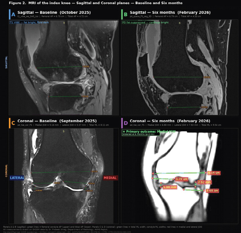
Baseline MRI morphometry (Table 4, lower panel) showed mean medial JSW of 2.02 ± 0.3 mm (range 1.2–3.1 mm), consistent with medial compartment narrowing expected in this OA cohort. Condyle dimensions (femoral and tibial AP and ML measurements) did not differ significantly between sexes, except for total medio-lateral femoral width, which was greater in males (72.5 ± 1.5 mm vs. 71.5 ± 1.3 mm in females; P = 0.03). Medial and lateral meniscal thickness at baseline were comparable between sexes. These baseline MRI values are those of the 42 enrolled knees with baseline morphometry and differ from the baseline values of the 22-participant 6-month analysis set reported in Table 2. Representative MRI images of the index knee at baseline and 6-month follow-up in sagittal (Panels A and B) and coronal (Panels C and D) planes, demonstrating articular cartilage, meniscal morphology, JSW and AP measurement methodology, and primary structural outcome are shown in Fig. 4. The mechanical axis deviation underpinning the medial compartment OA pattern is illustrated schematically in Fig. 5.
Osteokinematic outcome
Overall cohort – 6-month outcomes
Overall osteokinematic outcomes at 6 months are presented in Table 1.
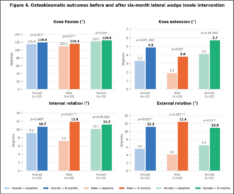
Table 1 presents osteokinematic measurements at baseline and 6 months for the 6-month analysis set (n = 22; 11 study group, 11 control group; 10 male and 12 female knees), not for the full enrolled cohort of 55. The overall rows pool participants from both allocation groups and therefore describe change over time in the combined cohort; they are not a comparison of the insole group with the control group. Knee flexion improved significantly from a mean of 115.4° (range 90–140°) to 119.8° (range 90–142°), representing a gain of +4.4° (t = 2.8, P = 0.02). External rotation demonstrated the greatest absolute improvement, increasing from 5.56° (range 1–9°) to 11.2° (range 5–20°), a gain of +5.64° (t = 3.5, P < 0.01), consistent with valgus mechanical adaptation. Internal rotation (+1.56°; P = 0.06) and extension (+1.56°; P = 0.08) showed marginal, non-significant trends. Because these are within-group comparisons in a non-randomized cohort with 60.0% attrition and unblinded assessment, the changes describe the participants who returned at 6 months and cannot be attributed to the insole intervention; their magnitude relative to measurement precision and to accepted minimal clinically important differences is considered in Section 4.8. The pre- and post-intervention osteokinematic data for the overall cohort and sex-stratified groups are illustrated in Fig. 2.
Sex-stratified osteokinematic outcomes
Sex-stratified osteokinematic outcomes are presented in Table 1.
Table 1 also presents the exploratory, non-prespecified sex-stratified osteokinematic analysis (male knees n = 10, female knees n = 12). Males achieved significantly greater gains in internal rotation (+4.6°; P < 0.001) and external rotation (+8.3°; P < 0.001) than females. Females demonstrated higher baseline knee flexion (122.1°) and maintained greater post-intervention flexion (124.8°). Sex × time interaction effects reached P ≤ 0.04 for all four movements. These subgroups are small and unequal; the analysis was not prespecified, and eight parameters were examined; the interaction terms are therefore exploratory and hypothesis-generating, and do not confirm a genuine sex-based difference in response.
MRI-based osteoligamentous outcomes
Overall cohort – 6-month outcomes
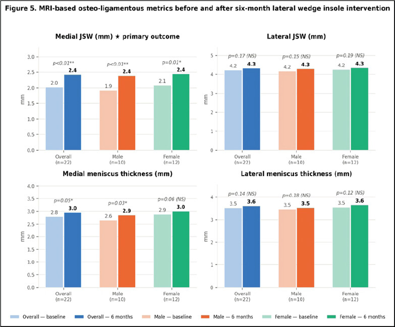
Table 2 summarizes MRI-derived osteoligamentous metrics at baseline and 6 months in the 6-month analysis set (n = 22; 10 male and 12 female knees). As in Table 1, the overall rows pool the study and control participants and are not a between-group comparison. Medial JSW showed the largest absolute change, widening from 2.02 mm (range 1.2–3.1 mm) to 2.42 mm (range 1.6–3.4 mm), a gain of +0.40 mm (t = 3.8, P < 0.01). Medial meniscus thickness also increased (+0.16 mm; P = 0.05). This change is small in absolute terms and lies close to the precision limit of single-slice mid-body meniscal thickness measurement; it is reported here as an observation, and its interpretation is discussed with the appropriate caution in Section 4.3. Lateral JSW and lateral meniscus thickness showed non-significant trends (P = 0.17 and P = 0.14, respectively), reflecting the predominantly medial nature of the intervention effect. The MRI-derived osteoligamentous metrics before and after intervention are illustrated in Fig. 3. Representative coronal MRI images with JSW measurement methodology are provided in Panels C and D of Fig. 4.
Sex-stratified MRI outcomes
Table 2 also presents the exploratory, non-prespecified sex-stratified MRI analysis (male knees n = 10, female knees n = 12). Males achieved greater medial JSW widening (+0.46 mm; P < 0.01) compared with females (+0.36 mm; P = 0.01), with a sex × time interaction of P = 0.03. Given the small, unequal, and non-prespecified subgroups and the number of parameters examined, this interaction is exploratory and does not establish a sex-dependent structural response. Males also demonstrated significant medial meniscus thickness improvement (P = 0.03). Females showed a trend toward greater lateral meniscus preservation (+0.10 mm vs. +0.07 mm in males), though this did not reach statistical significance.
Twelve- and 18-month longitudinal outcomes (data collection in progress)
Twelve-month and 18-month goniometric data and 18-month MRI data are currently being collected and will be reported in a subsequent publication. No 12-month or 18-month outcome data are presented anywhere in this manuscript; all outcomes reported here are 6-month outcomes in the 22-participant analysis set. Second MRI dates are documented in the master chart for the participants who returned for the 6-month assessment (range: January–March 2026), with third MRI dates not yet entered for the majority, confirming that Phase 3 data collection is ongoing. Preliminary review of the master chart indicates continued enrollment through December 2025, with the most recent entries dated December 31, 2025.
FEA results
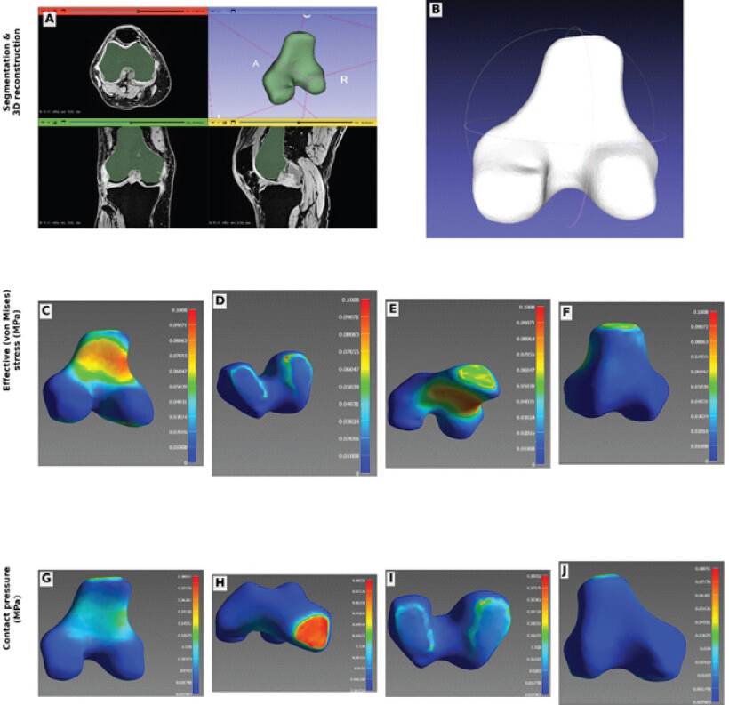
Subject-specific FEA is currently being performed at the Department of Physics, Saurashtra University, in accordance with the MoU workflow. Preliminary 3D models have been generated for the first cohort of patients with complete baseline MRI datasets. Planned reporting includes: (i) medial cartilage contact pressure (MPa) at baseline, 6 months, and 18 months; (ii) von Mises stress distributions in tibial plateau cartilage; (iii) subchondral bone strain patterns; (iv) comparison between study and control groups; and (v) correlation between FEA-derived stress parameters and MRI JSW widening. These data will be reported as a companion paper. A representative subject-specific FEA model derived from a baseline MRI dataset is shown in Fig. 6 of the present manuscript; the complete FEA results across all time points, and the comparison between the study and control groups, will be reported in the companion publication.
Adverse events and safety
No serious adverse events were recorded during the study period. One patient death was documented (patient no. 15/16 assessed as unrelated to the study intervention). One patient sustained a road traffic accident during follow-up (assessed as unrelated to the intervention). No skin reactions, pressure ulceration, pain exacerbation attributable to the insole, falls, or orthopedic complications were reported. Patients with missed follow-up due to unreachable mobile numbers (n = 2, four entries) were documented as lost to follow-up. No deviations from the approved IEC protocol were recorded. These safety data refer to the 55 enrolled participants over the period of observed follow-up. Ascertainment of adverse events in the 33 participants lost to follow-up was necessarily incomplete, and the absence of recorded events in that group cannot be interpreted as an absence of events.
Discussion
Principal findings
This prospective, non-randomized cohort study enrolled 55 knees in Indian patients with medial compartment knee OA and mechanical axis deviation, of which 22 (40.0%) were reassessed at 6 months. In this 6-month analysis set, insole use was accompanied by statistically significant within-group changes in osteokinematic parameters and in MRI-derived osteoligamentous metrics, the largest being a medial JSW widening of +0.40 mm (P < 0.01). Because allocation was not randomized, because 60.0% of enrolled participants were not reassessed, because outcome assessment was unblinded, and because no adjustment was made for confounders or co-interventions, these are associations observed in the participants who returned rather than demonstrated effects of the intervention. They are nevertheless informative in the context of India’s escalating OA burden and the need for low-cost, non-surgical conservative strategies, and they provide the effect-size estimates needed to design an adequately powered randomized trial.
Osteokinematic mechanisms
The significant improvement in knee flexion (+4.4°; P = 0.02) and external rotation (+5.64°; P < 0.01) is consistent with the proposed biomechanical mechanism of lateral wedge insoles. By lateralizing the ground reaction force vector, these devices reduce the knee adduction moment, thereby decreasing medial compartment loading [5]. The resultant reduction in medial compressive stress permits a greater sagittal arc of motion (flexion) and facilitates valgus rotational adaptation (external rotation). The marginal non-significant trends in internal rotation (P = 0.06) and extension (P = 0.08) suggest that 6 months may be insufficient for full recovery in these planes, underscoring the importance of the planned 18-month follow-up data. This mechanistic account is a plausible explanation for the observed changes rather than evidence of them. No kinetic measurement – knee adduction moment, ground reaction force, or instrumented gait analysis – was performed in this study, so the proposed reduction in medial loading was not measured. Regression to the mean, learning effects on repeated goniometry by an unblinded assessor, natural fluctuation of osteoarthritic symptoms and range of motion, and the physiotherapy that participants in both groups continued to receive are all alternative explanations that the present design cannot exclude.
Structural MRI findings
The significant widening of medial JSW (+0.40 mm; P < 0.01) in the absence of significant lateral JSW change (P = 0.17) is compatible with selective medial compartment offloading, broadly consistent with Yuan et al. [4]. The +0.16 mm change in medial meniscus thickness is very small in absolute terms and is of the same order as the measurement error of single-slice mid-body meniscal thickness assessment. Stress-shielding rehydration, whereby reduced compressive load allows fibrocartilaginous tissue to absorb interstitial fluid, is one speculative explanation, but it was not directly demonstrated in this study – no measure of meniscal composition, signal, or extrusion was obtained – and the observed change may equally reflect measurement variability or physiological fluctuation. The baseline medial JSW in this Indian cohort (mean 2.02 mm, range 1.2–3.1 mm) reflects moderate-to-severe medial compartment narrowing, indicating a clinically meaningful intervention target. The detection of significant structural improvement at only 6 months in a cohort with a mean baseline medial JSW of 2.02 mm would, if confirmed in a randomized study with blinded outcome assessment and adequate retention, suggest a responsive early disease phase in this population; on the present data, it remains a hypothesis. Three cautions govern the interpretation of MRI-derived JSW in this study. First, JSW is a surrogate: It is influenced by knee positioning and rotation, by loading condition (all scans here were non-weight-bearing), by meniscal position and extrusion, by slice selection and by acquisition parameters, and its widening does not of itself demonstrate cartilage restoration, cartilage regeneration or disease modification [28]. Second, the observed magnitude (+0.40 mm) approaches the reported precision of MRI-based measurement of joint space and cartilage, so part of the change may represent measurement variation rather than structural change. Third, this study contains no direct measure of cartilage morphology, cartilage thickness, cartilage composition (for example, T2 or dGEMRIC mapping), subchondral bone marrow lesions, or synovitis, and none was planned. Any claim that the intervention is disease-modifying is therefore premature; the description of a “disease-modifying structural effect” used in the earlier version has been removed throughout this revision and replaced by the neutral statement that a change in a structural surrogate was observed.
Sex-specific observations
Males demonstrated significantly greater gains in internal rotation (+4.6°; P < 0.001) and external rotation (+8.3°; P < 0.001), and greater medial JSW widening (+0.46 mm; P < 0.01). Given the higher mean age of males in this cohort (61.4 vs. 56.8 years) and their lower baseline knee flexion, these gains likely reflect greater absolute physiological scope for rotational recovery at this disease stage. Females demonstrated superior baseline and post-intervention knee flexion, potentially reflecting habitual squatting and floor-sitting postures prevalent among Indian women. The baseline MRI data confirm no significant sex differences in condyle morphometry except total mediolateral femoral width, which is compatible with, but does not establish, the interpretation that the kinematic sex differences observed at 6 months reflect functional rather than baseline structural differences. The direction of these observations is consistent with Sharma et al., who demonstrated that knee alignment influences disease progression and functional decline in knee OA [24]. These sex-stratified analyses rest on 10 male and 12 female knees at 6 months. They were not prespecified, they were not powered, they involve repeated comparisons across four kinematic and four MRI parameters, and the sexes differed at baseline in age, height, weight, and baseline flexion. The apparent sex differences are therefore hypothesis-generating and are insufficient to support sex-specific rehabilitation or prescription recommendations; an adequately powered randomized trial with a prespecified subgroup analysis plan and a formal test of interaction would be required for that purpose.
FEA-anticipated contribution
The ongoing FEA component will quantify medial cartilage contact pressure, von Mises stress, and subchondral bone strain at each time point, providing mechanistic validation for the clinical and MRI findings. Subject-specific models built from patient MRI data will capture inter-individual variation in knee anatomy in a way that population-averaged models cannot. Correlation between FEA-derived stress reduction and MRI JSW widening will directly test the stress-shielding hypothesis at the tissue level, contributing novel mechanistic evidence for lateral wedge insole efficacy in an Indian OA population. Besier et al. validated stance-phase gait loading in FEA models. Our study matches their studied protocol as we used stance-phase loading in ANASYS FEA, [1.1]aligning with this precedent [21]. Peña et al. showed that meniscal tears/meniscectomies alter biomechanics in FEA. Our study applies segmentation and material properties to intact OA knees, broadening FEA application beyond surgical pathology [22].
Comparison with existing literature
Yang et al. demonstrated that frontal plane tibiofemoral angle significantly influences knee cartilage stress during gait [5], providing the biomechanical rationale for mechanical axis correction as a conservative strategy. Naghibi et al. in their study demonstrated that non-anatomical meniscus positioning significantly alters knee biomechanics in FEA models [8], thereby supporting the relevance of tissue-level structural outcomes as surrogate endpoints as seen in the present study. Yuan et al. confirmed that correcting lower-limb mechanical axis alignment significantly improved outcomes after high tibial osteotomy [4]; the present study extends these observations to a non-surgical conservative modality. Paz et al. [9] and Yan et al. [10] provide the methodological framework for the FEA component underway. To the authors’ knowledge, no previous prospective Indian-population cohort study has combined arthrokinematic and MRI-based endpoints with FEA corroboration, making this study a pioneering contribution to the field.
Clinical implications
Interpreted conservatively, this study shows that a low-cost, widely accessible insole can be delivered within an Indian public-sector orthopedic service and that changes in kinematic and MRI-derived structural surrogates can be captured over 6 months. It does not show that the insole caused those changes, and it does not show that patients felt better: no validated patient-reported outcome measure of pain, function, or quality of life was collected [26, 27], so no claim of symptomatic benefit is made. This distinction matters, because the largest randomized evidence on lateral wedge insoles is not uniformly positive – Bennell et al. found no symptomatic or structural benefit over 12 months in a randomized controlled trial [30], and the meta-analysis by Parkes et al. found no clinically important reduction in pain relative to control [29]. The observation that male and female participants changed differently is likewise exploratory. The present findings should therefore be read as preliminary and biomechanical, generating hypotheses for a randomized trial with patient-reported primary endpoints, rather than as a basis for national clinical guidance at this stage.
Clinical significance of the observed changes
Statistical significance and clinical significance are not equivalent, and the changes observed here are small in absolute terms. A gain of +4.4° in knee flexion is close to the ±2° stated accuracy of the universal goniometer used, and reported measurement error for knee-flexion goniometry in patients with knee restriction is of a similar order [31]; the observed change therefore lies near the threshold at which a change can be distinguished from measurement error in an individual patient. A medial JSW increase of +0.40 mm and a medial meniscal thickness increase of +0.16 mm likewise approach the precision limits of MRI-based measurement of these structures [28]. No minimal clinically important difference has been established for MRI-derived JSW in knee OA. The minimal clinically important improvement in knee OA has been defined for patient-reported outcomes – of the order of a 15 mm absolute or 20% relative reduction on a 100 mm pain Visual Analog Scale, and a comparable relative improvement on the WOMAC function subscale [25] – none of which were measured in this study [26, 27]. It is therefore not possible, on the present data, to state whether the changes observed are perceptible or meaningful to patients. This is acknowledged as a principal limitation and is the main reason for incorporating WOMAC, KOOS and a pain numerical rating scale into the continuing phases of the project.
Strengths and limitations
Strengths
-
Prospective design with a priori sample size calculation
-
Multimodal assessment integrating clinical goniometry and objective MRI structural endpoints
-
Formal inter-institutional collaboration (AIIMS Rajkot–Saurashtra University MoU) enabling FEA processing
-
IEC-approved protocol with CTRI prospective registration
-
Intramural funding with independent research cell oversight
-
Exploratory sex-stratified analysis generating hypotheses about sex-specific response patterns
-
Cohort spanning a broad age and BMI range, recruited consecutively from a public-sector orthopedic outpatient service in western India.
Limitations
-
Non-randomized design: allocation to the intervention or control group was at the treating clinician’s discretion and was informed by the severity of mechanical axis deviation, introducing selection bias and confounding by indication. No allocation concealment was used, and no adjustment for confounders was performed, so all findings are associations rather than causal effects
-
High loss to follow-up: 33 of 55 enrolled participants (60.0%) did not attend the 6-month assessment. All 6-month outcomes are complete-case analyses in 22 participants; no imputation was performed, and an intention-to-treat analysis was not possible. Attrition bias may be substantial, and the analyzed sample may not represent the enrolled cohort
-
Single-center design may limit generalizability
-
No healthy non-OA control group was included
-
Full blinding of the principal investigator performing statistical analysis was not achieved
-
MRI field strength and scanner model were not specified in the master chart and will need to be reported
-
Twelve-month and 18-month longitudinal data and the FEA results are pending and are not included in the present report, which is confined to the 6-month follow-up data collected during the 1st project year
-
No between-group comparison was performed. The 6-month results pool the 11 study-group and the 11 control-group participants who returned, so they describe change over time in the combined cohort and cannot isolate any effect attributable to the lateral wedge insole. A group-wise comparison of change, with the between-group difference and its confidence interval, is required and is marked for insertion in Tables 1 and 2
-
The sample size calculation assumed 55 participants with complete data; the analyzed sample of 22 is underpowered for the primary outcome and substantially underpowered for subgroup and interaction analyses
-
Baseline comparability between the intervention and control groups could not be demonstrated: radiographic OA grade, mechanical axis deviation angle, pain, functional score, range of motion, analgesic use, physiotherapy exposure, and baseline MRI findings were not compared group-wise in the case record form
-
No validated patient-reported outcome measure (WOMAC, KOOS, Oxford Knee Score, pain VAS or NRS, or a quality-of-life instrument) was collected, so the clinical significance of the biomechanical and structural changes reported is unknown, and no claim of symptomatic benefit can be made
-
The magnitude of the observed changes approaches the measurement precision of the instruments used, and no minimal clinically important difference has been established for MRI-derived JSW; statistical significance should not be read as clinical significance
-
MRI-derived JSW is a surrogate influenced by knee position, loading condition, meniscal extrusion, slice selection, and acquisition parameters. Its widening does not establish cartilage restoration or disease modification, and no direct measure of cartilage morphology or composition was obtained
-
The insole intervention is incompletely specified in the available records (wedge angle, material, thickness, footwear, fabrication method and fitting protocol), which limits reproducibility; the missing specifications are marked for completion from the project file in the Methods (Study Group: Lateral Wedge Insole Intervention)
-
Adherence to insole wear was assessed by verbal self-report only, with no objective monitoring of wear time, so the relationship between actual intervention exposure and outcome cannot be examined
-
Both groups continued to receive physiotherapy, bracing and analgesic or anti-inflammatory medication, the intensity, frequency and adherence of which were not quantified; co-intervention bias cannot be excluded
-
Goniometric assessments were made by a single unblinded assessor who also performed the statistical analysis, and no intra-observer or inter-observer reliability testing (ICCs) was undertaken; observer and expectation bias cannot be excluded
-
Sex-stratified analyses were not prespecified, involve small and unequal subgroups (10 male and 12 female knees at 6 months) and multiple comparisons; they are hypothesis-generating only and do not support sex-specific clinical recommendations
-
No kinetic or gait measurement (knee adduction moment, ground reaction force) was obtained, so the proposed offloading mechanism was inferred rather than measured.
Conclusion
Among the 22 of 55 initially recruited Indian patients with medial compartment knee OA and mechanical axis deviation who were reassessed at 6 months, use of a customized lateral wedge insole was associated with a small widening of the medial tibiofemoral joint space (+0.40 mm; P < 0.01) and with improvement in knee flexion and external rotation. Because allocation of the patients was of a non-randomized nature, 60.0% of enrolled participants were lost to follow-up. In addition, the outcome assessment was unblinded, and the co-interventions were not quantified; further, no validated patient-reported outcome measure was collected; hence, these findings are associations rather than demonstrated treatment effects, and the clinical importance of changes of this magnitude, which approach the precision limits of the measurement methods, remains undetermined. The apparent sex differences are derived from small, unpowered, non-prespecified subgroups and are hypothesis-generating only; they do not yet justify sex-specific rehabilitation protocols. The small increase in medial meniscal thickness should not be interpreted as evidence of disease modification. Subject-specific FEA and 12- and 18-month follow-up are ongoing, and an adequately powered randomized controlled trial incorporating validated patient-reported outcomes, objective adherence monitoring, and blinded outcome assessment will be required before conclusions about efficacy can be drawn.
Clinical Message
Customized lateral wedge insoles are a low-cost and widely accessible adjunct to conservative care, and in this non-randomized cohort their use over 6 months was associated with modest widening of the medial joint space and improvement in knee range of motion. These are surrogate biomechanical measures obtained in 22 of 55 enrolled patients, without blinded assessment and without validated patient-reported outcomes, so they should inform the design of a randomized trial rather than immediate changes in practice. Clinicians may reasonably offer a lateral wedge insole alongside standard conservative care with 3-monthly review, while recognizing that the best available randomized evidence has not shown consistent symptomatic benefit and that no disease-modifying effect has been demonstrated.
Conflict of Interest:
Nil
Source of Support:
Nil
Consent:
The authors confirm that informed consent was obtained from the patient for publication of this article
How to Cite this Article
Charmode SH, Shah N, Mishra AK, Sihag P, Mehra S, Solanki P, Raval N. Arthrokinematic and Magnetic Resonance Imaging-Based Outcomes of Customized Lateral Wedge Insoles in Medial Compartment Knee Osteoarthritis: Six-Month Interim Findings of a Prospective Cohort Study. Journal of Orthopaedic Case Reports 2026 October;16(10): 580-598.
References
- Pandey P, Singh R, Srivastava S, Mishra MK. A review on osteoarthritis and its eradication approaches: An update. Curr Drug Res Rev 2025;17:153-9. [Google Scholar] | [PubMed]
- Singh A, Das S, Chopra A, Danda D, Paul BJ, March L. Burden of osteoarthritis in India and its states, 1990-2019: Findings from the global burden of disease study 2019. Osteoarthritis Cartilage 2022;30:1070-8. [Google Scholar] | [PubMed]
- Lakkireddy M, Kiran Kumar KK, Karra M, Palthi S, Patnala C. Analysis of total knee replacements in a South Indian institute. Int J Res Orthop 2018;4:187-92. [Google Scholar] | [PubMed]
- Yuan L, Niu S, Zhai C, Li S, Bian J, Zhao X. Influence of lower-limb mechanical axis on the curative effect of medial high tibial osteotomy for knee osteoarthritis. BMC Surg 2022;22:177. [Google Scholar] | [PubMed]
- Yang NH, Nayeb-Hashemi H, Canavan PK, Vaziri A. Effect of frontal plane tibiofemoral angle on the stress and strain at the knee cartilage during the stance phase of gait. J Orthop Res 2010;28:1539-47. [Google Scholar] | [PubMed]
- Chokhandre S, Schwartz A, Klonowski E, Landis B, Erdemir A. Open knee(s): A free and open source library of specimen-specific models and related digital assets for finite element analysis of the knee joint. Ann Biomed Eng 2023;51:10-23. [Google Scholar] | [PubMed]
- Falcinelli C, Valente F, Vasta M, Traini T. Finite element analysis in implant dentistry: State of the art and future directions. Dent Mater 2023;39:539-56. [Google Scholar] | [PubMed]
- Naghibi H, Janssen D, Van Den Boogaard T, Van Tienen T, Verdonschot N. The implications of non-anatomical positioning of a meniscus prosthesis on predicted human knee joint biomechanics. Med Biol Eng Comput 2020;58:1341-55. [Google Scholar] | [PubMed]
- Paz A, Orozco GA, Korhonen RK, García JJ, Mononen ME. Expediting finite element analyses for subject-specific studies of knee osteoarthritis: A literature review. Appl Sci 2021;11:11440. [Google Scholar] | [PubMed]
- Yan M, Liang T, Zhao H, Bi Y, Wang T, Yu T. Model properties and clinical application in the finite element analysis of knee joint: A review. Orthop Surg 2024;16:289-302. [Google Scholar] | [PubMed]
- Von Elm E, Altman DG, Egger M, Pocock SJ, Gøtzsche PC, Vandenbroucke JP. The strengthening the reporting of observational studies in epidemiology (STROBE) statement: Guidelines for reporting observational studies. Ann Intern Med 2007;147:573-7. [Google Scholar] | [PubMed]
- Von Elm E, Altman DG, Egger M, Pocock SJ, Gøtzsche PC, Vandenbroucke JP. The STROBE reporting checklistHarwood J, Albury C, De Beyer J, Schlüssel M, Collins G. The EQUATOR Network Reporting Guideline Platform.. Oxford: The UK EQUATOR Centre; 2025. [Google Scholar] | [PubMed]
- World Medical Association. World Medical Association Declaration of Helsinki: Ethical principles for medical research involving human subjects. JAMA 2013;310:2191-4. [Google Scholar] | [PubMed]
- Indian Council of Medical Research. National Ethical Guidelines for Biomedical and Health Research Involving Human Participants. New Delhi: ICMR; 2017. [Google Scholar] | [PubMed]
- Pal CP, Singh P, Chaturvedi S, Pruthi KK, Vij A. Epidemiology of knee osteoarthritis in India and related factors. Indian J Orthop 2016;50:518-22. [Google Scholar] | [PubMed]
- Dhand NK, Khatkar MS. Statulator: An Online Statistical Calculator. Sample Size Calculator for Comparing two Independent Proportions 2014. Available from: https://statulator.com/samplesize/ss2p.html. [Last accessed on 2024 Aug 24]. [Google Scholar] | [PubMed]
- American Academy of Orthopaedic Surgeons. Joint Motion: Method of Measuring and Recording. Chicago: AAOS; 1965. [Google Scholar] | [PubMed]
- World Health Organization. WHO Child Growth Standards: Length/Height-for-Age, Weight-for-Age, Weight-for-Length, Weight-for-Height, Body Mass Index-for-Age. Geneva: WHO; 2006. Available from: https://www.who.int/tools/child-growth-standards. [Last accessed on 15 Mar 2026].. [Google Scholar] | [PubMed]
- Medixant. RadiAnt DICOM Viewer [Computer Program. Version 2023. Poznań (Poland): Medixant 2023. Available from: https://www.radiantviewer.com. [Last accessed on 15 Mar 2026].. [Google Scholar] | [PubMed]
- Peterfy CG, Guermazi A, Zaim S, Tirman PF, Miaux Y, White D. Whole-organ magnetic resonance imaging score (WORMS) of the knee in osteoarthritis. Osteoarthritis Cartilage 2004;12:177-90. [Google Scholar] | [PubMed]
- Besier TF, Gold GE, Delp SL, Fredericson M, Beaupré GS. The influence of femoral internal and external rotation on cartilage stresses within the patellofemoral joint. J Orthop Res 2008;26:1627-35. [Google Scholar] | [PubMed]
- Peña E, Calvo B, Martínez MA, Palanca D, Doblaré M. Finite element analysis of the effect of meniscal tears and meniscectomies on human knee biomechanics. Clin Biomech (Bristol) 2005;20:498-507. [Google Scholar] | [PubMed]
- IBM Corp. IBM SPSS Statistics for Windows, Version 16.0. Armonk, NY: IBM Corp; 2017. [Google Scholar] | [PubMed]
- Sharma L, Song J, Felson DT, Cahue S, Shamiyeh E, Dunlop DD. The role of knee alignment in disease progression and functional decline in knee osteoarthritis. JAMA 2001;286:188-95. [Google Scholar] | [PubMed]
- Tubach F, Ravaud P, Baron G, Falissard B, Logeart I, Bellamy N. Evaluation of clinically relevant changes in patient reported outcomes in knee and hip osteoarthritis: The minimal clinically important improvement. Ann Rheum Dis 2005;64:29-33. [Google Scholar] | [PubMed]
- Bellamy N, Buchanan WW, Goldsmith CH, Campbell J, Stitt LW. Validation study of WOMAC: A health status instrument for measuring clinically important patient relevant outcomes to antirheumatic drug therapy in patients with osteoarthritis of the hip or knee. J Rheumatol 1988;15:1833-40. [Google Scholar] | [PubMed]
- Roos EM, Lohmander LS. The knee injury and osteoarthritis outcome score (KOOS): From joint injury to osteoarthritis. Health Qual Life Outcomes 2003;1:64. [Google Scholar] | [PubMed]
- Eckstein F, Cicuttini F, Raynauld JP, Waterton JC, Peterfy C. Magnetic resonance imaging (MRI) of articular cartilage in knee osteoarthritis (OA): Morphological assessment. Osteoarthritis Cartilage 2006;14 Suppl A:A46-75. [Google Scholar] | [PubMed]
- Parkes MJ, Maricar N, Lunt M, LaValley MP, Jones RK, Segal NA. Lateral wedge insoles as a conservative treatment for pain in patients with medial knee osteoarthritis: A meta-analysis. JAMA 2013;310:722-30. [Google Scholar] | [PubMed]
- Bennell KL, Bowles KA, Payne C, Cicuttini F, Williamson E, Forbes A. Lateral wedge insoles for medial knee osteoarthritis: 12 month randomised controlled trial. BMJ 2011;342:d2912. [Google Scholar] | [PubMed]
- Brosseau L, Balmer S, Tousignant M, O'Sullivan JP, Goudreault C, Goudreault M. Intra- and intertester reliability and criterion validity of the parallelogram and universal goniometers for measuring maximum active knee flexion and extension of patients with knee restrictions. Arch Phys Med Rehabil 2001;82:396-402. [Google Scholar] | [PubMed]
© 2026 Journal of Orthopaedic Case Reports - Published by Indian Orthopaedic Research Group
About the Authors
-

Sundip Hemant Charmode
-

Nikesh Shah
-

Abhishek Kumar Mishra
-

Prateek Sihag
-

Simmi Mehra
-

Piyush Solanki
Related Articles in Journal of Orthopaedic Case Reports
 October 1, 2026 Association between Coronal Mechanical Axis Correction and Early Functional Outcomes after Primary Total Knee Arthroplasty: A Retrospective Observational Study
 October 1, 2026 Morphometric Variants of the Coccyx in an Eastern Indian Cohort: Analysis from a Tertiary Care Center
 October 1, 2026 Efficacy of Intra-Articular Injection of Platelet-Rich Plasma and Growth Factor Concentrate in Degenerative Meniscal Lesions
 October 1, 2026 Comparative Efficacy of Leukocyte-Poor Platelet-Rich Plasma, Leukocyte-Rich Platelet-Rich Plasma, Injectable Platelet-Rich Fibrin, and Corticosteroid Injections in Kellgren–Lawrence Grade 2–3 Knee Osteoarthritis: A Prospective Comparative Study