


Introduction
Distal radius fractures are among the most frequently encountered fractures in orthopedic practice and are reported across a wide age spectrum. Their epidemiology is typically bimodal, with high-energy trauma more often affecting younger adults and low-energy falls affecting older patients with compromised bone quality [1,2]. Intra-articular and unstable distal radius fractures are clinically important because even small residual deformities may alter wrist biomechanics and reduce grip strength, range of motion, and patient-reported function [3,4].
The therapeutic goal in unstable distal radius fractures is restoration of the radiocarpal and distal radioulnar joint relationships through correction of volar tilt, radial inclination, radial length, ulnar variance, and articular congruity. Loss of reduction, dorsal comminution, metaphyseal instability, and intra-articular displacement are established factors that can compromise outcomes if anatomical alignment is not maintained [3,4,5]. Although non-operative treatment remains appropriate for stable patterns, operative fixation is commonly required for displaced intra-articular fractures to obtain stable reduction and permit functional rehabilitation [5,6]. Classical treatment texts and current-concept reviews emphasize that treatment selection should be individualized according to fracture stability, articular involvement, bone quality, and soft-tissue considerations, with external fixation reserved mainly for selected unstable or highly comminuted patterns [7,8,9].
Volar locking plate fixation has become a widely used method for unstable distal radius fractures because it provides angular stability and allows early mobilization while avoiding many extensor tendon complications associated with dorsal plating [6,10,11]. The conventional volar Henry approach is safe and familiar, but it may limit visualization of the dorsal metaphysis, radial styloid fragments, and the volar ulnar corner in highly comminuted intra-articular fractures. Inadequate visualization of these key fragments can contribute to imperfect reduction, residual articular incongruity and distal radioulnar joint dysfunction [10,11,12,13].
The extended flexor carpi radialis (eFCR) approach is an extensile modification of the standard volar approach. By extending the dissection along the FCR sheath, releasing the radial septum and brachioradialis insertion and allowing controlled pronation of the proximal radial fragment, this approach improves access to the radial column, dorsal fragments, and volar ulnar corner [11,14,15]. The present study evaluated functional, radiological, and safety outcomes after open reduction and internal fixation of closed intra-articular distal radius fractures using the eFCR approach and volar locking plate fixation.
Methodology
Study design and setting
This prospective observational study was conducted in the Department of Orthopedics, People’s College of Medical Sciences and Research Center, Bhopal, from April 2024 to June 2025. The study was designed to evaluate functional and radiological outcomes of distal end radius fractures treated surgically using the eFCR approach. Institutional ethical approval by Peoples University, Bhopal was obtained before commencement of the study (Ethics code no. IEC- 2024/59) on March 26, 2024.
Participants
Thirty consecutive patients with distal end radius fractures requiring operative management were enrolled after written informed consent. Adults aged 18 years and above with closed intra-articular distal radius fractures, injury duration <4 weeks, medical fitness for surgery, and willingness to comply with follow-up were included.
Patients were excluded if they had age below 18 years, open fracture, extra-articular fracture, pathological fracture, active infection around the wrist, delayed presentation beyond 4 weeks, previous surgery on the ipsilateral upper limb, associated neurovascular injury, residual neurological deficit after cerebrovascular accident, cognitive impairment affecting assessment or compliance, medical unfitness for surgery, or unwillingness to participate.
Pre-operative assessment
All patients underwent detailed history and clinical examination, including mechanism of injury, hand dominance, time since trauma, deformity, swelling, skin condition, and neurovascular status. Standard posteroanterior and lateral wrist radiographs were obtained, and fractures were classified using the AO/OTA classification system.
Surgical technique
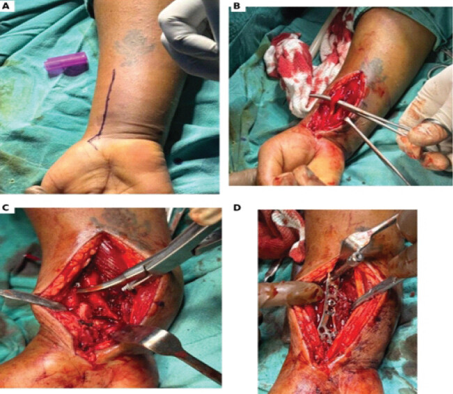
Surgery was performed with the patient supine and the involved limb placed on a radiolucent hand table under regional or general anesthesia. A pneumatic tourniquet was applied to the upper arm. A longitudinal volar incision was made along the FCR tendon and extended distally toward the wrist. The FCR sheath was incised, the FCR tendon was retracted ulnarly and the palmar cutaneous branch of the median nerve was protected. The flexor pollicis longus was retracted ulnarly to expose the pronator quadratus, which was released in an inverted L-shaped fashion while preserving a radial cuff for repair.
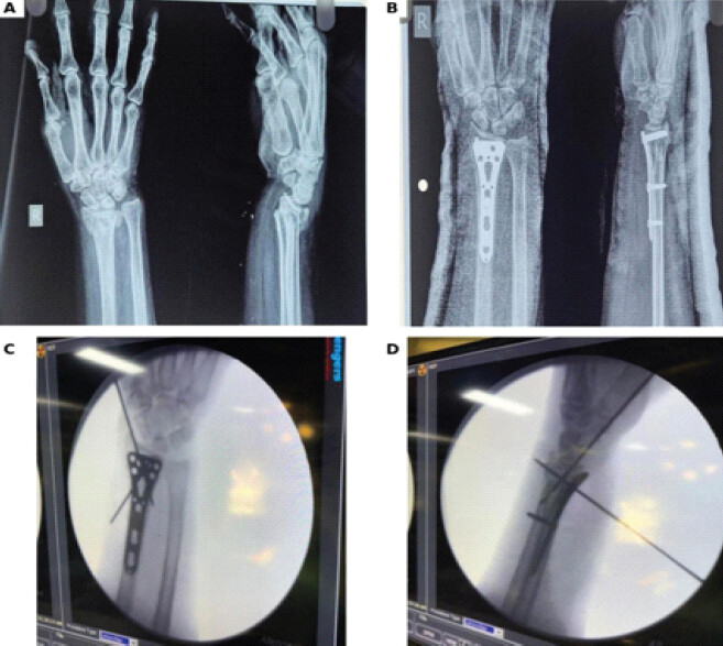
The approach was extended by releasing the radial septum and the brachioradialis insertion, followed by subperiosteal elevation of the radial and dorsal periosteum. This allowed controlled pronation of the proximal radial fragment and improved visualization of the dorsal and radial aspects of the distal radius and the volar ulnar corner. Reduction was achieved using manual manipulation, K-wires and plate-assisted techniques as required. Fixation was performed with a volar locking plate under fluoroscopic guidance. The pronator quadratus was repaired over the plate wherever feasible, the wound was closed in layers and a below-elbow volar splint was applied (Fig. 1).
Post-operative protocol and outcome assessment
Sutures were removed between the 10th and 14th post-operative day. Gradual wrist mobilization exercises were initiated after splint removal, and physiotherapy was advised as required. Operative time was recorded from skin incision to final closure. Intraoperative blood loss was estimated from suction bottle volume and blood absorbed in gauze.
Functional outcome was assessed using the disabilities of the arm, shoulder, and hand (DASH) score at 2 weeks, 4 weeks, 6 weeks, 3 months, and 6 months postoperatively. Wrist and forearm range of motion, including flexion, extension, supination, and pronation, was measured at follow-up using a manual goniometer [16]. Radiographs were independently assessed by two observers using standardized measurement techniques for volar tilt, radial inclination, radial length, and ulnar variance. Each observer performed the measurements independently, and the mean of the two measurements was used for the final analysis. Complications such as surgical site infection, hardware irritation, tenosynovitis, tendon rupture, neurovascular complication, implant failure, delayed union, non-union, and need for secondary procedure were recorded.
Statistical analysis
Categorical variables were summarized as frequencies and percentages. Continuous variables were expressed as mean ± standard deviation. Pre-operative and post-operative continuous variables were compared using t-tests as appropriate, and P < 0.05 was considered statistically significant.
Ethical considerations
Institutional ethical approval by Peoples University, Bhopal was obtained before commencement of the study (Ethics code no. IEC- 2024/59) on March 26, 2024. Written informed consent was obtained from all participants, and patient confidentiality was maintained.
Results
Thirty patients with closed intra-articular distal radius fractures were treated with eFCR exposure and volar locking plate fixation. All patients completed 6 months of follow-up (Table 1).
Baseline demographic, injury, and operative characteristics (n=30)
| Characteristic | Category | Value |
|---|---|---|
| Age, mean±SD | 45.83±14.99 | years |
| Age group | 18–30 years | 3 (10.0%) |
| 31–40 years | 10 (33.3%) | |
| 41–50 years | 9 (30.0%) | |
| 51–60 years | 3 (10.0%) | |
| >60 years | 5 (16.7%) | |
| Sex | Male | 12 (40.0%) |
| Female | 18 (60.0%) | |
| Side involved | Right | 14 (46.7%) |
| Left | 16 (53.3%) | |
| Mechanism of injury | Road traffic accident | 12 (40.0%) |
| Fall on outstretched hand | 10 (33.3%) | |
| Fall from stairs | 3 (10.0%) | |
| Slip and fall | 3 (10.0%) | |
| Fall from height | 2 (6.7%) | |
| AO/OTA | Type B | 8 (26.7%) |
| fracture type | Type C | 22 (73.3%) |
| AO/OTA subtype | 2R3B2/2R3B3 | 4 (13.3%)/4 (13.3%) |
| 2R3C1/2R3C2 / 2R3C3 | 4 (13.3%)/11 (36.7%)/7 (23.3%) | |
| Operative time | Mean±SD | 89.23±11.01 min |
| Intraoperative blood loss | Mean±SD | 96.33±11.89 mL |
Continuous variables are presented as mean ± SD; categorical variables are presented as number (%)
The 31–40 year age group represented the largest subgroup. AO/OTA type C fractures predominated, accounting for nearly three-fourths of cases. Road traffic accident was the most frequent mechanism of injury, followed by fall on an outstretched hand (Fig. 2).
Functional outcome
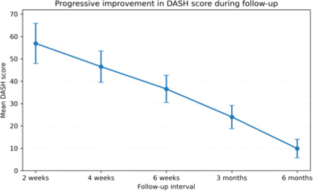
DASH scores improved progressively over the entire follow-up period. The mean DASH score decreased from 56.92 ± 8.98 at 2 weeks to 46.51 ± 6.98 at 4 weeks, 36.55 ± 6.09 at 6 weeks, 24.03 ± 5.18 at 3 months, and 9.92 ± 4.20 at 6 months. The improvement from 2 weeks to 6 months was statistically significant (P < 0.001) (Fig. 3).
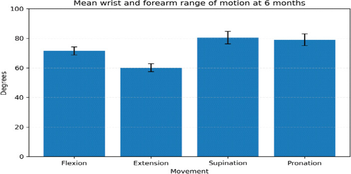
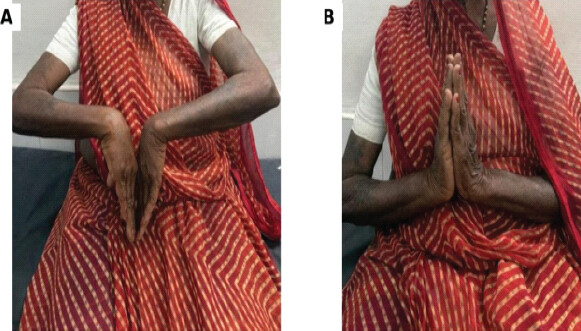
At 6 months, the mean flexion was 71.50 ± 2.70°, extension was 60.10 ± 2.77°, supination was 80.47 ± 4.31°, and pronation was 79.03 ± 3.94°. Functional movement thresholds were achieved by most patients, including flexion of at least 70° in 23 patients (76.7%) and extension of at least 60° in 18 patients (60.0%) (Fig. 4, 5 and Table 2).
Final clinical outcome, union, and complication profile (n=30)
| Endpoint | Value (%) |
|---|---|
| DASH score at 6 months | 9.92±4.20 |
| Flexion >70° | 23 (76.7) |
| Extension >60° | 18 (60.0) |
| Supination >75° | 27 (90.0) |
| Pronation >75° | 26 (86.7) |
| Clinical and radiological union | 30 (100.0) |
| No post-operative complication | 28 (93.3) |
| Hardware irritation | 1 (3.3) |
| Transient tenosynovitis | 1 (3.3) |
| Surgical site infection | 0 (0.0) |
| Tendon rupture/neurovascular injury/implant failure/non-union | 0 (0.0) |
DASH: Disabilities of the arm, shoulder, and hand
Radiological outcome
Radiological assessment demonstrated significant restoration of distal radial alignment following fixation. Mean pre-operative volar tilt improved from −15.90 ± 3.48° to 9.81 ± 0.59° at 6 months (P < 0.001). Radial inclination increased from 13.12 ± 2.24° preoperatively to 24.74 ± 1.20° at 6 months (P = 0.007), while radial length increased from 6.18 ± 1.25 mm to 12.06 ± 1.31 mm (P = 0.001). Ulnar variance improved from +4.04 ± 1.07 mm preoperatively to 0.65 ± 0.58 mm at 6 months (P = 0.0001). The progressive post-operative measurements also demonstrated maintenance of alignment during follow-up (Table 3).
Comparison of pre-operative and post-operative radiological parameters
| Parameter | Pre-operative mean±SD | Post-operative mean±SD | Mean change | P-value |
|---|---|---|---|---|
| Volar tilt (degrees) | –15.90±3.48 | 9.81±0.59 | +25.72° | <0.001 |
| Radial inclination (degrees) | 13.12±2.24 | 24.74±1.20 | +11.62° | <0.001 |
| Radial length (mm) | 6.18±1.25 | 12.06±1.31 | +5.88 mm | <0.001 |
| Ulnar variance (mm) | +4.04±1.07 | +0.65±0.58 | –3.39 mm | <0.001 |
Radiological parameters showed significant correction after surgery. Volar tilt, radial inclination, radial length, and ulnar variance all improved significantly from pre-operative, indicating restoration of distal radial alignment and wrist biomechanics.
Discussion
This prospective observational study found that fixation of closed intra-articular distal radius fractures through the eFCR approach was associated with substantial short-term functional recovery, restoration and maintenance of measured radiological alignment, reliable union, and a low observed rate of early complications at 6 months.
The demographic profile was consistent with the known epidemiology of distal radius fractures. Court-Brown and Caesar described distal radius fractures as common injuries across a broad age spectrum, while Chung and Spilson also emphasized their frequent occurrence among hand and forearm fractures [1,2]. In the current cohort, the mean age was 45.83 ± 14.99 years and both road traffic accidents and falls were important mechanisms, reflecting the typical overlap of higher-energy injuries in younger adults and fall-related injuries in older patients. This injury distribution is clinically relevant because high-energy intra-articular fractures and osteoporotic metaphyseal collapse both require careful restoration of distal radial geometry to avoid persistent deformity and disability.
The cohort included both AO/OTA type B and type C intra-articular fractures, with type C fractures predominating. Fracture morphology and articular complexity may independently influence reduction difficulty and functional recovery. Knirk and Jupiter demonstrated that residual articular incongruity after intra-articular distal radius fractures is associated with poor long-term outcome, and Mackenney et al. identified instability-related features that can lead to loss of reduction if anatomical alignment is not adequately restored [4,5]. In this context, the eFCR approach is particularly useful because it improves access to the radial column, dorsal metaphysis, and volar ulnar corner, all of which are difficult to control in comminuted articular fractures.
The surgical rationale for the eFCR approach is supported by the principles of volar fixed-angle fixation described by Orbay and Fernandez, who highlighted the value of stable volar constructs for unstable distal radius fractures [6,11]. In comparison with the conventional volar Henry approach, Akgun et al. reported that the extended FCR approach provides improved exposure while maintaining comparable functional and radiological outcomes [14]. Alrefai et al. similarly reported favorable clinical outcomes and a low complication profile following fixation through the extended FCR approach [15]. The present findings are in agreement with these studies, particularly because satisfactory reduction and recovery were achieved despite the high proportion of complete intra-articular fractures.
Functional improvement was substantial. The mean DASH score improved from 56.92 ± 8.98 at 2 weeks to 9.92 ± 4.20 at 6 months, indicating minimal residual disability at final follow-up. This is comparable with Alrefai et al., who reported excellent functional recovery after eFCR fixation, with a final DASH score in the low-disability range [15]. It is also consistent with volar locking plate studies such as Arora et al., where operative fixation of displaced and unstable distal radius fractures produced favorable functional recovery compared with non-operative management in elderly patients [17]. The progressive decline in DASH score across follow-up suggests that stable fixation permitted gradual rehabilitation and return to daily activities.
The range-of-motion outcomes also compare favorably with published eFCR and volar plating results. At 6 months, mean wrist flexion was 71.50 ± 2.70°, extension was 60.10 ± 2.77°, supination was 80.47 ± 4.31°, and pronation was 79.03 ± 3.94°. These values closely resemble the post-operative motion reported by Alrefai et al., who described satisfactory wrist flexion, extension, and forearm rotation after eFCR fixation [15]. The near-normal supination and pronation are especially important because distal radioulnar joint mechanics are sensitive to restoration of radial length and ulnar variance, as emphasized in biomechanical studies by Palmer and Werner and by Short et al [18,19].
Radiological correction was statistically significant for every measured parameter. Volar tilt corrected from dorsal angulation to positive volar tilt, radial inclination, and radial length increased, and positive ulnar variance was reduced. These radiological improvements are consistent with Akgun et al., who reported satisfactory volar tilt, radial inclination, radial height, and ulnar variance after fixation through the extended FCR approach [14]. They also support Medoff’s emphasis on accurate radiographic evaluation and restoration of distal radius alignment as a foundation for successful management [3]. The maintained correction in this cohort suggests that the approach allowed adequate fragment mobilization and stable plate placement.
The importance of this radiological restoration is reinforced by biomechanical evidence. Short et al. showed that distal radius deformity alters wrist mechanics, while Palmer and Werner demonstrated the importance of the distal radioulnar joint and ulnar-sided load transmission in forearm rotation [18,19]. The correction of ulnar variance and radial length in this cohort therefore provides a plausible explanation for the favorable rotational arc observed at final follow-up. Similarly, Harness et al. reported that dorsal comminution can influence functional outcome after distal radius fixation, supporting the value of an approach that improves dorsal metaphyseal and radial column visualization without requiring routine dorsal exposure [12].
The intraoperative profile was acceptable for complex intra-articular fracture fixation. Although an extensile volar exposure may theoretically increase operative morbidity, operative duration and blood loss remained within a practical range. This aligns with the experience reported by Akgun et al., who found that improved exposure with the extended FCR approach did not translate into excessive surgical morbidity [14]. The finding also complements the concept of volar fixation described by Orbay and Fernandez, in which stable fixation can be achieved through relatively soft-tissue-friendly volar dissection [11].
The safety profile was favorable. Although only two minor complications were observed during the 6-month follow-up, the small sample size and limited follow-up may have resulted in under-detection of uncommon or delayed complications, including flexor tendon irritation or rupture, median nerve symptoms, symptomatic hardware, and subsequent implant removal. The absence of these complications in this series should therefore not be interpreted as evidence that they cannot occur. This low complication rate is comparable with the favorable outcomes reported by Alrefai et al. for the extended FCR approach [15]. It also compares favorably with concerns associated with dorsal plating, where Ruch and Papadonikolakis discussed tendon-related complications in intra-articular distal radius fracture fixation [10]. Arora et al. have also described implant-related complications after palmar locking-plate fixation, highlighting the need for careful plate placement and post-operative surveillance [20].When viewed against older treatment modalities, the current results support the role of eFCR-based volar plating in selected complex fractures. External fixation based on ligamentotaxis can help restore radial length, but Agee noted that it has limitations related to fragment control and technique-dependent reduction [8]. In contrast, the eFCR approach allows direct visualization and manipulation of important fragments while preserving the advantages of volar fixation. Compared with dorsal exposure, it avoids routine dorsal soft-tissue dissection and may reduce extensor tendon irritation while still permitting reduction of dorsal and radial fragments through an extensile volar window [10,14].
Overall, the findings indicate that the eFCR approach balances extensile exposure with soft-tissue preservation. The results are consistent with comparative and clinical literature showing improved visualization without compromising recovery or increasing complications [14,15]. In selected intra-articular distal radius fractures, AO/OTA type C and type B patterns, the approach facilitates anatomical reduction, stable fixation, early functional recovery, and reliable short-term radiological maintenance.
The present study was not designed to compare eFCR with the conventional volar Henry approach. Therefore, the observed functional and radiological improvements cannot be attributed exclusively to the eFCR approach or interpreted as evidence of superiority. The principal potential advantage of eFCR in the present context is its extensile exposure, particularly for visualization and manipulation of the radial column, dorsal metaphysis, and volar ulnar corner in selected complex fracture patterns.
Limitations
The relatively small sample size of 30 patients and single-center setting may limit statistical power and generalizability. The prospective, non-randomized, single-arm design may be subject to selection bias and confounding and does not permit causal or superiority comparisons with the conventional volar Henry approach. Although both AO/OTA type B and C fractures were included, the sample was not sufficiently large for reliable subgroup analysis according to fracture subtype or specific articular involvement. DASH and wrist/forearm range of motion were assessed serially, but PRWE, grip strength, patient satisfaction, return-to-work status, and standardized contralateral wrist measurements were not prospectively recorded. Early DASH improvement therefore reflects overall post-operative recovery and cannot be attributed specifically to the eFCR approach. Intraoperative blood loss was estimated from suction volume and gauze absorption, while manual goniometry may be subject to measurement variability. Finally, the 6-month follow-up primarily evaluates short-term outcomes and cannot adequately assess late complications or long-term functional deterioration. Nevertheless, the prospective design, predefined eligibility criteria, consecutive recruitment, quantitative radiological assessment by two observers, and serial functional follow-up provide a structured assessment of early outcomes. Larger multicenter comparative studies with longer follow-up and comprehensive functional assessment are needed.
Conclusion
In this prospective observational series of closed intra-articular distal radius fractures, fixation through the eFCR approach was associated with favorable short-term functional and radiological outcomes, reliable union, and a low rate of early complications at 6 months. The extensile exposure may be particularly useful when visualization and manipulation of the radial column, dorsal metaphysis, and volar ulnar corner and facilitates reduction of complex fragments, restores key radiological parameters, and supports functional recovery. At 6 months, patients achieved significant DASH improvement, satisfactory motion, and few minor complications. The eFCR approach may be considered a useful extensile volar exposure for selected intra-articular distal radius fractures. Comparative studies are required to determine whether its use provides advantages over the conventional volar Henry approach.
Clinical Message
The extended flexor carpi radialis (eFCR) approach provides an extensile volar exposure that facilitates visualization and reduction of complex intra-articular distal radius fractures, with favorable short-term functional and radiological outcomes following volar locking plate fixation.
Conflict of Interest:
Nil
Source of Support:
Nil
Consent:
The authors confirm that informed consent was obtained from the patient for publication of this article
How to Cite this Article
Gupta N, Chatterji G, Mahajan AV, Mehrotra R, Ashwinbhai GY, Sahu P. Evaluation of Functional Outcome of Extended Flexor Carpi Radialis Approach for Fracture Fixation of Distal End Radius Fracture. Journal of Orthopaedic Case Reports 2026 October;16(10): 500-507.
References
- Court-Brown CM, Caesar B. Epidemiology of adult fractures: A review. Injury 2006;37:691-7. [Google Scholar] | [PubMed]
- Chung KC, Spilson SV. The frequency and epidemiology of hand and forearm fractures in the United States. J Hand Surg Am 2001;26:908-15. [Google Scholar] | [PubMed]
- Medoff RJ. Essential radiographic evaluation for distal radius fractures. Hand Clin 2005;21:279-88. [Google Scholar] | [PubMed]
- Knirk JL, Jupiter JB. Intra-articular fractures of the distal end of the radius in young adults. J Bone Joint Surg Am 1986;68:647-59. [Google Scholar] | [PubMed]
- Mackenney PJ, McQueen MM, Elton R. Prediction of instability in distal radial fractures. J Bone Joint Surg Am 2006;88:1944-51. [Google Scholar] | [PubMed]
- Orbay JL. The treatment of unstable distal radius fractures with volar fixation. Hand Surg 2000;5:103-12. [Google Scholar] | [PubMed]
- Fernandez DL, Jupiter JB. Fractures of the Distal Radius: A Practical Approach to Management. Berlin: Springer; 1996. [Google Scholar] | [PubMed]
- Agee JM. External fixation: Technical advances based upon multiplanar ligamentotaxis. Orthop Clin North Am 1993;24:265-74. [Google Scholar] | [PubMed]
- Cooney WP. Fractures of the distal radius: Current concepts. J Hand Surg Am 1989;14:342-9. [Google Scholar] | [PubMed]
- Ruch DS, Papadonikolakis A. Volar versus dorsal plating in the management of intra-articular distal radius fractures. J Hand Surg Am 2006;31:9-16. [Google Scholar] | [PubMed]
- Orbay JL, Fernandez DL. Volar fixed-angle plate fixation for unstable distal radius fractures. J Hand Surg Am 2002;27:205-15. [Google Scholar] | [PubMed]
- Harness NG, Ring D, Zurakowski D, Harris GJ, Jupiter JB. The influence of dorsal comminution on functional outcome after distal radius fracture fixation. J Hand Surg Am 2006;31:1453-9. [Google Scholar] | [PubMed]
- Ring D, Jupiter JB. Treatment of osteoporotic distal radius fractures. Osteoporos Int 2005;16 Suppl 2:S80-4. [Google Scholar] | [PubMed]
- Akgun U, Canbek U. Comparison of extended FCR approach and standard volar Henry approach in distal radius fractures. Acta Orthop Traumatol Turc 2018;52:1-6. [Google Scholar] | [PubMed]
- Alrefai S, Hsu D, Persitz J, Paul R. Clinical outcomes of extended flexor carpi radialis approach in distal radius fractures. Eur J Orthop Surg Traumatol 2020;30:1045-52. [Google Scholar] | [PubMed]
- Fan S, Cepek J, Symonette C, Ross D, Chinchalkar S, Grant A. Variation of grip strength and wrist range of motion with forearm rotation in healthy young volunteers aged 23 to 30. J Hand Microsurg 2019;11:88-93. [Google Scholar] | [PubMed]
- Arora R, Lutz M, Deml C, Krappinger D, Haug L, Gabl M. A prospective randomized trial comparing nonoperative treatment with volar locking plate fixation for displaced and unstable distal radial fractures in patients sixty-five years of age and older. J Bone Joint Surg Am 2011;93:2146-53. [Google Scholar] | [PubMed]
- Palmer AK, Werner FW. Biomechanics of the distal radioulnar joint. Clin Orthop Relat Res 1984;187:26-35. [Google Scholar] | [PubMed]
- Short WH, Palmer AK, Werner FW, Murphy DJ. A biomechanical study of distal radial fractures. J Hand Surg Am 1987;12:529-34. [Google Scholar] | [PubMed]
- Arora R, Lutz M, Hennerbichler A, Krappinger D, Espen D, Gabl M. Complications following internal fixation of unstable distal radius fracture with a palmar locking-plate. J Orthop Trauma 2007;21:316-22. [Google Scholar] | [PubMed]
© 2026 Journal of Orthopaedic Case Reports - Published by Indian Orthopaedic Research Group
About the Authors
-

Nilesh Gupta
-

Gautam Chatterji
-

Ajinkya Vinod Mahajan
-

Ravi Mehrotra
-

Gohel Yuvraj Ashwinbhai
-

Prateek Sahu
Related Articles in Journal of Orthopaedic Case Reports
 August 1, 2025 A Case Series of Functional and Radiological Outcome of Comminuted Distal Radius Fractures Treated with Bridging External Fixator with Optional Percutaneous K-wires
 May 1, 2025 Functional Outcome of Distal Radius Fractures Managed by Minimally Invasive Plate Osteosynthesis: A Prospective Study of 20 Patients
 October 1, 2024 A Study of Functional Outcome and Assessment of Role of Proximal Humerus Internal Locking System (Philos) Plating in Elderly Population with Proximal Humerus Fracture: A Case Series
 March 1, 2026 Pull Out Suture Technique for Fixing Coronoid Fractures using a Single Lateral Extensor Digitorum Communis Split Approach and Fiberwire Endobutton Fixation by Retrograde Drilling – A Case Series