


Introduction
The knee is one of the most commonly injured joints and is particularly susceptible to traumatic and sports-related injuries. Accurate identification of intra-articular pathology is essential for appropriate treatment planning and optimal clinical outcomes. Arthroscopy was initially developed and extensively applied in the knee, and many of its fundamental principles and techniques were later adopted for other joints. Although early knee arthroscopy developed in Japan and Europe, the technique later gained widespread acceptance in India and has evolved considerably over the past several decades [1,2].
Clinical examination remains fundamental in the evaluation of traumatic knee injuries; however, imaging plays an important role in establishing the diagnosis and determining the extent of structural damage [3]. Magnetic resonance imaging (MRI) has become an important non-invasive modality for evaluating traumatic knee injuries. MRI can accurately demonstrate abnormalities involving intra-articular structures, particularly the menisci and cruciate ligaments, and has been shown to influence diagnostic and therapeutic decision-making in patients with acute knee injuries [4,5,6].
Several imaging modalities are available for the evaluation of knee pathology. Conventional radiography is primarily used for the initial assessment of osseous injuries, whereas computed tomography provides detailed evaluation of complex fractures. MRI offers superior soft-tissue contrast and is particularly useful for assessing intra-articular structures such as the menisci and cruciate ligaments [7].
MRI has particular advantages in evaluating the knee due to its excellent soft-tissue contrast and multiplanar imaging capability. These characteristics allow detailed visualization of the menisci, cruciate and collateral ligaments, articular cartilage, bone marrow, and other periarticular structures [8,9]. The ability to assess multiple anatomical structures in different planes makes MRI particularly valuable in patients with suspected knee internal derangement.
However, diagnostic performance may vary according to the type, severity, chronicity, and complexity of the injury, and discrepancies between MRI findings and arthroscopic findings may occur, particularly in subtle or complex lesions.
Multiple associated injuries may further complicate interpretation and cause discrepancies between MRI findings and intra-articular pathology identified at surgery [2,3,4,5,6]. Therefore, MRI findings should be interpreted alongside clinical examination and the overall clinical presentation.
Knee arthroscopy provides direct visualization of intra-articular structures and has an important role as both a diagnostic and therapeutic modality in patients with internal derangement of the knee [10,11]. It permits direct assessment of the menisci, anterior cruciate ligament (ACL), posterior cruciate ligament (PCL), articular cartilage, and other intra-articular structures while allowing simultaneous treatment of appropriate lesions. However, arthroscopy is invasive and primarily provides direct assessment of intra-articular pathology. In contrast, MRI provides a non-invasive assessment of both intra-articular and extra-articular structures, while clinical examination remains complementary in the assessment of ligamentous and meniscal injuries.
The present study aimed to evaluate the diagnostic accuracy of pre-operative MRI for detecting selected traumatic intra-articular knee injuries by comparing MRI findings with arthroscopic findings in patients undergoing therapeutic knee arthroscopy. Specifically, the study assessed the diagnostic performance of MRI for ACL injury, PCL injury, medial meniscal injury, lateral meniscal injury, and osteochondral defects using arthroscopic findings as the reference standard.
Materials and Methods
Study design and setting
This was a single-center diagnostic accuracy study conducted at Shri Balaji Institute of Medical Sciences, Raipur, a tertiary referral institution in Central India. The study was approved by the Institutional Ethics Committee (IEC) (approval number: SBIMS/IEC/Certi./171/2025, dated September 16, 2025) and was conducted in accordance with the ethical principles of the Declaration of Helsinki.
Study population
Medical records of consecutive patients who underwent knee arthroscopy for traumatic internal derangement between January 2024 and December 2025 were retrospectively reviewed. The study population consisted of patients with traumatic knee injuries who had persistent symptoms or clinical findings suggestive of internal derangement and underwent therapeutic knee arthroscopy. As only patients undergoing arthroscopy were included, the cohort represented a selected population with a relatively high pretest probability of intra-articular pathology.
Inclusion and exclusion criteria
We included patients aged 15–60 years who presented with traumatic knee injuries, had clinical features suggestive of internal derangement, underwent pre-operative MRI, and subsequently underwent therapeutic knee arthroscopy. Only patients with complete documentation of pre-operative MRI findings and corresponding arthroscopic findings were included in the final diagnostic accuracy analysis.
We excluded patients with concomitant fractures involving the femur, tibia, or patella; open knee injuries; non-traumatic internal derangement; inadequate or unavailable MRI evaluation; or incomplete arthroscopic documentation.
Clinical and MRI evaluation
All patients underwent a detailed clinical history and physical examination. Clinical assessment included McMurray, Lachman, anterior drawer, posterior drawer, and varus-valgus stress tests, as clinically indicated. Anteroposterior and lateral radiographs of the affected knee were obtained to exclude associated fractures.
Clinical examination findings were recorded as part of the routine pre-operative assessment and were presented descriptively. Individual clinical tests were not independently subjected to diagnostic accuracy analysis or directly compared with MRI because the present study was primarily designed to evaluate MRI findings against arthroscopic findings.
MRI was generally performed after the acute phase of injury, typically at least 3 weeks after trauma. MRI examinations were performed using either institutional or external imaging facilities. Institutional MRI examinations were performed using a 1.5-T system with standard knee protocols, including multiplanar T1-weighted and fluid-sensitive fat-suppressed sequences. External MRI examinations were performed using 1.5-T or 3.0-T systems according to the protocols of the respective imaging centres.
MRI assessment included sagittal, coronal, and axial images for evaluation of the menisci, cruciate ligaments, articular cartilage, and osteochondral abnormalities. For the primary diagnostic accuracy analysis, MRI findings were obtained from the original formal radiological reports generated before arthroscopy. No post hoc reinterpretation of MRI images with knowledge of arthroscopic findings was performed for the primary analysis. When MRI examinations had been performed externally, the available radiological reports and imaging records were reviewed retrospectively. The predefined diagnostic categories were ACL injury, PCL injury, medial meniscal injury, lateral meniscal injury, and osteochondral defects.
Independent blinded re-evaluation of the MRI examinations by a separate radiologist was not performed due to the retrospective study design. Therefore, the primary analysis relied on the original clinical radiological reports available before arthroscopy.
MRI examinations were interpreted by the radiologists at the respective imaging facilities as part of routine clinical care, and the original reports available before arthroscopy were used for the primary analysis.
Arthroscopic evaluation
All patients underwent therapeutic knee arthroscopy under spinal anesthesia as part of routine clinical management. A systematic arthroscopic examination was performed using standard portals, with additional portals created when required. The medial and lateral menisci, ACL, PCL, articular cartilage, and other relevant intra-articular structures were systematically evaluated. The presence, absence, location, and characteristics of intra-articular pathology were documented at the time of surgery, and appropriate therapeutic procedures were performed as indicated. Arthroscopic findings were subsequently used as the reference standard for comparison with the pre-operative MRI findings for the evaluated intra-articular lesions.
MRI–arthroscopy correlation
We retrospectively compared pre-operative MRI findings with the corresponding arthroscopic findings for each predefined diagnostic category, including ACL injury, PCL injury, medial meniscal injury, lateral meniscal injury, and osteochondral defects. For each lesion, we classified MRI findings as true-positive, true-negative, false-positive, or false-negative, using arthroscopic findings as the reference standard.
We assessed MRI diagnostic performance by calculating sensitivity, specificity, positive predictive value (PPV), negative predictive value (NPV), and overall diagnostic accuracy. Agreement between MRI and arthroscopic findings was assessed using Cohen’s kappa (κ) statistic.
Definition of diagnostic categories
For each predefined diagnostic category, MRI and arthroscopic findings were classified as either positive or negative. ACL and PCL injuries were considered present when a ligament tear or injury was documented in the corresponding MRI report or arthroscopic record. Medial and lateral meniscal injuries were considered present when a meniscal tear was documented. Osteochondral defects were considered present when a focal osteochondral lesion was documented. Arthroscopic findings served as the reference standard for classification of each evaluated lesion.
Statistical analysis
Data were analysed using IBM Statistical Package for the Social Sciences Statistics version 23.0 (IBM Corp., Armonk, NY, USA). Continuous variables were expressed as mean ± standard deviation or median with range, as appropriate, whereas categorical variables were expressed as frequencies and percentages.
MRI diagnostic performance was assessed using 2 × 2 contingency tables, with arthroscopic findings as the reference standard. For each predefined diagnostic category, sensitivity, specificity, PPV, NPV, and overall diagnostic accuracy were calculated. Agreement between MRI and arthroscopic findings was assessed using Cohen’s kappa (κ) statistic. Where appropriate, 95% confidence intervals were calculated for diagnostic performance estimates.
Results
A total of 35 patients with traumatic knee injuries were included. The cohort comprised 28 males (80.0%) and seven females (20.0%), with an age range of 15–60 years. The most represented age group was 15–20 years (31.4%), followed by 21–30 years (28.6%), 31–40 years (25.7%), and 41–50 years (14.3%). Sports-related trauma was the predominant mechanism of injury (54.3%), followed by falls (42.9%). The right knee was involved in 21 patients (60.0%) and the left knee in 14 (40.0%). No patients were aged 51–60 years (Table 1).
Demographic and clinical characteristics of the study population
| Characteristic | n (%) |
|---|---|
| Sex | |
| Male | 28 (80.0) |
| Female | 7 (20.0) |
| Age group (years) | |
| 15–20 | 11 (31.4) |
| 21–30 | 10 (28.5) |
| 31–40 | 9 (25.7) |
| 41–50 | 5 (14.2) |
| 51–60 | 0 (0.0) |
| Mechanism of injury | |
| Sports-related | 19 (54.3) |
| Fall | 15 (42.9) |
| Other | 1 (2.9) |
| Side involved | |
| Right | 21 (60) |
| Left | 14 (40) |
Distribution of knee injuries
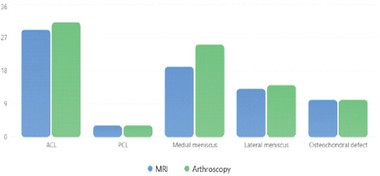
Clinical examination identified ACL injury in 29 patients (82.9%), PCL injury in three (8.6%), medial meniscal injury in 19 (54.3%), lateral meniscal injury in 10 (28.6%), and osteochondral defects in nine (25.7%). MRI identified ACL injury in 29 patients (82.9%), PCL injury in three (8.6%), medial meniscal injury in 19 (54.3%), lateral meniscal injury in 13 (37.1%), and osteochondral defects in 10 (28.6%). Arthroscopy identified 31 ACL injuries (88.6%), three PCL injuries (8.6%), 25 medial meniscal injuries (71.4%), 14 lateral meniscal injuries (40.0%), and 10 osteochondral defects (28.6%) (Table 2).
Distribution of injuries identified by clinical examination, MRI, and arthroscopy
| Injury | Clinical examination, n (%) | MRI, n (%) | Arthroscopy, n (%) |
|---|---|---|---|
| ACL | 29 (82.9) | 29 (82.9) | 31 (88.6) |
| PCL | 3 (8.6) | 3 (8.6) | 3 (8.6) |
| Medial meniscus | 19 (54.3) | 19 (54.3) | 25 (71.4) |
| Lateral meniscus | 10 (28.6) | 13 (37.1) | 14 (40.0) |
| Osteochondral defect | 9 (25.7) | 10 (28.6) | 10 (28.6) |
ACL: Anterior cruciate ligament, PCL: Posterior cruciate ligament, MRI: Magnetic resonance imaging
Clinical examination findings are presented descriptively in Table 2. Because the study was designed primarily to assess MRI findings against arthroscopic findings, diagnostic accuracy measures were not calculated separately for individual clinical examination tests.
The distribution of traumatic intra-articular knee injuries identified by MRI and arthroscopy is shown in Fig. 1. ACL injury was the most frequently identified lesion on both MRI and arthroscopy, followed by medial meniscal injury.
MRI versus arthroscopy
MRI findings were compared with arthroscopic findings for each predefined diagnostic category, with arthroscopy serving as the reference standard. Table 3 summarizes the numbers of true-positive, true-negative, false-positive, and false-negative MRI findings.
MRI findings compared with arthroscopic findings
| Injury | True positive | True negative | False positive | False negative |
|---|---|---|---|---|
| ACL | 29 | 4 | 0 | 2 |
| PCL | 3 | 32 | 0 | 0 |
| Medial meniscus | 19 | 10 | 0 | 6 |
| Lateral meniscus | 13 | 21 | 0 | 1 |
| Osteochondral defect | 10 | 25 | 0 | 0 |
ACL: Anterior cruciate ligament, PCL: Posterior cruciate ligament, MRI: Magnetic resonance imaging
Diagnostic performance of MRI
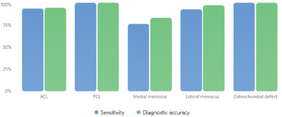
MRI demonstrated high diagnostic performance across the evaluated diagnostic categories compared with arthroscopy as the reference standard. Diagnostic accuracy ranged from 82.86% for medial meniscal injuries to 100.00% for PCL injuries and osteochondral defects (Table 4). The perfect diagnostic estimates for PCL injuries and osteochondral defects should be interpreted cautiously due to the small number of positive cases in these subgroups.
Diagnostic performance of MRI compared with arthroscopy
| Injury | Sensitivity (%) | Specificity (%) | PPV (%) | NPV (%) | Accuracy (%) |
|---|---|---|---|---|---|
| ACL | 93.55 | 100 | 100 | 66.67 | 94.29 |
| PCL | 100 | 100 | 100 | 100 | 100 |
| Medial meniscus | 76 | 100 | 100 | 62.5 | 82.86 |
| Lateral meniscus | 92.86 | 100 | 100 | 95.45 | 97.14 |
| Osteochondral defect | 100 | 100 | 100 | 100 | 100 |
ACL: Anterior cruciate ligament, PCL: Posterior cruciate ligament, PPV: Positive predictive value, NPV: Negative predictive value, MRI: Magnetic resonance imaging
As shown in Fig. 2, sensitivity was highest for PCL injuries and osteochondral defects (100.0%), followed by ACL injuries (93.6%) and lateral meniscal injuries (92.9%). Sensitivity was lowest for medial meniscal injuries (76.0%). Diagnostic accuracy ranged from 82.9% for medial meniscal injuries to 100.0% for PCL injuries and osteochondral defects.
Agreement between MRI and arthroscopy
Agreement between MRI and arthroscopy was assessed using Cohen’s kappa statistic. Agreement was substantial for ACL injuries (κ = 0.768) and medial meniscal injuries (κ = 0.644), and almost perfect for PCL injuries (κ = 1.000), lateral meniscal injuries (κ = 0.940), and osteochondral defects (κ = 1.000) (Table 5). The perfect agreement observed for PCL injuries and osteochondral defects should be interpreted cautiously due to the limited number of positive cases, particularly for PCL injuries.
Agreement between MRI and arthroscopy for detection of knee joint injuries
| Injury | Cohen’s κ | Interpretation |
|---|---|---|
| ACL | 0.768 | Substantial |
| PCL | 1 | Almost perfect |
| Medial meniscus | 0.644 | Substantial |
| Lateral meniscus | 0.89 | Almost perfect |
| Osteochondral defect | 1 | Almost perfect |
κ: Cohen’s kappa coefficient. Interpretation based on the Landis-Koch classification: 0.61–0.80, substantial agreement; 0.81–1.00, almost-perfect agreement. MRI: Magnetic resonance imaging, ACL: Anterior cruciate ligament, PCL: Posterior cruciate ligament
Discussion
MRI has an established role in pre-operative evaluation of traumatic knee injuries because it provides non-invasive visualization of the menisci, cruciate ligaments, articular cartilage, and associated soft-tissue structures. Arthroscopy allows direct visualization of intra-articular pathology and simultaneous treatment. The clinical usefulness of MRI depends on how well its findings correspond with arthroscopic pathology and the clinical significance of discrepancies. In this study, MRI demonstrated particularly high sensitivity for ACL, PCL, lateral meniscal, and osteochondral pathology in this cohort. Medial meniscal injury represented the principal diagnostic limitation.
Earlier meta-analytic evidence similarly demonstrated high diagnostic accuracy for MRI in meniscal tears, with pooled sensitivities of 92% for medial and 80% for lateral meniscal tears [12]. Recent arthroscopy-correlated studies have reported comparable variability. El-Hagrasy et al. reported MRI sensitivity and specificity of 94.29% and 78.75%, respectively, for medial meniscal tears and 76.74% and 94.03% for lateral meniscal tears [13]. A 2024 Cureus study also showed strong diagnostic performance of MRI for traumatic meniscal injuries when arthroscopy was the reference standard [14]. Another recent study of isolated meniscal tears reported sensitivity of 90% and specificity of 83% for medial meniscal tears and sensitivity of 65% and specificity of 88% for lateral meniscal tears [15]. Variability among studies likely reflects differences in patient selection, tear morphology, concomitant injuries, imaging protocols, and interpretation criteria.
The lower sensitivity and NPV for medial meniscal injury suggest a negative MRI may not fully exclude a clinically relevant tear when suspicion remains high. The high specificity and PPV indicate that positive MRI findings corresponded closely with arthroscopic findings in this cohort. The lower NPV suggests some lesions may remain occult on imaging. Differences in tear morphology and location may contribute to false-negative MRI findings. Kim et al. showed MRI diagnostic performance varies by tear type and location, especially with associated ACL injury [16]. Contemporary artificial intelligence studies suggest automated systems are more reliable at detecting a meniscal tear than accurately determining its precise location [17].
MRI demonstrated 100% sensitivity, specificity, PPV, NPV, and diagnostic accuracy for PCL injuries in this cohort, with complete observed agreement with arthroscopy. However, this finding should be interpreted cautiously because only three PCL injuries were identified. These findings are consistent with contemporary arthroscopy-correlated literature. Zeb et al. reported a sensitivity of 98.57%, a specificity of 87.50%, and a diagnostic accuracy of 96.51% for MRI in ACL injury [18]. Yadav et al. similarly demonstrated MRI’s high diagnostic performance for ACL and PCL injuries compared with arthroscopy [19]. The two ACL injuries missed by MRI in this cohort demonstrate that false-negative MRI examinations can occur.
MRI and arthroscopy showed complete agreement for PCL injuries, with 100% sensitivity, specificity, PPV, NPV, and diagnostic accuracy. This finding is consistent with the high diagnostic performance of MRI for PCL injury reported in recent studies [19]. Nevertheless, these perfect diagnostic estimates should be interpreted cautiously due to the limited number of PCL injuries. Only three PCL injuries were identified, so these results should not be generalized to larger populations.
MRI also demonstrated complete agreement with arthroscopy for osteochondral defects, with 100% sensitivity, specificity, PPV, NPV, and diagnostic accuracy. However, these findings should be interpreted cautiously because only 10 osteochondral defects were identified, and lesions were not further characterised according to size, depth, grade, or anatomical location. A 2024 systematic review and meta-analysis found that quantitative and three-dimensional MRI techniques provide greater sensitivity for chondral lesions than conventional two-dimensional MRI [20]. Accordingly, the excellent agreement observed in this cohort should not be extrapolated to subtle superficial cartilage abnormalities or early degenerative changes.
Emerging three-dimensional MRI techniques and artificial intelligence-based approaches may further improve detection and characterization of subtle intra-articular abnormalities. However, variability in datasets, algorithms, reporting methods, and external validation currently limits their routine clinical application [17,21,22,23].
Clinical examination remains an important component of the evaluation of traumatic knee injuries. Although MRI provides detailed anatomical information, imaging findings should be interpreted in the context of the patient’s symptoms and physical examination. Previous studies have demonstrated the diagnostic value of structured clinical examination for meniscal and ligamentous injuries [24,25,26]. However, the present study was primarily designed to evaluate MRI–arthroscopy correlation and did not independently assess the diagnostic accuracy of individual clinical examination tests.
An important consideration in interpreting these findings is the selected nature of the study population. All included patients had sufficient clinical suspicion of internal derangement to undergo therapeutic arthroscopy, resulting in a cohort with a relatively high prevalence of intra-articular pathology. This may influence predictive values and limits direct extrapolation of the findings to patients with lower pretest probability or those managed non-operatively. Nevertheless, the use of arthroscopy as the reference standard allowed direct lesion-level comparison of pre-operative MRI findings with intra-articular pathology.
Limitations
This study has several limitations. The small sample size (n = 35), single-center retrospective design, and uneven distribution of lesions – particularly the small number of PCL injuries and osteochondral defects – limited statistical precision and generalizability. Only patients undergoing therapeutic arthroscopy were included, resulting in a selected cohort with a relatively high pretest probability of intra-articular pathology; therefore, the findings may not be applicable to patients with lower clinical suspicion or those managed non-operatively. MRI examinations were performed using 1.5-T and 3.0-T systems, and interpretation was based on routine radiological reports from different imaging facilities; standardized MRI protocols, independent blinded reinterpretation, and control of radiologist variability were not possible. Lesions were classified primarily as present or absent without detailed assessment of morphology, grade, size, or location, particularly for osteochondral defects. Arthroscopy, although used as the reference standard, may itself be influenced by lesion visualization, surgical technique, and surgeon experience. Clinical examination tests were not independently evaluated for diagnostic accuracy, and the analysis was limited to five predefined intra-articular lesion categories. Finally, the findings, particularly the apparently perfect diagnostic estimates for PCL injuries and osteochondral defects, require validation in larger prospective multicenter studies.
Conclusion
MRI demonstrated high diagnostic performance for most traumatic intra-articular knee lesions, with substantial to almost-perfect agreement with arthroscopy. Medial meniscal injuries showed comparatively lower sensitivity and NPV, indicating that a negative MRI does not completely exclude clinically relevant pathology when clinical suspicion remains high. MRI remains an important component of pre-operative evaluation, but its findings should be interpreted alongside the clinical history and physical examination, particularly when clinical and radiological findings are discordant. The retrospective, single-center design, small sample size, and limited number of certain lesions warrant cautious interpretation and validation in larger prospective multicenter studies.
Clinical Message
MRI demonstrated high diagnostic performance for most evaluated traumatic intra-articular knee injuries in this selected cohort. However, the lower sensitivity for medial meniscal injuries indicates that a negative MRI finding may not completely exclude clinically suspected medial meniscal pathology. MRI findings should therefore be interpreted in conjunction with the clinical examination and overall clinical presentation, particularly when clinical suspicion remains high.
Conflict of Interest:
Nil
Source of Support:
Nil
Consent:
The authors confirm that informed consent was obtained from the patient for publication of this article
How to Cite this Article
Qureshi A, Faatima N, Agrawal SS. Magnetic Resonance Imaging-Arthroscopy Correlation in Traumatic Knee Injuries: A Retrospective Diagnostic Accuracy Study. Journal of Orthopaedic Case Reports 2026 October;16(10): 440-447.
References
- Ishibashi Y, Yamamoto Y. The history of arthroscopyIn: Miller MD, Cole BJ, editors. Textbook of Arthroscopy. Philadelphia, PA: Saunders; 2004. 3-7. [Google Scholar] | [PubMed]
- Alioto RJ, Browne JE, Barnthouse CD, Scott AR. The influence of MRI on treatment decisions regarding knee injuries. Am J Knee Surg 1999;12:91-7. [Google Scholar] | [PubMed]
- Bui-Mansfield LT, Youngberg RA, Warme W, Pitcher JD, Nguyen PL. Potential cost savings of MR imaging obtained before arthroscopy of the knee: Evaluation of 50 consecutive patients. AJR Am J Roentgenol 1997;168:913-8. [Google Scholar] | [PubMed]
- Maurer EJ, Kaplan PA, Dussault RG, Diduch DR, Schuett A, McCue FC. Acutely injured knee: Effect of MR imaging on diagnostic and therapeutic decisions. Radiology 1997;204:799-805. [Google Scholar] | [PubMed]
- Ruwe PA, Wright J, Randall RL, Lynch JK, Jokl P, McCarthy S. Can MR imaging effectively replace diagnostic arthroscopy? Radiology 1992;183:335-9. [Google Scholar] | [PubMed]
- Vincken PW, Ter Braak BP, Van Erkell AR, De Rooy TP, Mallens WM, Post W. Effectiveness of MR imaging in selection of patients for arthroscopy of the knee. Radiology 2002;223:739-46. [Google Scholar] | [PubMed]
- Grey SD, Kaplan PA, Dussault RG. Imaging of the knee. Current status. Orthop Clin North Am 1997;28:643-58. [Google Scholar] | [PubMed]
- Kaplan PA, Walker CW, Kilcoyne RF, Brown DE, Tusek D, Dussault RG. Occult fracture patterns of the knee associated with anterior cruciate ligament tears: Assessment with MR imaging. Radiology 1992;183:835-8. [Google Scholar] | [PubMed]
- Kean DM, Worthington BS, Preston BJ, Roebuck EJ, McKim-Thomas H, Hawkes RC. Nuclear magnetic resonance imaging of the knee: Examples of normal anatomy and pathology. Br J Radiol 1983;56:355-64. [Google Scholar] | [PubMed]
- Boden SD, Labropoulos PA, Vailas JC. MR scanning of the acutely injured knee: Sensitive, but is it cost effective? Arthroscopy 1990;6:306-10. [Google Scholar] | [PubMed]
- Nguyen JC, Yaya-Quezada C, Lerebo WT, Patel VS, Chalian M, Flores DV. MRI diagnosis of meniscus tears in the knee: An updated systematic review and meta-analysis. Radiology 2026;319:e252288. [Google Scholar] | [PubMed]
- Wang W, Li Z, Peng HM, Bian YY, Li Y, Qian WW. Accuracy of MRI diagnosis of meniscal tears of the knee: A meta-analysis and systematic review. J Knee Surg 2021;34:121-9. [Google Scholar] | [PubMed]
- El-Hagrasy AM, Theckayil AJ, Khan MA, Khan HN, Butt AJ. Magnetic resonance imaging is an effective first-line noninvasive tool for meniscal tear detection: A retrospective comparative analysis with knee arthroscopy. Arthrosc Sports Med Rehabil 2025;7:101065. [Google Scholar] | [PubMed]
- Khalid D, Iqbal J, Mustafa K, Altaf R, Fatima R. Diagnostic accuracy of magnetic resonance imaging in the detection of meniscal injury in patients with knee trauma: Keeping arthroscopy as a gold standard. Cureus 2024;16:e72343. [Google Scholar] | [PubMed]
- Bottomley J, Al-Dadah O. Diagnostic accuracy of magnetic resonance imaging in meniscal tears. Cureus 2025;17:e92155. [Google Scholar] | [PubMed]
- Kim SH, Lee HJ, Jang YH, Chun KJ, Park YB. Diagnostic accuracy of magnetic resonance imaging in the detection of type and location of meniscus tears: Comparison with arthroscopic findings. J Clin Med 2021;10:606. [Google Scholar] | [PubMed]
- Zhao Y, Coppola A, Karamchandani U, Amiras D, Gupte CM. Correction: Artificial intelligence applied to magnetic resonance imaging reliably detects the presence, but not the location, of meniscus tears: A systematic review and meta-analysis. Eur Radiol 2024;34:5954-64. [Google Scholar] | [PubMed]
- Zeb J, Chaudary MI, Zeb M, Mersal M, Ahmad B, Alsonbaty M. Diagnostic accuracy of non-invasive tests versus arthroscopy in anterior cruciate ligament (ACL) injuries. Cureus 2024;16:e60925. [Google Scholar] | [PubMed]
- Yadav V, Chandrashekhar CK, Ram RP, Mohammed R. Precision of magnetic resonance imaging in detection of anterior cruciate ligament and posterior cruciate ligament ruptures and subsequent arthroscopic correlations: An investigation. Rev Bras Ortop (Sao Paulo) 2024;59:e707-11. [Google Scholar] | [PubMed]
- Chen X, You M, Liao K, Zhang M, Wang L, Zhou K. Quantitative magnetic resonance imaging had greater sensitivity in diagnosing chondral lesions of the knee: A systematic review and meta-analysis. Arthroscopy 2024;40:2760-73.e14. [Google Scholar] | [PubMed]
- Santomartino SM, Kung J, Yi PH. Systematic review of artificial intelligence development and evaluation for MRI diagnosis of knee ligament or meniscus tears. Skeletal Radiol 2024;53:445-54. [Google Scholar] | [PubMed]
- Botnari A, Kadar M, Patrascu JM. A comprehensive evaluation of deep learning models on knee MRIs for the diagnosis and classification of meniscal tears: A systematic review and meta-analysis. Diagnostics (Basel) 2024;14:1090. [Google Scholar] | [PubMed]
- Prabakar S, Jawaharlal P, Narayanan S. Diagnostic efficacy of fat-suppressed 3D volume isotropic turbo spin-echo acquisition imaging versus conventional 2D sequences in the detection of cruciate and meniscal tears using a 3-tesla magnetic resonance imaging scanner: A comparative study. Cureus 2025;17:e86841. [Google Scholar] | [PubMed]
- Rana R, Mehan N, Hassan R, Gohil Y, Tyagi A, Maffulli N. Diagnostic accuracy of clinical examination versus MRI for meniscal tears: A systematic review and meta-analysis. Br Med Bull 2026;159:ldag023. [Google Scholar] | [PubMed]
- Mannan M, Khalil S, Iqbal MA, Karim F, Hameed MH, Shrivastava N. Evaluation of the diagnostic accuracy of clinical examination for diagnosing medial meniscal injuries using arthroscopy. Cureus 2025;17:e89026. [Google Scholar] | [PubMed]
- Nakasa T, Ishikawa M, Nekomoto A, Nakata K, Omoto T, Kamei G. Needle arthroscopy as a promising alternative to MRI for the diagnosis of meniscus injury. Cureus 2023;15:e48671. [Google Scholar] | [PubMed]
© 2026 Journal of Orthopaedic Case Reports - Published by Indian Orthopaedic Research Group
About the Authors
-

Aminuddin Qureshi
-

Nishat Faatima
-

Sandesh Subhash Agrawal
Related Articles in Journal of Orthopaedic Case Reports
 September 1, 2026 Diagnostic Accuracy of Magnetic Resonance Imaging Signs for Detecting Meniscal Root Tears: A Prospective Comparison with Arthroscopic Findings
 October 1, 2026 Incidence, Arthroscopic Detection, and Clinical Outcomes of Ramp Lesions in Patients Undergoing Anterior Cruciate Ligament Reconstruction: A Prospective Observational Study
 April 10, 2021 Arthroscopic Removal of Tenosynovial Giant-Cell Tumors of the Cruciate Ligaments. Presentation of Two Cases
 October 1, 2026 Arthroscopic Posterior Cruciate Ligament Tibial Spine Avulsion Fixation Using a Suture Pull-Through Single-Tunnel Technique