


Introduction
The glenohumeral joint is the most mobile yet inherently unstable large joint in the human body, accounting for approximately 50% of all major joint dislocations. Anatomically, these injuries are classified into anterior, posterior, and inferior dislocations, representing approximate incidences of 97%, 2.5%, and 0.5%, respectively [1]. Inferior shoulder dislocation is classically termed luxatio erecta humeri (LEH) because the affected extremity is held rigidly in abduction over 90°. This gives a distinctive, unmistaken presentation: The “Statue of Liberty” or “salute” posture in unilateral cases and the “hands-up” position in bilateral cases [2]. LEH is typically precipitated by high-energy trauma. The two primary pathomechanical mechanisms include direct axial loading onto a fully abducted extremity, or an indirect hyperabduction force that leverages the humeral head against the acromion, causing severe inferior capsular disruption [3].
Standard management of acute LEH consists of emergent closed reduction utilizing traction-countertraction maneuvers, under intravenous sedation or general anesthesia. However, closed reduction can fail if the humeral head becomes entrapped—”buttonholed”—through a tear in the inferior glenohumeral capsule or the inferior glenohumeral ligament complex, necessitating open surgical reduction [4]. Concomitant injuries are frequent, including rotator cuff tears, greater tuberosity fractures, and transient brachial plexus neurapraxias. While these associated injuries frequently resolve spontaneously following successful glenohumeral reduction, prolonged or unreduced dislocations drastically elevate the risk of permanent neurovascular compromise and irreversible soft-tissue contractures [5].
While simultaneous bilateral LEH from high-energy trauma is occasionally documented [6], an asynchronous bilateral presentation is extraordinarily unique. This report details a case of bilateral, asynchronous LEH in an institutionalized male with severe, multi-system developmental disabilities, including profound intellectual disability, cerebral palsy, epilepsy, total blindness, and chronic self-injurious behavior. Notably, visual impairment is ten times more prevalent among individuals with intellectual disabilities, and severe self-injury frequently manifests in this population as a maladaptive mechanism for self-stimulation to compensate for sensory deficits [7]. This case highlights the profound diagnostic and therapeutic challenges encountered when managing rare, high-acuity orthopedic trauma within vulnerable, non-verbal institutionalized populations.
Case Report
Patient history and baseline status
The patient was a 46-year-old male born prematurely at 28 weeks gestation as a product of a twin pregnancy. His neonatal course required 5 weeks of mechanical ventilation and oxygen therapy, which was later complicated by retrolental fibroplasia and dense bilateral cataracts, resulting in total blindness. He exhibited severe developmental delay and was subsequently diagnosed with spastic cerebral palsy, profound intellectual disability, epilepsy, and severe self-injurious behavior. His medical history was further notable for primary hypothyroidism, glucose-6-phosphate dehydrogenase deficiency, gastroesophageal reflux disease, and bilateral cryptorchidism.
Despite his neurodevelopmental deficits, the patient possessed normal motor power, had no generalized ligamentous laxity, and was capable of independent ambulation. He remained non-verbal, incontinent, and entirely dependent in all activities of daily living. Due to severe self-injurious behavior requiring psychotropic polypharmacy, his family transitioned him to a government-run, long-term institutional care facility at age seven. To prevent self-harm, the patient’s limbs were routinely secured with physical restraints while in bed.
Timeline of right shoulder pathology (age 42)
In 2021, at age 42, institutional medical records noted that the patient’s right shoulder had become fixed and locked in a prominent abducted position without a documented history of acute trauma. He was referred to a tertiary orthopedic hospital where a diagnosis of acute right inferior shoulder dislocation was confirmed. Closed reduction under sedation was attempted but failed, likely secondary to soft-tissue entrapment or buttonholing of the humeral head through the inferior joint capsule [4]. The patient’s legal guardians declined consent for a subsequent open surgical reduction, citing concerns over high-risk general anesthesia and perioperative complications. Consequently, the patient was discharged against medical advice with a chronic, irreducible right shoulder dislocation. Complete radiological data and operative notes from this admission were unavailable within the institutional file.
Clinical presentation of left shoulder pathology (age 46)
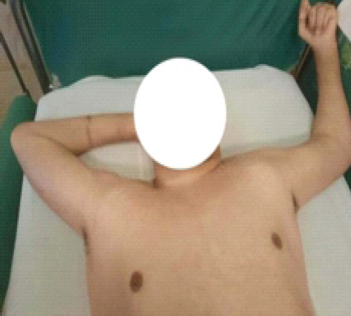
The institutional medical staff evaluated the patient in late 2025 when he was 46 years old. Upon physical examination, the patient was noted to be heavy-set (weight: 90 kg) and exhibited a new, matching deformity: His left shoulder was now similarly locked in fixed abduction (Fig. 1). No acute traumatic events or fall history had been reported by staff. When questioned regarding the delayed reporting of the abnormal limb posture, institutional caregivers stated they had attributed the fixed abduction to a new stereotypical behavioral preference or posturing by the patient.
Diagnostic workup and imaging
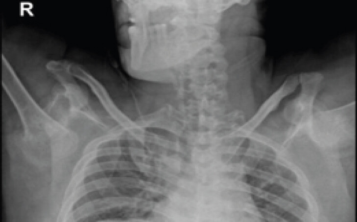
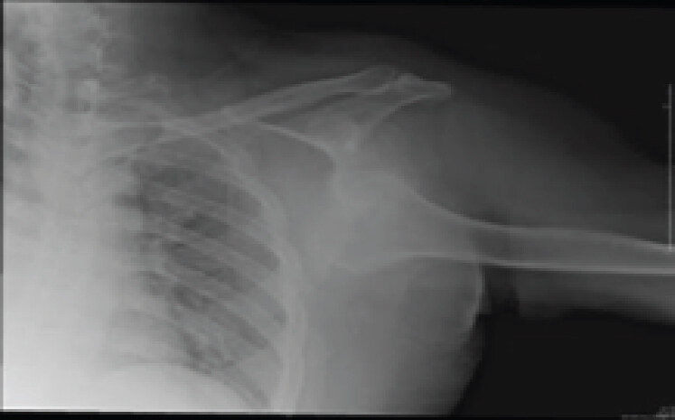
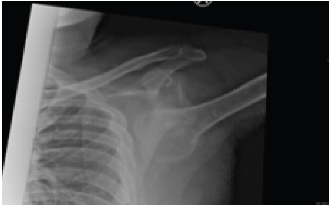
The patient was urgently referred to the orthopedic hospital for comprehensive evaluation. All pulses of both upper limbs were present. Anteroposterior plain radiographs of both shoulders were taken (Fig. 2) as well as multiplanar computed tomography (CT) reconstructions. CT of the left shoulder revealed a chronic, neglected left inferior glenohumeral dislocation. The left humeral head was displaced immediately inferior and slightly medial to the scapular glenoid cavity. Significant chronic changes were present, including advanced glenohumeral osteoarthritis, extensive osteophytosis, marginal humeral head irregularity, subchondral sclerosis of both the humerus and scapula, and subcortical cystic changes indicative of a well-established pseudoarthrosis. Joint effusion and capsular and synovial soft-tissue thickening were also visualized (Fig. 3). CT of the right shoulder showed similar, but more advanced, changes (Fig. 4).
Electrodiagnostic evaluation through nerve conduction studies demonstrated bilateral median neuropathies at the wrists, consistent with severe carpal tunnel syndrome, but no acute brachial plexus avulsion. Given the chronic, fixed nature of the bilateral deformities and the advanced structural pseudoarthrosis, total shoulder arthroplasty was the only remaining treatment option. However, due to the patient’s severe cognitive impairment, total lack of cooperation with post-operative restrictions, and high risk of implant dislocation or failure, surgical intervention was deemed contraindicated. The patient was managed conservatively with supportive positioning, and further orthopedic disability was permanently compounded onto his existing neurological deficits.
Discussion
LEH represents an exceptionally rare clinical entity, making it worthwhile to report. Given that LEH accounts for fewer than 0.5% of all shoulder dislocations, the statistical probability of a patient presenting with bilateral LEH is mathematically estimated at roughly 0.25%. Since its initial description in 1859 [8], approximately 200 cases of unilateral LEH have appeared in the literature [2]. Since the first report of bilateral, synchronous LEH was made in 1972 [9], roughly 50 cases have been reported [6]. To the authors’ knowledge, this report represents the first documented case of bilateral, asynchronous LEH, adding a novel clinical scenario to the sparse global literature on this injury pattern.
The classic clinical presentation of unilateral LEH is striking and readily identifiable. The affected limb is typically locked in an abducted position with the humerus aligned parallel to the spine of the scapula. The elbow remains flexed with the forearm pronated, placing the hand over or behind the head. The unaffected hand supports the arm in order to stabilize the affected arm and alleviate the pain. Physical examination typically reveals a prominent acromion, an empty glenoid fossa, a palpable humeral head within the axilla adjacent to the lateral chest wall, and prominent superior skin creasing. Because any manual attempt to lower the arm causes excruciating pain, the presentation is rarely missed in neurotypical individuals. However, in institutionalized patients with severe developmental disabilities, this dramatic physical posture can be masked or misattributed, leading to catastrophic delays in care.
Patients with spastic cerebral palsy inherently suffer from muscle imbalances that elevate their baseline risk for joint subluxations and dislocations. While spasticity typically predisposes patients to anterior dislocations due to the adduction-internal rotation vectors of the compromised musculature, and generalized seizures typically induce posterior dislocations [10], the etiology of this patient’s asynchronous bilateral LEH remains complex.
Because the patient resided in a highly monitored, 24-h long-term facility with no recorded falls or macro-trauma, a subtle or repeated micro-traumatic mechanism must be considered. On one hand, the patient lacked generalized ligamentous laxity, and his epilepsy was controlled through anticonvulsants, making a standard seizure-induced injury unlikely since epileptic forces characteristically adduct the limbs. On the other hand, the patient’s severe, chronic self-injurious behavior and stereotypic movements are highly relevant. Attempting to scratch hard-to-reach areas of the back or neck can induce forceful, involuntary hyperabduction.
Furthermore, the intermittent use of physical restraints to manage self-harm may have inadvertently altered his upper extremity biomechanics, causing the patient to leverage his upper arms against the bed frame or bedding to shift his weight, creating a severe indirect axial or hyperabduction force. Non-traumatic or atraumatic inferior glenohumeral subluxations have been previously reported in neuromuscular disorders, where chronic soft-tissue laxity, persistent joint effusion, and profound muscular imbalance combine to destabilize the capsuloligamentous complex over time [11].
This case tragically illustrates the severe consequences of chronic, unreduced LEH. Over a 4-year period, the untreated right glenohumeral joint developed advanced structural pseudoarthrosis, severe osteophytosis, and subchondral cystic breakdown. When the contralateral left shoulder subsequently sustained an asynchronous inferior dislocation, it went completely unrecognized by caregivers, who assumed the overhead “hands up” arm position was merely a new stereotypic behavioral posturing. By the time an orthopedic evaluation was requested, the left shoulder had similarly transitioned into a chronic, fixed, neglected state, leaving total shoulder arthroplasty as the sole theoretical anatomical salvage option. Given the patient’s profound cognitive impairment and inability to adhere to critical post-operative movement restrictions, surgery was contraindicated, permanently stacking a severe orthopedic deformity onto his pre-existing neurological deficits.
Conclusion
While the diagnosis and mechanical reduction of acute LEH are relatively straightforward in mainstream emergency medicine, the diagnostic pathway is frequently obstructed in patients with complex neurodevelopmental profiles. This clinical failure is heavily driven by diagnostic overshadowing, a cognitive bias wherein specialized clinicians or primary institutional caregivers erroneously misattribute new physical signs, asymmetric deformities, or structural changes to a patient’s baseline intellectual disability, spasticity, or behavioral stereotypes.
To safeguard this highly vulnerable population, structured training must be implemented for institutional healthcare staff and non-medical caregivers. Institutional protocols should mandate that any acute, semi-acute, or persistent alteration in a resident’s baseline limb positioning or joint symmetry be treated as an urgent orthopedic event requiring immediate plain radiography. Prompt specialist assessment is vital; missing the narrow therapeutic window for closed reduction can lead to permanent structural pseudoarthrosis, irreversible neurovascular degradation, and an avoidable, profound decline in the patient’s quality of life.
Clinical Message
Clinicians and institutional caregivers must maintain an exceptionally high index of suspicion for subtle musculoskeletal changes in vulnerable, non-verbal populations. Prompt radiographic evaluation and urgent orthopedic intervention are mandatory to prevent catastrophic, irreversible functional decline in patients with pre-existing neurological deficits.
Conflict of Interest:
Nil
Source of Support:
Nil
Consent
The authors confirm that informed consent was obtained from the patient for publication of this article
How to Cite this Article
Ali FE, Ali YF. Bilateral, Asynchronous, Inferior Shoulder Dislocation in a Multidisabled Patient: A Case Report and Review of the Literature. J Orthop Case Rep. 2026 November;16(11): 104-108. doi: 10.13107/jocr.2026.v16.i11.8404.
References
- Mallon WJ, Bassett FH 3rd, Goldner RD. Luxatio erecta: The inferior glenohumeral dislocation. J Orthop Trauma 1990;4:19-24. [Google Scholar] | [PubMed]
- Nambiar M, Owen D, Moore P, Carr A, Thomas M. Traumatic inferior shoulder dislocation: A review of management and outcome. Eur J Trauma Emerg Surg 2018;44:45-51. [Google Scholar] | [PubMed]
- Wolf O, Ekholm C. Luxatio erecta of the humerus: The spectrum of injury of inferior shoulder dislocation and analysis of injury mechanisms. JSES Rev Rep Tech 2022;2:497-504. [Google Scholar] | [PubMed]
- Khedr H, Al-Zahrani A, Al-Zahrani A, Al-Qattan MM. Bilateral irreducible inferior shoulder dislocation: A case report. Int J Surg Case Rep 2017;31:124-7. [Google Scholar] | [PubMed]
- Groh GI, Wirth MA, Rockwood CAJr. Results of treatment of luxatio erecta (inferior shoulder dislocation). J Shoulder Elbow Sur 2010;19:423-6. [Google Scholar] | [PubMed]
- Ntourantonis D, Mousafeiris V, Pantazis K, Iliopoulos I, Kaspiris A, Korovessis P. The 'Holy Grail' of shoulder dislocations: A systematic review on traumatic bilateral luxatio erecta; Is it in reality a once-in-a-lifetime experience for an orthopaedic surgeon?. Arch Orthop Trauma Surg 2024;144:205-17. [Google Scholar] | [PubMed]
- Carvill S. Sensory impairments, intellectual disability and psychiatry. J Intellect Disabil Res 2001;45:467-83. [Google Scholar] | [PubMed]
- Middeldorpf M, Scharm B. De nova humeri luxationis specie. Clin Eur 1859;2:12-6. [Google Scholar] | [PubMed]
- Murard J. Un cas de luxatio erecta de l'epaule, double et symetrique. Rev Orthop 1920;7:23-9. [Google Scholar] | [PubMed]
- Gosens T, Poels PJ, Rondhuis JJ. Posterior dislocation fractures of the shoulder in seizure disorders--two case reports and a review of literature. Seizure 2000;9:446-8. [Google Scholar] | [PubMed]
- Sonanis SV, Das S, Deshmukh N, Wray C. A true traumatic inferior dislocation of shoulder. Injury 2002;33:842-4. [Google Scholar] | [PubMed]
© 2026 Journal of Orthopaedic Case Reports - Published by Indian Orthopaedic Research Group
About the Authors
-

Fawzi E Ali
-

Yasmina F Ali
Related Articles in Journal of Orthopaedic Case Reports
 April 10, 2022 Reviewers Acknowledgement & Photo Gallery April 2022
 July 10, 2021 A 6-month-old neglected obturator inferior variety of anterior hip dislocation in a young male managed with an intermediary salvage measure without the need for osteotomy or excision arthroplasty with a resultant ambulatory natural hip a case report
 April 7, 2014 Scalloping Sacral Arachnoid Cyst as a Cause of Perianal Pain- A Case Report
 November 10, 2023 Knee Extension Lag Treated with Posterior Capsulotomy of the Knee: A Case Report and Review of the Literature