


Introduction
Congenital muscular torticollis (CMT) is the third most common pediatric orthopedic diagnosis in children [1]. Benign sternocleidomastoid mass causes asymmetric posture, restricted cervical range of motion (ROM), plagiocephaly, facial asymmetry, and cranial molding. CMT resolves spontaneously in 80–90% of cases [2]; if it persists beyond 1 year of age, sternocleidomastoid muscle lengthening should be considered. Untreated CMT is rare until adolescence [3] since CMT has better treatment outcomes if administered in the youth and shows a small change in quality of life even after surgery [4].
Accurate assessment of cervical motion is essential for evaluating the effects of CMT treatment. Torticollis was evaluated using a videotape [5,6] or goniometer [7]. Although visual estimation has acceptable reliability for some movements, the complex movement of the head and neck in all directions requires more robust assessment tools [6].
Cervical movement is difficult to evaluate because it involves multidirectional movements, such as flexion, extension, lateral bending, and rotation. Although a three-dimensional (3D) motion analysis device allows for detailed and accurate evaluation of neck posture and movement [8], there are no reports of CMT evaluating the effect of surgery or bracing using this analytical method. In a few reports on adolescents with CMT, the effectiveness of surgery for neck motion was described based on clinical assessment alone [9,10].
This report determined the effect of surgery on neck movement in adolescents with CMT. A detailed evaluation was conducted to check changes in resting posture and active ROM by comparing neck movements before and after surgery with those of healthy controls.
Case Report
A 21-year-old female presented with torticollis and right-sided neck and shoulder pain. She was diagnosed with right CMT in her childhood at a local hospital and received rehabilitation treatment until 18 years old. The torticollis worsened with growth, associated with right neck and shoulder pain without limitation in activities of daily living.
On examination, a hard mass was identified in the right sternocleidomastoid muscle. The right shoulder was slightly elevated, and the neck was in torticollis, tilted 10° to the right with left rotation. Facial asymmetry was also noted (Fig. 1a and b). Active neck ROM, as measured by a goniometer, revealed 60°/35°(right/left) in lateral bending and 65°/80° in rotation.
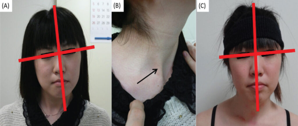
The patient received bipolar tenotomy (i.e., transection of the proximal and distal ends) of the right sternocleidomastoid muscle. Postoperatively, the patient had a polyethylene cervical collar for 5 weeks and underwent cervical ROM self-training for 1 year after surgery. Although the right neck and shoulder pain alleviated and the right neck tilting at rest has improved to 5° (Fig. 1c) and neck active ROM changed to 45°/35°in lateral bending and 70°/70° in rotation, the facial asymmetry persisted.
Three-dimensional motion analysis
We measured the neck movement of the participant using 3D motion analysis 3 times (before surgery, 6 months, and 1 year after surgery) as described in a previous study [8]. To compare neck movements, we recruited healthy controls without a history of neck disorders and measured the neck movement once.
Measurements of neck movements were obtained while the participants performed six types of movements for five trials per direction in the sitting position: flexion, extension, left and right lateral bending, and left and right rotation. For analysis, the trunk-head angle was calculated; 0° represents the head segment in the same direction as the trunk segment. The trunk-head angles in the resting position were calculated by averaging the angles before starting neck movements in all 30 trials. The active neck ROMs were the difference of trunk-head angles between the start and maximum postures. We calculated the main plane neck ROM and other plane ROMs, accompanying ROM.
We also calculated the onset timing of neck movement in each plane to follow the change in motion pattern before and after surgery. The timing of the onset was defined as the moment when the angle was equal to one-twentieth of the full ROM, and the zero point was set at the onset time of the main movement. The onset timing of the accompanying movements was calculated by averaging five trials for each movement.
Calculations were performed using MATLAB R2024a (MathWorks, Natick, MA, U.S.A.). We performed one-way analysis of variance with a Bonferroni post hoc test, an unpaired t-test, and a paired t-test for statistics using the Statistical Package for the Social Sciences version 21 (IBM Corp., Armonk, NY, U.S.A.). The significance level was set at P<0.05.
The backgrounds of the patient preoperatively and the healthy controls are shown in Table 1.
Background of the participants
| Patient (n=1)* | Control (n=9) | |
|---|---|---|
| Female | 1 | 3 |
| Age (y.o.) | 21 | 35.3 (9.4) |
| Height (cm) | 158 | 165.4 (9.7) |
| Body weight (kg) | 45 | 63.4 (12.1) |
| Body mass index (kg/m2) | 17.9 | 23.0 (2.2) |
*
Data at pre-surgery measurement. Control: Average (standard deviation)
Resting position
For the resting position, the trunk-head angle was tilted to the right with left rotation. The left rotation position showed slight improvement after surgery (Table 2).
Trunk-neck angle at resting position
| Patient | Control (n=9) | |||
|---|---|---|---|---|
| Pre-surgery | Post-surgery | |||
| 6 months | 1 year | |||
| Sagittal plane | −16.5° (1.6°) | −16.0° (2.7°) | −28.1° (2.8°)*† | −12.8° (13.3°) |
| Coronal plane | 7.3° (1.0°) | 7.0° (1.5°) | 7.7° (1.2°)† | 0.7° (2.6°) |
| Transverse plane | −3.9° (1.2°) | −2.2° (2.1°)* | −2.7° (1.2°)* | 1.8° (3.8°) |
Average (standard deviation). Trunk-head angle 0° represent the head segment is in the same direction as the trunk segment. Positive value means flexion for sagittal plane, right (affected-side) tilting for coronal plane, and right rotation for transverse plane.
*
Compare to pre-surgery P<0.05,
†
Compare to 6 months after surgery P<0.05
Active neck ROMs for the main direction (Table 3)
Active neck range of motions in main direction and ranges of accompanying movement
| Patient | Control (n=9) | |||
|---|---|---|---|---|
| Pre-surgery | Post-surgery | |||
| 6 months | 1 year | |||
| Flexion | ||||
| Sagittal plane | 55.8° (1.5°) | 56.1° (2.5°) | 57.7° (1.1°) | 59.8° (8.2°) |
| Coronal plane | −1.7° (0.9°) | −2.2° (1.0°) | −3.2° (0.7°) | −0.6° (3.9°) |
| Transverse plane* | −6.1° (1.0°) | −7.5° (1.8°) | −4.4° (0.4°)† | 0.1° (3.9°) |
| Extension | ||||
| Sagittal plane* | −61.9° (2.5°) | −72.8° (2.0°)⁑ | −75.3° (2.2°)⁑ | −64.5° (10.6°) |
| Coronal plane* | 1.9° (0.7°) | −1.9° (0.4°)⁑ | −6.2° (1.3°)⁑† | 0.6° (3.8°) |
| Transverse plane* | 6.3° (1.2°) | 7.9° (1.3°) | 9.0° (1.7°)⁑ | 1.7° (4.3°) |
| Right bending | ||||
| Sagittal plane | −19.7° (1.8°) | −14.8° (2.4°)* | −18.2° (5.1°) | −6.1° (5.3°) |
| Coronal plane | 38.2° (1.7°) | 40.5° (1.2°) | 40.4° (1.9°) | 36.1° (7.6°) |
| Transverse plane* | 16.1° (2.1°) | 9.8° (0.9°)⁑ | 25.8° (1.9°)⁑† | 14.9° (10.3°) |
| Left bending | ||||
| Sagittal plane | 4.4° (1.9°) | 3.3° (3.2°) | −5.3° (1.1°)*† | −2.2° (6.4°) |
| Coronal plane* | −34.5° (2.1°)‡ | −38.7° (1.0°)⁑‡ | −43.1° (2.0°)⁑† | −38.7° (6.8°) |
| Transverse plane* | −11.3° (1.2°) | −4.6° (2.5°)⁑ | -13.1° (4.1°)† | −15.8° (6.6°) |
| Right rotation | ||||
| Sagittal plane* | −5.7° (2.5°) | −16.7° (2.4°)⁑ | −9.7° (1.7°)† | 5.7° (6.7°) |
| Coronal plane* | −20.3° (2.2°) | −15.7° (1.9°)⁑ | −31.8° (1.1°)⁑† | −12.2° (6.0°) |
| Transverse plane* | 57.5° (4.0°) | 67.6° (3.2°)⁑ | 73.1° (1.5°)⁑† | 63.2° (7.5°) |
| Left rotation | ||||
| Sagittal plane* | 11.1° (1.8°) | -7.7° (2.5°)⁑ | 11.5° (2.3°)† | 6.8° (8.6°) |
| Coronal plane* | 18.5° (0.5°) | 6.7° (1.3°)⁑ | 17.8° (1.2°)† | 12.8° (6.4°) |
| Transverse plane* | -67.8° (2.6°)§ | -74.5° (3.6°)⁑§ | -79.5° (0.6°)⁑†§ | −71.5° (8.3°)§ |
Average (standard deviation). Positive value means flexion for the sagittal plane, right (affected-side) bending for the coronal plane, and right rotation for the transverse plane. Bolded data indicate range of main movement.
*
ANOVA shows significant difference date, ⁑Compare to pre-surgery P<0.05,
†
Compare to 6 months after surgery P<0.05,
‡
Compared to right bending P<0.05,
§
Compared to right rotation P<0.05
Preoperatively, the patient with CMT showed limited extension, left bending, and bilateral rotation ROM compared to healthy controls, which increased after surgery. Compared to the opposite side, the ROM for left bending was significantly limited before surgery and 6 months after surgery, and the ROM for right rotation was limited across all time points. In contrast, there was no difference between the left and right bending ROM in the healthy controls.
Neck ROM for accompanying movement (Table 3)
During flexion movements, the patient showed accompanying movement in left bending and left rotation before surgery. One year postoperatively, left bending significantly increased, whereas left rotation decreased. During extension movements, the patient showed right bending and rotation before surgery, which changed to left bending and right rotation 1 year postoperatively. In contrast healthy controls exhibited almost no accompanying lateral bending or rotation during flexion or extension.
During lateral bending, the same direction rotation was observed in both the patient and the controls, but extension accompanying movement during right bending was observed in the patient.
During rotation, contralateral lateral bending was observed in both the healthy controls and the patient, and neck flexion occurred in the healthy control, whereas extension occurred in the patient. In the patient, compared with pre-surgery, left bending during right rotation decreased significantly at 6 months postoperatively but increased again at 1 year. During left rotation, right bending decreased significantly at 6 months postoperatively.
Onset timing (Fig. 2)
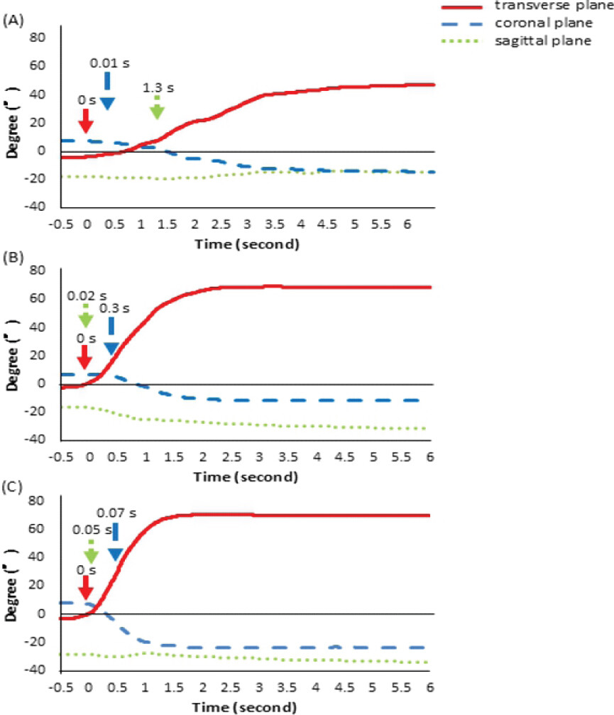
The onset timing patterns in the three directions during neck movements showed large variations. For right rotation movements, the left bending onset timing was delayed at 6 months postoperatively compared to pre-surgery or 1 year after surgery.
Discussion
In this study, we measured the precise neck movements in the patient with CMT and confirmed that the resting posture, active neck ROM, accompanying movements, and onset timing changed after surgery. Torticollis assessment, relying on photographs or goniometers, is expected to be more precise [6]. In this study, 3D motion analysis allowed more precise ROM measurement and the evaluation of associated movements and differences in movement timing.
Surgery partially corrected the patient’s preoperative resting posture, improving rotation ROM but not lateral tilting. Unilateral shortening of the sternocleidomastoid in the CMT causes ipsilateral lateral tilting and contralateral rotation, and post-operative lengthening of this muscle likely enabled the observed shift toward ipsilateral rotation [11]. Although the clinical examination suggested an improvement in affected-side tilting, precise 3D motion measurements showed no clear change. The clinical assessment was performed with the patient consciously instructed to face forward, whereas 3D motion measurements were taken between task trials and likely represented a more natural resting posture. Moreover, persistent facial asymmetry may reflect habituation to a lateral tilting position.
Even if there is no awareness of restrictions in ROM or activities of daily living, ROM limitations were observed in adolescents with CMT. CMT limits lateral bending toward the unaffected side and rotation toward the affected side [7], which is due to the shortening of the sternocleidomastoid muscle. During activities of daily living, the neck undergoes large movements: descending stairs, putting on socks, and tying shoelaces require approximately 20° of flexion; squatting requires approximately 30° of extension; shaving and face washing require roughly 25° of lateral bending; and checking behind while parking requires approximately 90° of rotation [12,13]. In this patient, restrictions on flexion–extension and lateral bending ROM were relatively small, whereas rotation was limited on both sides, which may have interfered with tasks such as looking backward. The patient reported no restriction in activities of daily living because she had ROM limitations since early childhood and had developed compensatory movements using the trunk or the rest of the body.
ROM limitations of adolescent CMT may improve with surgery in multiple directions. Even in adults, surgery for CMT improves the restriction of unaffected-side bending and affected-side rotation [14,15,16]. Surgical treatment aims to reduce side-to-side ROM asymmetry with targets such as a lateral bending ROM difference of <20° [17] and rotational ROM of <30° [18]. For the patient in this study, in addition to improvements in extension, unaffected-side bending, and affected-side rotation, restrictions on unaffected-side rotation also improved. The former three are likely to be due to lengthening of the sternocleidomastoid, and the latter might be attributed to reduced cervical muscle tension following the relief of neck pain after surgery and post-operative cervical ROM training.
The accompanying movements during neck motion observed only in the patient, which change preoperatively, might be due to multiple factors, including not only shortening of the sternocleidomastoid muscle but also the effects of sensory development. During flexion, the patient with CMT showed non-affected-side bending and non-affected-side rotation, which cannot be explained solely by shortening of the right sternocleidomastoid and its post-operative correction, as shortening of the sternocleidomastoid may cause non-affected-side bending, affected-side rotation, and improvement after surgery. Possible causes include disturbed body balance, semicircular canal (vestibular) dysfunction, and visual factors. Sensory development in patients with CMT is delayed compared to healthy controls [19], and development of the semicircular canals, and thus vestibular function, is influenced by vestibular stimulation received up to adolescence [20,21]. In adults with CMT, facial asymmetry is not resolved by surgery [11]. Therefore, because this patient lived in a torticollis posture throughout adolescence, her body image, such as the perception of the midline, may differ from that of healthy individuals.
Analyzing the perioperative change in timing of neck movements may help understand the mechanisms of motor learning in patients with motor limitations. It is known that compensatory movements occur in other parts of the body when moving body parts with restricted ROM or pain, such as compensatory movement during exercise for patients with back pain [22]. However, no studies have investigated the timing at which these compensatory movements occur. In the present study, we suspected that accompanying movements occurred to compensate for the restricted neck motion or body image. Using a 3D motion analysis system may help elucidate the mechanisms of the accompanying movements and could potentially contribute to suppressing unnecessary compensation or effectively utilizing compensatory strategies.
This study had several limitations. First, it summarizes a detailed neck motion assessment in a single adult patient with CMT; its findings may not be generalizable to other adult CMT cases. Future studies should include a larger sample. Second, we did not record muscle activity using electromyography or collect kinetic data. Therefore, muscle function was inferred only from kinematic measurements. Incorporating direct assessments of muscle activity in future studies may reveal more detailed disease and treatment-related changes.
Conclusion
Surgical intervention for CMT resulted in improved neck ROM even in adolescence. In contrast to the goniometers currently used in clinical practice, 3D motion analysis systems enable the continuous and highly precise evaluation of neck movements. With further technological advancements in motion analysis systems, clinical implementation of these technologies may allow for a more accurate assessment of therapeutic outcomes.
Clinical Message
CMT may induce different neck movement patterns compared with healthy participants, and surgery in adolescence partially improves neck ROM and abnormal neck movement patterns.
Conflict of Interest:
Nil
Source of Support:
Nil
Consent
The authors confirm that informed consent was obtained from the patient for publication of this article
How to Cite this Article
Inokuchi H, Tojima M, Haga N, Ogata T. Three-Dimensional Changes of Neck Motion Following Surgery for Adolescent Congenital Muscular Torticollis: A Case Report. J Orthop Case Rep. 2026 November;16(11):109-115. doi: 10.13107/jocr.2026.v16.i11.8406.
References
- Antares JB, Jones MA, Chak NT, Chi Y, Li H, Li M. Efficacy of non-surgical, non-pharmacological treatments for congenital muscular torticollis: A systematic review and meta-analysis. BMC Musculoskelet Disord 2025;26:178. [Google Scholar] | [PubMed]
- Do TT. Congenital muscular torticollis: Current concepts and review of treatment. Curr Opin Pediatr 2006;18:26-9. [Google Scholar] | [PubMed]
- Petronic I, Brdar R, Cirovic D, Nikolic D, Lukac M, Janic D. Congenital muscular torticollis in children: Distribution, treatment duration and out come. Eur J Phys Rehabil Med 2010;46:153-7. [Google Scholar] | [PubMed]
- Høiness PR, Medbøe A. Surgical treatment of congenital muscular torticollis: Significant improvement in health-related quality of life among a 2-year follow-up cohort of children, adolescents, and young adults. J Pediatr Orthop 2023;43:e769-74. [Google Scholar] | [PubMed]
- Kassaye SG, De Pauw J, De Waele S, De Hertogh W, Gudina EK, Crosiers D. Interdisciplinary consensus in evaluating the severity subscale of the original and revised Toronto western spasmodic torticollis rating scale through video-based assessment: An inter-rater reliability study. Mov Disord Clin Pract 2024;11:1587-91. [Google Scholar] | [PubMed]
- Seager A, Meldrum D, Conroy R, French HP. Congenital muscular torticollis: The reliability of visual estimation in the assessment of cervical spine active rotation and head tilt by physiotherapists and the impact of clinical experience. Eur J Pediatr 2020;179:1823-32. [Google Scholar] | [PubMed]
- Lee JY, Koh SE, Lee IS, Jung H, Lee J, Kang JI. The cervical range of motion as a factor affecting outcome in patients with congenital muscular torticollis. Ann Rehabil Med 2013;37:183-90. [Google Scholar] | [PubMed]
- Inokuchi H, Tojima M, Mano H, Ishikawa Y, Ogata N, Haga N. Neck range of motion measurements using a new three-dimensional motion analysis system: Validity and repeatability. Eur Spine J 2015;24:2807-15. [Google Scholar] | [PubMed]
- Monreal J. Long-term results after percutaneous myotomies and enriched fat grafting for congenital muscular torticollis treatment. Aesthetic Plast Surg 2025;49:3132-42. [Google Scholar] | [PubMed]
- Chang SH, Ohtori S, Okawa A, Kawamura K, Saiki H, Nakada I. A surgical treatment for adult muscular torticollis. Case Rep Orthop 2013;2013:965693. [Google Scholar] | [PubMed]
- Ippolito E, Tudisco C. Idiopathic muscular torticollis in adults. Results of open sternocleidomastoid tenotomy. Arch Orthop Trauma Surg (1978) 1986;105:49-54. [Google Scholar] | [PubMed]
- Bennett SE, Schenk RJ, Simmons ED. Active range of motion utilized in the cervical spine to perform daily functional tasks. J Spinal Disord Tech 2002;15:307-11. [Google Scholar] | [PubMed]
- Bible JE, Biswas D, Miller CP, Whang PG, Grauer JN. Normal functional range of motion of the cervical spine during 15 activities of daily living. J Spinal Disord Tech 2010;23:15-21. [Google Scholar] | [PubMed]
- Yuan B, Qu F, Zhao G, Wang J, Shen X, Liu Y. Arthroscopic surgical treatment for neglected congenital muscular torticollis in adults. J Craniofac Surg 2015;26:512-5. [Google Scholar] | [PubMed]
- Cheng JC, Tang SP. Outcome of surgical treatment of congenital muscular torticollis. Clin Orthop Relat Res 1999;362:190-200. [Google Scholar] | [PubMed]
- Lim KS, Shim JS, Lee YS. Is sternocleidomastoid muscle release effective in adults with neglected congenital muscular torticollis? Clin Orthop Relat Res. 2014;472:1271-8. [Google Scholar] | [PubMed]
- Ippolito E, Tudisco C, Massobrio M. Long-term results of open sternocleidomastoid tenotomy for idiopathic muscular torticollis. J Bone Joint Surg Am 1985;67:30-8. [Google Scholar] | [PubMed]
- Canale ST, Griffin DW, Hubbard CN. Congenital muscular torticollis. A long-term follow-up. J Bone Joint Surg Am 1982;64:810-6. [Google Scholar] | [PubMed]
- Zorlular R, Burak SE, Beyoglu R, Elbasan B. Motor development and sensory processing skills in infants with congenital muscular torticollis: A cross-sectional study. Eur J Pediatr 2025;184:630-2. [Google Scholar] | [PubMed]
- Božanić Urbančič N, Battelino S, Vozel D. Appropriate vestibular stimulation in children and adolescents-a prerequisite for normal cognitive, motor development and bodily homeostasis-a review. Children (Basel) 2023;11:2. [Google Scholar] | [PubMed]
- O'Reilly R, Grindle C, Zwicky EF, Morlet T. Development of the vestibular system and balance function: Differential diagnosis in the pediatric population. Otolaryngol Clin North Am 2011;44:251-71. vii. [Google Scholar] | [PubMed]
- Sellmann A, Wagner D, Holtz L, Eschweiler J, Diers C, Williams S. Detection of typical compensatory movements during autonomously performed exercises preventing low back pain (LBP). Sensors (Basel) 2021;22:111. [Google Scholar] | [PubMed]
© 2026 Journal of Orthopaedic Case Reports - Published by Indian Orthopaedic Research Group
About the Authors
-

Haruhi Inokuchi
-

Michio Tojima
-

Nobuhiko Haga
-

Toru Ogata
Related Articles in Journal of Orthopaedic Case Reports
 April 13, 2013 Traumatic Fracture in a patient of Osteopoikilosis with Review of Literature
 June 10, 2024 Lambrinudi Triple Arthrodesis with Posterior Tibial Tendon Transfer in Adult Fixed Equinus Deformity: Case Report
 December 10, 2023 Conversion of First Metatarsal-Phalangeal Joint Arthrodesis to Arthroplasty: A Case Report
 October 10, 2016 Management of Infected Total Knee Arthroplasty by a New Innovative Customized Articulating Knee Spacer: An Early Experience