


Introduction
Iliac wing fractures primarily result from high-energy trauma such as road traffic accidents, crush injuries, or falls from height, often occurring with disruption of the pelvic ring [1,2]. Early treatment generally produce successful outcomes, but delayed management, commonly seen in resource-limited settings, can lead to significant long-term morbidity [3]. Progressive malunion, chronic pelvis instability, soft-tissue contracture or altered joint mechanics occur secondary to untreated iliac wing fractures and compound operative complexity [2,4].
Protrusio acetabuli is defined as medial displacement of the femoral head beyond the ilioischial (Köhler’s) line on anteroposterior pelvic radiographs, caused by failure of the medial acetabular wall under physiologic loading [5]. Chronic pelvic malunion compromises acetabular column integrity, gradually weakening the medial wall and predisposing to protrusio. This results in impingement, degenerative changes, and significant functional limitation [5,6].
Lumbar hernias are uncommon, representing <1.5% of abdominal wall hernias and representing protrusion of visceral/retroperitoneal contents through a defect in the posterior abdominal wall [7,8]. They occur through the inferior lumbar triangle of Petit or the superior lumbar triangle of Grynfeltt-Lesshaft. Post-traumatic lumbar hernias resulting from fascial dehiscence are uncommon, and their coexistence with pelvic instability further complicates management [7].
The presence of untreated iliac wing fracture with co-existing ipsilateral protrusio acetabuli and post-traumatic lumbar hernia following the same index trauma event is extremely rare. This case report documents the presentation, surgical management, and 1-year outcome of this exceptionally rare clinical trial, providing a potential approach for management of similar presentations.
Case Report
Patient history and examination
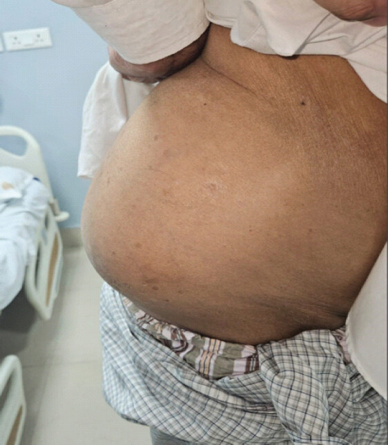
A 77-year-old male farmer presented with a 4-year history of progressive right lumbar swelling, chronic right hip pain and gait disturbance requiring a walking stick. He had sustained blunt trauma to the right hemipelvis in a road traffic accident 12 years ago, but received no medical treatment. Examination revealed a reducible, non-tender right lumbar swelling (8 × 6 cm) in the inferior lumbar triangle with a palpable fascial defect (Fig. 1), right-sided antalgic gait, pelvic obliquity, 2.5 cm right-sided limb length discrepancy and reduced right hip movements with flexion up to 60°, abduction to 15° and absent internal rotation. Patient reported Visual Analog Scale (VAS) pain score of 7/10 and Harris Hip Score of 56. The details of the initial injury and subsequent 12-year course were based on patient’s history, as records and imaging were not available and natural progression could not be assessed.
Imaging and planning
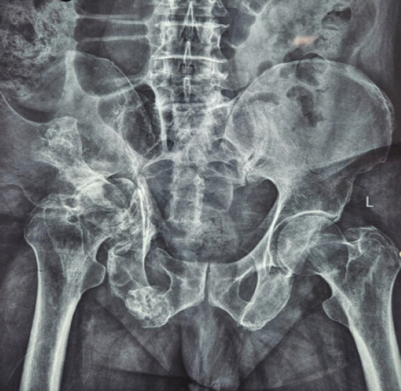
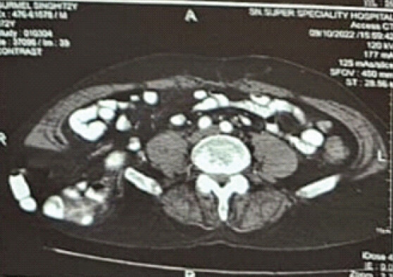
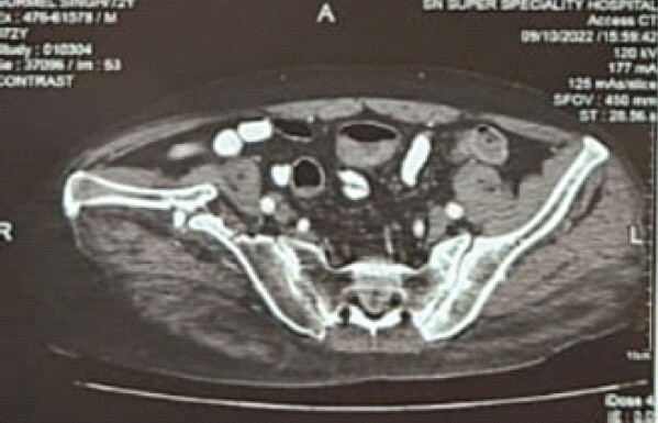
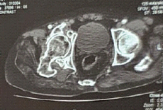
Plain anteroposterior pelvic radiograph demonstrated right iliac wing malunion with pelvic asymmetry and protrusio acetabuli (Fig. 2). Computed tomography confirmed chronic iliac wing malunion with medial femoral head migration and a well-defined posterior lumbar fascial defect measuring 6 × 4 cm, containing retroperitoneal fat without bowel or other visceral contents (Fig. 3, 4, 5). No pre-injury clinical records or imaging were available to assess for any pre-existing hip pathology. Bone density assessment revealed osteopenia. Following multidisciplinary evaluation, sequential operative management was planned due to altered pelvic biomechanics and risk of lumbar hernia incarceration. Total hip arthroplasty was offered in the same setting but declined by the patient. Given the chronic lumbar hernia, the potential risk of contamination was also considered. Arthroplasty remains a potential future option if clinically indicated.
Procedure
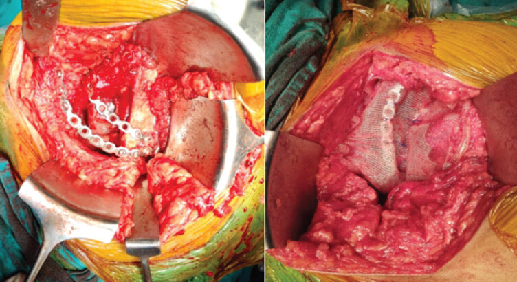
For lumbar hernioplasty, the patient was positioned in the left lateral decubitus position. An oblique skin incision was made 2 cm superior to the right iliac crest over the swelling. The dissection proceeded, and the external oblique fascia was incised. The hernia sac contained only retroperitoneal fat, which was reduced. The 6 × 4 cm posterior abdominal wall defect was reinforced using an onlay polypropylene mesh with at least 3 cm overlap and secured with interrupted 2–0 Prolene sutures at periphery. Through the same exposure, the right iliac wing was approached. The malunited iliac blade was osteotomized, and sclerotic fracture margins were freshened using an osteotome. Reduction was achieved under direct visualization with temporarily Kirschner wire fixation. Two 3.5 mm pre-contoured pelvic reconstruction plates were then placed along the inner table with bicortical screw fixation providing adequate purchase for stable fixation. An additional polypropylene mesh was placed over the construct and secured to the external oblique fascia to reinforce the iliac crest-abdominal wall interface (Fig. 6). The wound was irrigated and closed over a suction drain.
Post-operative management
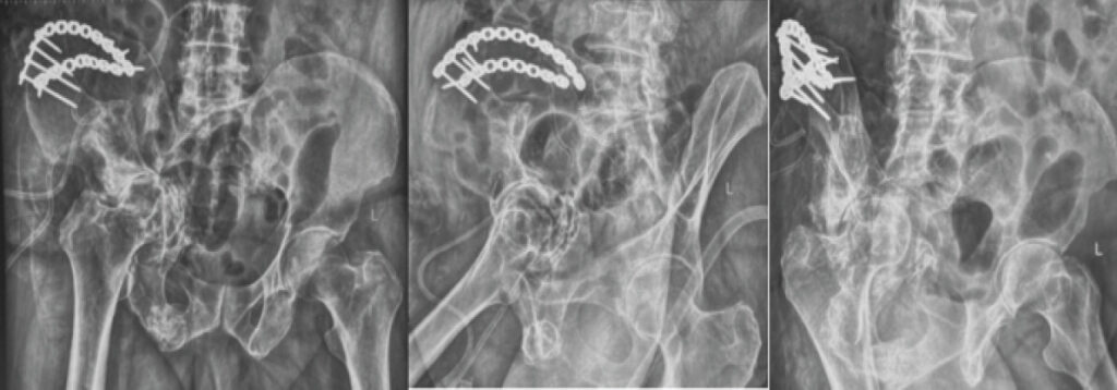
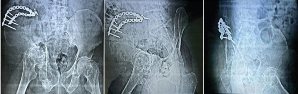
The procedure lasted 180 min without intraoperative complications. Postoperatively, the patient received intravenous ceftriaxone for 48 h, followed by oral cefuroxime for 5 days and low molecular weight heparin for 6 weeks. Supervised mobilization commenced on post-operative day 1, with partial weight bearing initiated on day 2. Post-operative radiographs (Fig. 7) confirmed satisfactory implant position and maintained fracture reduction. The patient was discharged on post-operative day 6. Follow-up evaluations were conducted at 6 weeks, 3 months, 6 months, and 12 months with clinical examination and serial radiographs. At 12 months, the lumbar swelling had completely resolved, and pain had improved. Functional improvement was assessed clinically with a VAS score of 5/10 and Harris Hip score of 68. There were no clinical features of mesh-related complications, including infection, seroma, mesh migration, erosion or chronic pain during follow-up. Final radiographs (Fig. 8) confirmed complete osseous integration of the iliac wing fracture, stable pelvic alignment, intact hardware, and no evidence of hernia recurrence.
Discussion
Pelvic ring stability depends primarily on the integrity of the osseoligamentous complex, which is responsible for transmitting physiologic load during weight bearing. Persistent malalignment of the pelvic ring therefore alters load distribution and may contribute to progressive secondary deformity and functional deterioration [9]. Untreated pelvic ring injuries have been shown to lead to progressive, long-term instability and degradation of pelvic structural integrity with secondary functional deterioration of the hip joint by Rommens et al., who demonstrated an observational cohort of pelvic fractures [6]. It emphasizes the importance of restoring pelvic stability to prevent long-term biomechanical deterioration. In the current case, the prolonged altered pelvic loading associated with the malunion may have contributed to progressive medial acetabular wall insufficiency and medial migration of the femoral head, which is consistent with Samanta et al.’s report of a similar mechanism for protrusio acetabuli, where mechanical overloading of the acetabular floor drives progressive femoral head displacement [5,10]. Pelvic ring injuries requiring operative intervention are typically associated with significant displacement, instability or persistent symptoms. In neglected injuries, correction of chronic deformity may be necessary to restore pelvic alignment and improve function [11].
The lumbar hernia likely resulted from fascial disruption during the original trauma, with gradual enlargement due to repetitive increases in intra-abdominal pressure and altered pelvic mechanics. The defect was located in the inferior lumbar (Petit’s) triangle, consistent with typical post-traumatic lumbar hernias as described by van Steensel et al. in their systematic review, which reported that posterior fascial disruptions following trauma can present decades after the causative event as the fascial defect gradually enlarges under physiological loading [7].
A combined operative strategy was decided to address both pathologies during the same anesthetic session. Repair of the lumbar hernia was performed first to restore posterior abdominal wall integrity and provide a stable soft-tissue envelope before pelvic reconstruction. Iliac wing malunion was then corrected using osteotomy and internal fixation with dual reconstruction plates placed along the inner table of the iliac blade, restoring bony contour and providing structural support to the posterior abdominal wall to reduce risk of recurrent herniation. It is consistent with the classification-based fixation strategies described by Prieto et al. and Villán-Gaona et al. for complex iliac wing fractures, which advocate stable fixation to address different planes of instability [12,13]. The literature also highlights that stable fixation restores pelvic alignment, facilitates early mobilization and minimizes risk of secondary displacement in complex iliac wing fractures [13]. Placement of an additional polypropylene mesh over the fixation reinforced the abdominal wall–iliac crest interface and provided soft-tissue support over the implant construct. This multidisciplinary approach was seen to be beneficial in minimizing aggregate peri-operative morbidity in the elderly patient, as proposed by Doke et al., which stated that a combined single-sitting multi-specialty procedure was more effective in reducing operative complication rate and hospital length of stay in the complex trauma patient [3]. Our approach enabled simultaneous reconstruction of the abdominal wall and pelvic ring, facilitating comprehensive management of both the osseous and soft-tissue components of this neglected injury. Such coordinated care is recommended in complex pelvic trauma [14].
This rare case suggests that operative intervention may be considered even in long-standing neglected pelvic injuries and provides a potential approach for similar complex presentations. Delayed presentation of pelvic trauma should prompt evaluation for associated abdominal wall defects with chronic pelvic deformity, and multidisciplinary management is essential for optimal outcomes. However, as this is a single case report, there was no comparative treatment group, and the findings cannot be generalized to all patients with neglected pelvic injuries.
Conclusion
This report demonstrates that a sequential, multidisciplinary approach can successfully address the combined osseous and soft-tissue sequelae of neglected pelvic trauma. Careful patient selection, comprehensive pre-operative planning and collaboration across specialities are essential to optimize functional outcomes in complex reconstructive cases. Clinicians should maintain a high index of suspicion for associated soft-tissue injuries in patients presenting with delayed pelvic trauma to ensure comprehensive evaluation and appropriate surgical planning.
Clinical Message
Neglected iliac wing fractures associated with lumbar hernia require comprehensive evaluation and individualized staged multidisciplinary management to restore pelvic stability, optimize functional outcomes and prevent long-term complications.
Conflict of Interest:
Nil
Source of Support:
Nil
Consent:
The authors confirm that informed consent was obtained from the patient for publication of this article
How to Cite this Article
Singla A, Rai A, Lalchandani T, Shergill J, Jabbal HS, Sharma PK. Neglected Iliac Wing Fracture with Protrusio Acetabuli and Lumbar Hernia – A Rare Case Report and Surgical Rationale. J Orthop Case Rep. 2026 November;16(11): 180-185. doi: 10.13107/jocr.2026.v16.i11.8432.
References
- Walters MM, Bergin PF, Russell GV, Morellato JT.. Geriatric acetabular fractures and femoral head protrusio: Methods of fixation and the option of total hip arthroplasty. J Orthop Trauma 2021;35 Suppl 5:S26-31. [Google Scholar] | [PubMed]
- Cruz JP, Cantrell CK, Johnson DJ, Gerlach EB, Butler BA.. Outcomes of iliac wing fractures: A systematic review of the literature. J Surg Orthop Adv 2023;32:139-47. [Google Scholar] | [PubMed]
- Doke S, Karnatapu S, Karnatapu S, Amalathasan T, Rodriguez N, Omeishi A. The significance of multidisciplinary approach to comprehensive trauma management. Ann Crit Care Emerg Med 2023;2:1018. [Google Scholar] | [PubMed]
- Reddy PM, Karthik M, John L.. A case report on how we managed a case of displaced comminuted right iliac wing fracture with associated undisplaced acetabulum fracture in an 83-year-old male. Int J Sci Res 2024;13:22-4. [Google Scholar] | [PubMed]
- Samanta J, Chattopadhyay A, Sharma A.. Protrusio acetabuli. J Clin Rheumatol 2020;26:e278. [Google Scholar] | [PubMed]
- Rommens PM, Arand C, Hopf JC, Mehling I, Dietz SO, Wagner D.. Progress of instability in fragility fractures of the pelvis: An observational study. Injury 2019;50:1966-73. [Google Scholar] | [PubMed]
- Van Steensel S, Bloemen A, Van Den Hil LC, Bos JS, Kleinrensink GJ, Bouvy ND.. Pitfalls and clinical recommendations for the primary lumbar hernia based on a systematic review of the literature. Hernia 2019;23:107-17. [Google Scholar] | [PubMed]
- Suárez S, Hernández J.. Laparoscopic repair of a lumbar hernia: Report of a case and extensive review of the literature. Surg Endosc 2013;27:3421-9. [Google Scholar] | [PubMed]
- Vrahas MS.. Classification and biomechanics of pelvic ring injuries. Oper Tech Orthop 1997;7:162-6. [Google Scholar] | [PubMed]
- Fahmy M, Abdelazeem H, Abdelazeem A, Shawky MA.. Delayed fixation of neglected acetabular fractures: Clinical challenges and mid-term outcomes of a prospective case series. J Orthop Surg Res 2025;20:872. [Google Scholar] | [PubMed]
- Halawi MJ.. Pelvic ring injuries: Surgical management and long-term outcomes. J Clin Orthop Trauma 2016;7:1-6. [Google Scholar] | [PubMed]
- Prieto E, Noboa E, Carrillo C, Endara F, Perez P, Castro A. Iliac wing fractures: Utilizing a novel classification system for tailored surgical management in three case studies. Trauma Case Rep 2025;58:101188. [Google Scholar] | [PubMed]
- Villán-Gaona J, Valderrama-Molina C, Altamirano-Cruz M, Montero-Oropeza Y.. Surgical management of iliac wing fractures: Proposal of a new classification system. Injury 2023;54 Suppl 6:110837. [Google Scholar] | [PubMed]
- Routt MLJr, Nork SE, Mills WJ.. High-energy pelvic ring disruptions. Orthop Clin North Am 2002;33:59-72.viii. [Google Scholar] | [PubMed]
© 2026 Journal of Orthopaedic Case Reports - Published by Indian Orthopaedic Research Group
About the Authors
-

Ayush Singla
-

Ankit Rai
-

Tushar Lalchandani
-

Jaspreet Shergill
-

Harmandeep Singh Jabbal
-

Pankaj K Sharma
Related Articles in Journal of Orthopaedic Case Reports
 September 1, 2026 Acetabular Giant Cell Tumor Presenting with Protrusio Acetabuli: A Rare Case Report
 September 10, 2021 Cutibacterium acnes Infection of Hip Bipolar Hemiarthroplasty Resulting in Protrusio Acetabuli: A Case Report and Review of Literature
 August 10, 2021 Patellofemoral Joint Auto-fusion after Patella Tendon Repair: A Case Report
 January 10, 2022 Late Metastasis of Renal Cell Carcinoma in Distal Femur Treated with Knee Joint Preservation: A Case Report